
Nygulgus evelinae Marcus 1954
|
publication ID |
https://doi.org/ 10.5281/zenodo.183668 |
|
DOI |
https://doi.org/10.5281/zenodo.5618728 |
|
persistent identifier |
https://treatment.plazi.org/id/B67787C9-3A2E-FFF0-FF7D-FE4D6C2BF92D |
|
treatment provided by |
Plazi |
|
scientific name |
Nygulgus evelinae Marcus 1954 |
| status |
|
Nygulgus evelinae Marcus 1954 View in CoL
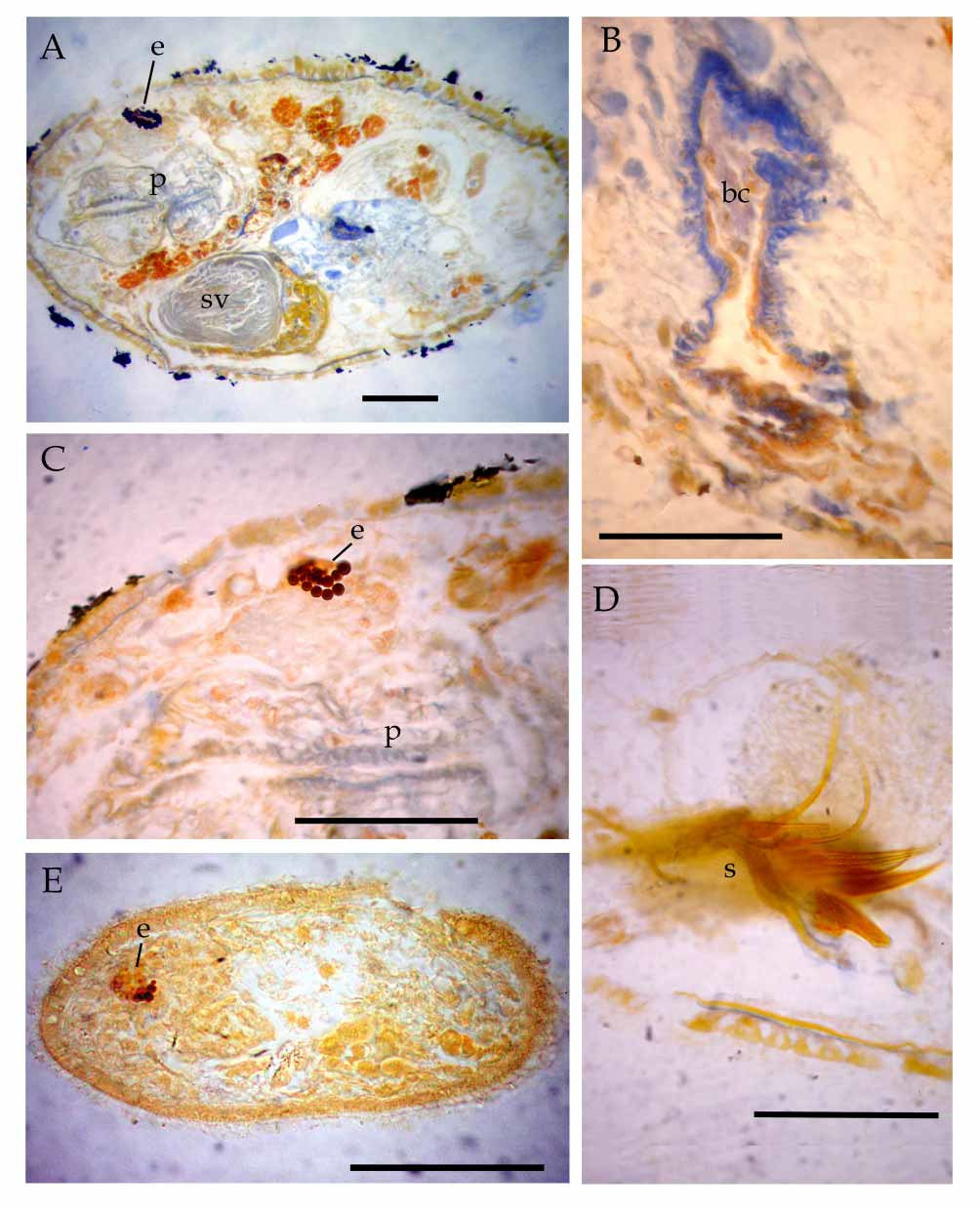
Figures 6 View FIGURE 6 , 8 View FIGURE 8 E.
Material and locality. S pecimens in vivo studied by squash method. Twelve specimens sectioned in the sagittal plane, MLP 5365, 5366, Punta Piedras (2800, 12201, 24801; 4402; 5303).
Comments. Live adult specimens were 480 µm long and 95–115 µm wide, and fixed specimens were 200 µm long. Marcus (1954) mentioned that the largest specimens were 500 µm long and 200 µm wide, which represents a slightly larger size compared to the specimens studied here.
The epithelium of the Río de la Plata specimens was 4 µm thick, with a well developed basal membrane and very numerous small rhabdites. The first two features are coincident with Marcus´description ( Marcus 1954); however, this author mentioned the absence of rhabdites in the epithelium. The parenchyma of live specimens contains symbiotic algae, greenish brown in color. Marcus (1954) mentioned that the distribution of the symbionts in the parenchyma is variable; however in the studied material the algae were concentrated on the dorsum, from the pharynx to the posterior body.The brain is large, almost the same size as the pharynx. The eyes are located in the anterior region, with large pigment granules arranged around three visual cells ( Figure 6 View FIGURE 6 C), in agreement with Marcus´description.
The mouth is subterminal, with a sphincter. The buccal tube has ciliated epithelium surrounded by muscles, expanded to form the pharyngeal bursa. The pharynx was 80 µm long and 40 µm wide in live Brazilian specimens ( Marcus 1954), while the pharynx of Argentine specimens is 44 µm long. In the same work, this author mentioned that the pharyngeal nuclei are located in the oesophagus, while the nuclei observed in the pharynx correspond to the pharyngeal glands and myoblasts. The pharynx presents a triradiate lumen, continued into the intestine. The latter bears a crown of “Minot’s gland cells” at its cephalic end.
The male reproductive system consists of two lateral testes located in posterior half of body (figure 6B); in contrast, Marcus (1954) mentioned that the testes are at pharynx level. One efferent duct issues from the caudal end of each testis, connecting with the seminal vesicle posterior to the pharynx. The ejaculatory duct crosses over the penis papilla, which is wrapped by a thin trumpetshaped sclerotic stylet. The base of the stylet is formed by two rings; it is 38 µm long and 16 µm wide at the level of these rings, and 7.5 µm wide at its expanded distal end (figure 6A). The stylet illustrated by Marcus (1954) is slighted shorter than the ones studied here (approximately 30 µm long and 12 µm wide proximally, after Marcus 1954, figure 35). The stylet reaches the male region of the atrium rostrally. The atrium opens to the exterior through a midventral gonopore, surrounded by a sphincter.
The morphology of the female reproductive system agrees with the original description ( Marcus 1954), comprising a pair of ovaries located near the caudal body, and partially covered by two dorsal vitellaria. The germinative region of the ovaries has rostroventral position, and the ovocites become mature towards the caudodorsal region. The oocytes are arranged biserially in the germination zone, and uniserially at mature stage. Two short ovovitelloducts open into the female region of atrium. The proximal atrium functions as uterus. Spermatozoids were frequently observed in both uterus and bursa; these structures are separated by a sphincter. The bursa is thin in young specimens and communicates with the intestine; this connection disappears in adults. The bursa functions as bursa resorbiens and the uterus acts as seminal receptacle ( Marcus 1954). A gland, sometimes lobulate, with eosinophilic secretions, opens on the ventrocaudal wall of the uterus.
Previously known distribution. Brazil: mangrove swamp at the estuary of Itanhaen River (50 km southwest from Santos) ( Marcus 1954). This is the first record of this species in Argentina, as well as the first citation after its original description.

| MLP |
Museo de La Plata |
No known copyright restrictions apply. See Agosti, D., Egloff, W., 2009. Taxonomic information exchange and copyright: the Plazi approach. BMC Research Notes 2009, 2:53 for further explanation.