
Oncylocotis (Oncylocotis) inexpectatus, Štys, Pavel, Ř, Petr Ba Ň A & Drescher, Jochen, 2010
|
publication ID |
https://doi.org/ 10.5281/zenodo.198828 |
|
DOI |
https://doi.org/10.5281/zenodo.6207423 |
|
persistent identifier |
https://treatment.plazi.org/id/03A487DC-FFAE-FFC6-FF36-FB49FEF4FBB4 |
|
treatment provided by |
Plazi |
|
scientific name |
Oncylocotis (Oncylocotis) inexpectatus |
| status |
sp. nov. |
Oncylocotis (Oncylocotis) inexpectatus View in CoL n. sp.
( Figs. 1–32 View FIGURES 1 – 6 View FIGURES 7 – 12 View FIGURES 13 – 20 View FIGURES 21 – 25 View FIGURES 26 – 32 )
Type material: Holotype, Ψ, ‘ Malaysia, Sabah / Poring Hot Springs, 6°02'41.78'' N, 116°42'01.97'' E / September – October 2008 / J. Drescher lgt’. The specimen is preserved in alcohol in a glass tube, together with locality label and red label: ‘ HOLOTYPE / Oncylocotis inexpectatus n. sp. / Štys, Baňař & Drescher det. 2010’. Holotype deposited in the collection of Moravian Museum (Brno). Paratypes, 14ɗ, 3Ψ, same data as holotype. Six paratypes (five males, one female) deposited in Moravian Museum (Brno), six (4 males, 2 females) in the collection of Pavel Štys (Charles University, Prague), one male in the collection of the National Museum (Prague), one male in the collection of the Oxford University Museum of Natural History (Oxford), one male in the collection of the Muséum national d’Histoire naturelle (Paris), one male in the collection of American Museum of Natural History (New York) and one male in the collection of Natural History Museum (London).Two unsexed specimens, locality data same as the holotype, deposited in University of Malaysia, Sabah ( Malaysia) by JD.
Description: Measurements (in mm); female holotype, male paratype (card-mounted specimen with right foreleg and right hindleg missing, figured in Figs. 1–3 View FIGURES 1 – 6 and 16 View FIGURES 13 – 20 , data in parentheses). Total body length —6.8 (6.3). Head (without neck). Total length—1.40 (1.29); posterior lobe, length—0.49 (0.47), posterior lobe, width—0.58 (0.58); distance of eye to apex of antennifer—0.40 (0.33); diatone (maximum width across eyes)—0.62 (0.58); dorsal synthlipsis (minimum interocular distance)—0.36 (0.33); ventral synthlipsis (minimum interocular distance)—0.31 (0.22); eye, length—0.20 (0.21); gena, length—0.22 (0.20); gena, minimum width—0.38 (0.33); interocellar distance—0.29 (0.27); minimum distance ocellus to eye—0.10 (0.10). Labium. Total length—1.20 (1.09); segment I, length—0.13 (0.11); segment II, length—0.16 (0.14); segment III, length—0.67 (0.65); segment III, maximum height—0.12 (0.11); segment IV, length—0.24 (0.22). Antenna. Segment I, length—0.24 (0.22); segment II, length—1.00 (0.98); segment III, length—0.74 (0.74); segment IV, length—0.82 (0.78), segment II, basal width—0.06 (0.06), segment II, distal width—0.11 (0.11). Pronotum. Total length (maximum)—1.18 (1.04); collum, length (median)—0.30 (0.29), maximum width—0.64 (0.64); midlobe, length (median)—0.42 (0.35), midlobe, width (maximum)—0.98 (0.94); hindlobe, length (maximum)—0.46 (0.40), hindlobe, length (mediane)—0.33 (0.31), hindlobe, width (maximum)—1.40 (1.35). Foreleg. Femur, length—1.36 (1.20), femur, maximum width—0.36 (0.28); tibia, length—1.13 (1.11), tibia, maximum width—0.36 (0.26); tarsus, length—0.29 (0.27), tarsus, maximum width—0.15 (0.12); anterior foreclaw, length—0.29 (0.26), posterior foreclaw, length—0.22 (0.20). Forewing. Maximum length—3.62 (3.36).
Coloration: Head, thorax and forewings light brown, antennae yellowish-brown, abdomen paler, (excluding pregenital and genital segments and sclerotized structures). Legs slightly bicolorous, generally yellowish-brown, distal halves of coxae, trochanters, distal parts (approximately 1/4) of all femora and basal parts (approximately 1/5) of all tibiae paler than the rest of legs (both sexes), i.e. ‘knees’ marked by pale colour.
Microsculpture: Semi-lustrous, hindlobe of pronotum distinctly rugulose, with numerous blackish granules. Rest of pronotum, head, antennae and legs finely rugulose, without conspicuous blackish granules. Ventral parts of thorax smooth.
Small setigerous tubercles present on head (dorsum of anterior lobe, all the posterior lobe - sparse, regularly distributed, minute), ventral face of fore femur (small and dense), prothoracic presupracoxale (sparse), and pronotum (midlobe - sparse on anterior and medial area: sparse, hindlobe - dense and regularly distributed all over, setae strikingly minute).
Ve s t it u re dense, setae yellowish. Dorsal and dorsolateral faces of anterior and posterior lobes of head densely covered with markedly curved setae, directed anterad. Lateral faces with semierect, slightly curved setae directed anterad. Basal third of ventral face of anterior lobe of head with long outstanding setae, directed caudad. Distal two thirds of ventral face of anterior lobe of head shorter than on basal third, directed anterad. Setae on ventral face of posterior lobe of head strikingly outstanding and long. All antennal segments densely covered with semierect setae directed towards apex; segments II–IV also with outstanding, long setae, longer than segment diameter; (II with 5 on anterior face; III with 15–18 and IV with approximately 15 on all faces). Labium covered with semierect setae directed towards apex.
Pronotum covered with dense, markedly curved setae. Setae on basal half of collum directed anterad, on the rest of pronotum directed caudad. Setae on lateral faces of pronotum and ‘proepimeral lobes’ less curved and more outstanding than on dorsum. Venter of thorax covered with fine, semierect setae directed caudad.
Setae on fore legs long, semierect, directed distad. Setae on ventral faces of trochanter, femur and tibia longer and less curved than on dorsal faces. Mid- and hindlegs uniformly covered with semierect setae.
Venter of abdomen (both sterna and ventral laterotergites) of males and females with dense, curved, ‘soft’, golden and very regular pubescence, becoming longer and provided with some outstanding long and strong setae on sides of segments 8 and 9; lateral margins of abdomen with a continuous dense fringe of setae, particularly conspicuous in females. Dorsum with sparse and short blackish pubescence, basiabdominal segments nearly bare.
Distribution of trichobothrium-like setae (long and thin, outstanding, erect to semierect): forefemur: 8–10 on ventral face; foretibia: 4 on dorsal face, 8–10 on ventral face; labium: segment II - 2 on ventral face, segment III - 6 on dorsal face and 2 on ventral face; segment IV - 4 on ventral face.
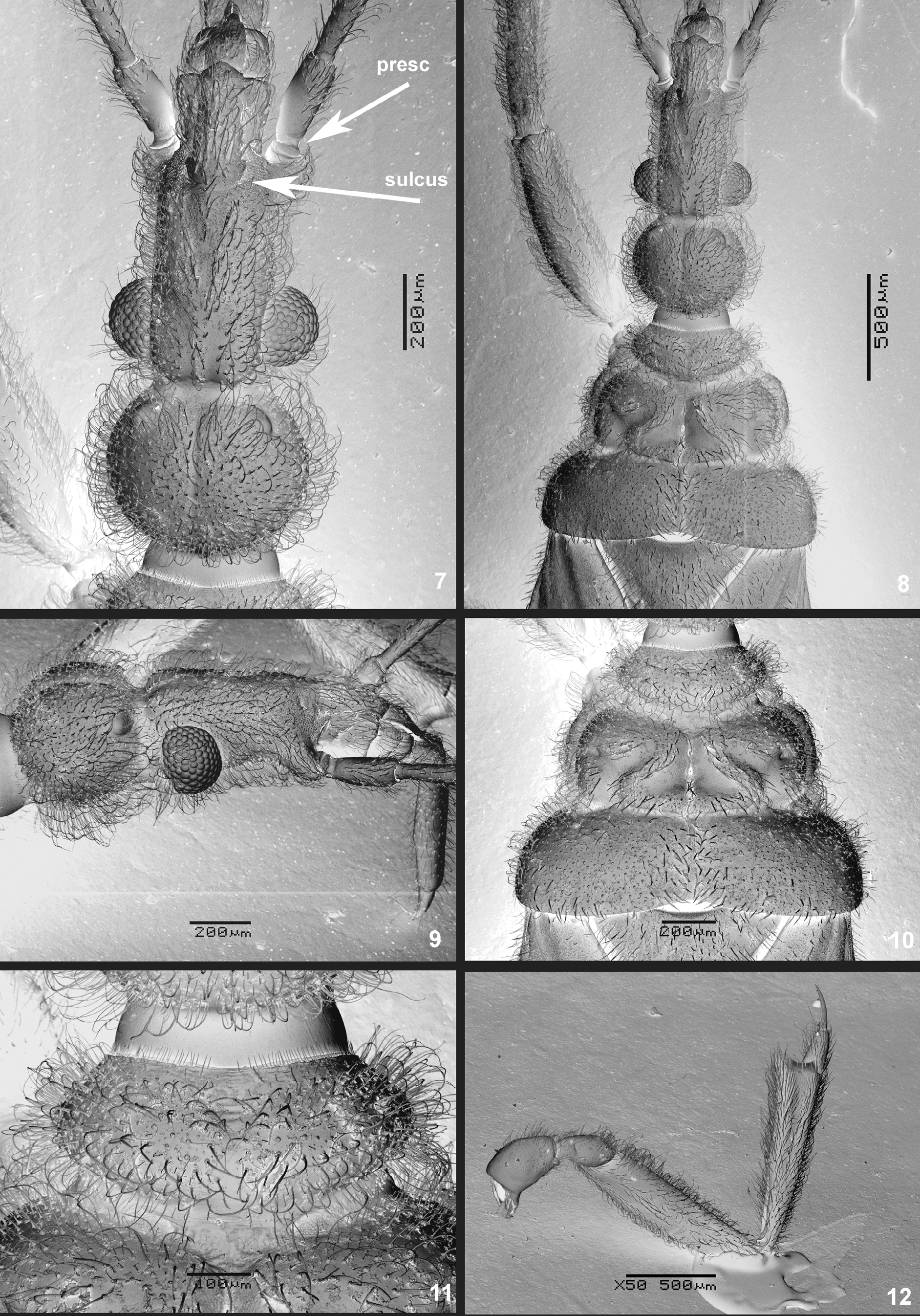
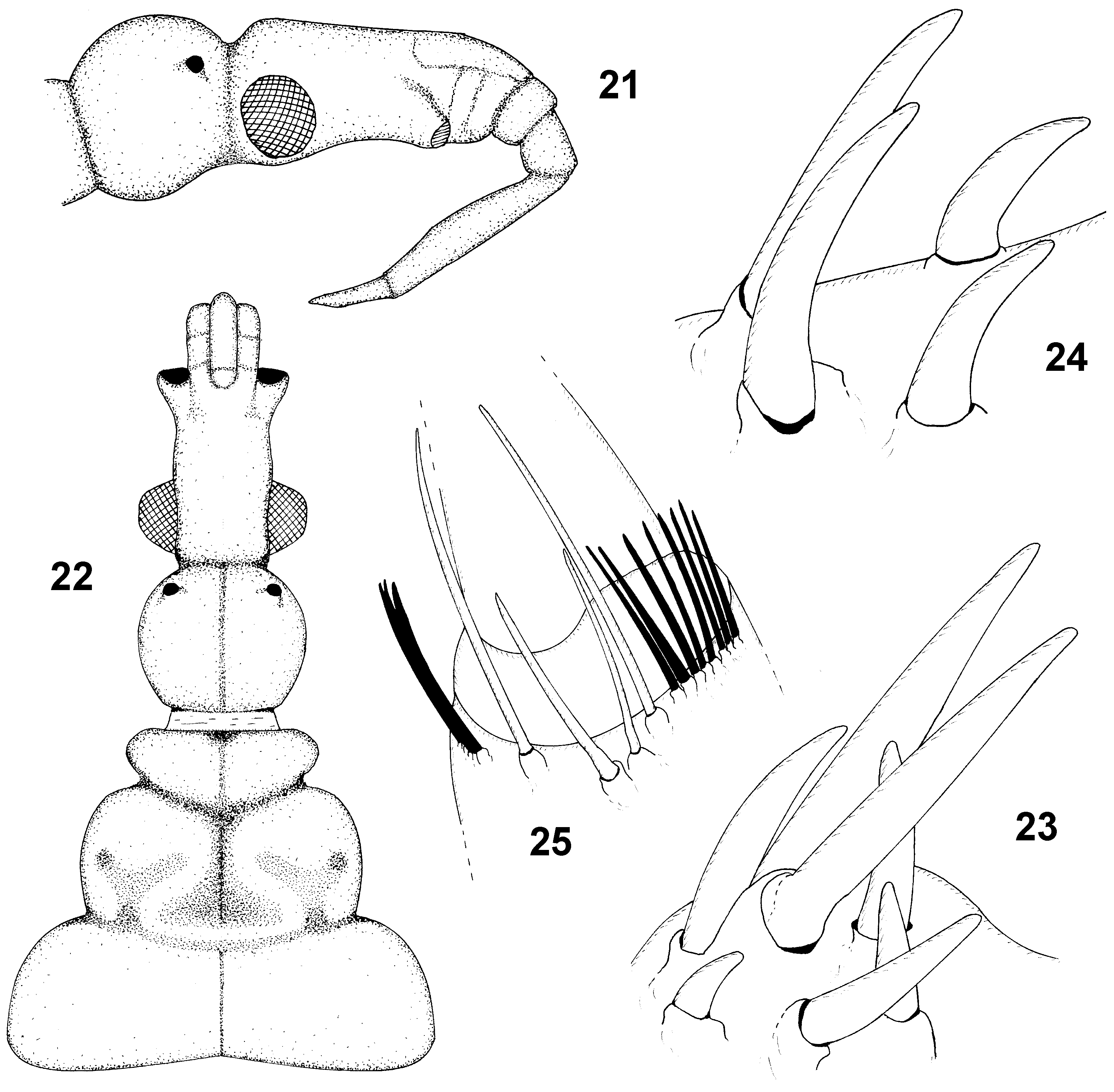
Head ( Figs. 2 View FIGURES 1 – 6 , 7–9 View FIGURES 7 – 12 , 21–22 View FIGURES 21 – 25 ) longer than pronotum, ratio 1.24 in males, 1.19 in females. Anterior lobe elongate, ratio of gena length to its minimum width 0.61 in males, 0.58 in females. Eyes medium sized, smaller in females, dorsal ocular index 2.64 in males, 2.77 in females; ventral ocular index 1.2 in males, 2.0 in females. Ratio of gena length to eye length 0.95 in males, 1.1 in females, ratio of distance eye - apex of antennifer to length of eye 1.57 in males, 2.0 in females. Postclypeus basally delimited by a transverse frontoclypeal sulcus; basis of anteclypeus marked by a pair of lateral notches. Constriction between anterior and posterior lobes deep and broad. Posterior lobe slightly transverse, ratio of its length to maximum width 0.81 in males, 0.84 in females; median with a shallow linear impression. Ocelli small, without ocellar tubercles, interocellar distance nearly 3 times as long as minimum distance ocellus to eye. Antennae longer than head and pronotum together (ratio 1.17 in males, 1.09 in females). Antennal formula (longest segment first): III:IV:II:I. Ratio of length of antennal segment II to length of antennal segment III 1.32 in males, 1.35 in females. Ratio of basal width of segment II to its distal width 0.55 in both sexes. Labial formula III:IV:II:I, labial segment III very long, ratio of length of labial segment III to length of labial segment II 4.64 in males, 4.19 in females. Ratio of length of labial segment III to its maximum height (lateral view) 5.91 in males, 5.58 in females.
Thorax: Pronotum ( Figs. 3 View FIGURES 1 – 6 , 8, 10–11 View FIGURES 7 – 12 , 22 View FIGURES 21 – 25 ). Collum ( Fig. 11 View FIGURES 7 – 12 ) robust with a broad V-shaped impression reaching from anterior margin to approximately one third of collum length, distal two thirds of its length with shallow linear impression. Precollum well developed. Constriction between collum and midlobe deep and long. Midlobe subrectangular, widest in 4/5 of its length. Ratio of midlobe maximum width to its median length 2.68 in males, 2.33 in females. Midlobe with broad and deep median impression ( Figs. 3 View FIGURES 1 – 6 , 8, 10 View FIGURES 7 – 12 , 22 View FIGURES 21 – 25 ) terminating by very broad triangular fossette, not reaching the posterior margin. Sublateral parts of midlobe with paired, deep and broad Y-shaped impressions, interrupting the posterior margin, the outer arm of each Y terminating in a conspicuous pronotal pit. Strictly lateral parts of midlobe with one isolated lateral pit. Hindlobe robust, with an inconspicuous longitudinal median ridge. Ratio of hindlobe width to its length 4.35 in males, 4.24 in females, widest in two thirds of its length. Hindlobe with universally present median ridge - low, sublinear, percurrent to disappearing shortly before reaching posterior margin. Posterior margin of hindlobe shallowly concave. Ratio of hindlobe width to midlobe width 1.44 in males, 1.43 in females. ‘Proepimeral lobes’ conspicuously exceeding midlobe in dorsal view, reaching about one third of the length of fore coxae. Eumesosternum with conspicuous impressed linear median, not reaching anterior and posterior margins. Eumetasternum with median wedge-like impression. Mesoscutellum triangular, rounded apically.
Forelegs ( Figs. 12–15 View FIGURES 7 – 12 View FIGURES 13 – 20 , 23–24 View FIGURES 21 – 25 ) slender and long, stouter in females. Femur 4.28 times as long as wide in males, 3.77 times in females, widest in the middle. Tibia 4.27 times as long as wide in males, 3.14 times in females. Apex of tibia with conspicuous process with apicitibial armature. Bristle comb on apex of tibia short, composed of 28 setae. Tarsus 2.25 times as long as wide in males, 1.93 in females. Anterior foretarsal claw longer than posterior one in both sexes. Apicitibial armature ( Figs. 13–14 View FIGURES 13 – 20 , 23 View FIGURES 21 – 25 ) consisting of seven spiniform setae, as illustrated. Tarsal armature ( Figs. 15 View FIGURES 13 – 20 , 24 View FIGURES 21 – 25 ) consisting of four spiniform setae, two basal longer, two distal short.
Mid- and hindlegs slender. Apices of tibiae with short combs of setae, only hindtibiae combs studied in detail: anterior comb consisting of eight short spiniform setae ( Fig. 25 View FIGURES 21 – 25 ), the posterior one of seven or eight such setae. Four long, spiniform setae, resembling those in apicitibial armature, present between the anterior and posterior combs on the ventralmost apex of hindtibia. Mid- and hindclaws ( Fig. 16 View FIGURES 13 – 20 ) isomorphic.
Forewings leaving the apex of abdomen exposed, reaching to basal 1/4 to 3/4 of dorsum 8 in males, to basis up to 1/2 of dorsum 7 in females. Shape, vestiture and venation as illustrated ( Figs. 4–6 View FIGURES 1 – 6 ). Macrotrichia present on all the longitudinal and transverse veins, absent from the wing membrane. Venation (for simplified notation see Štys 2002: fig. 1) as characteristic of the genus. Discal cell strikingly narrow, the both crossveins associated with the middle of the cell perpendicular to the cell and levelling mutually. One individual with the left forewing ( Fig. 5 View FIGURES 1 – 6 ) possessing an additional distal cu-an crossvein forming thus an additional closed cell between the discal cell and posterior wing margin. For the basis of clavus see Discussion.
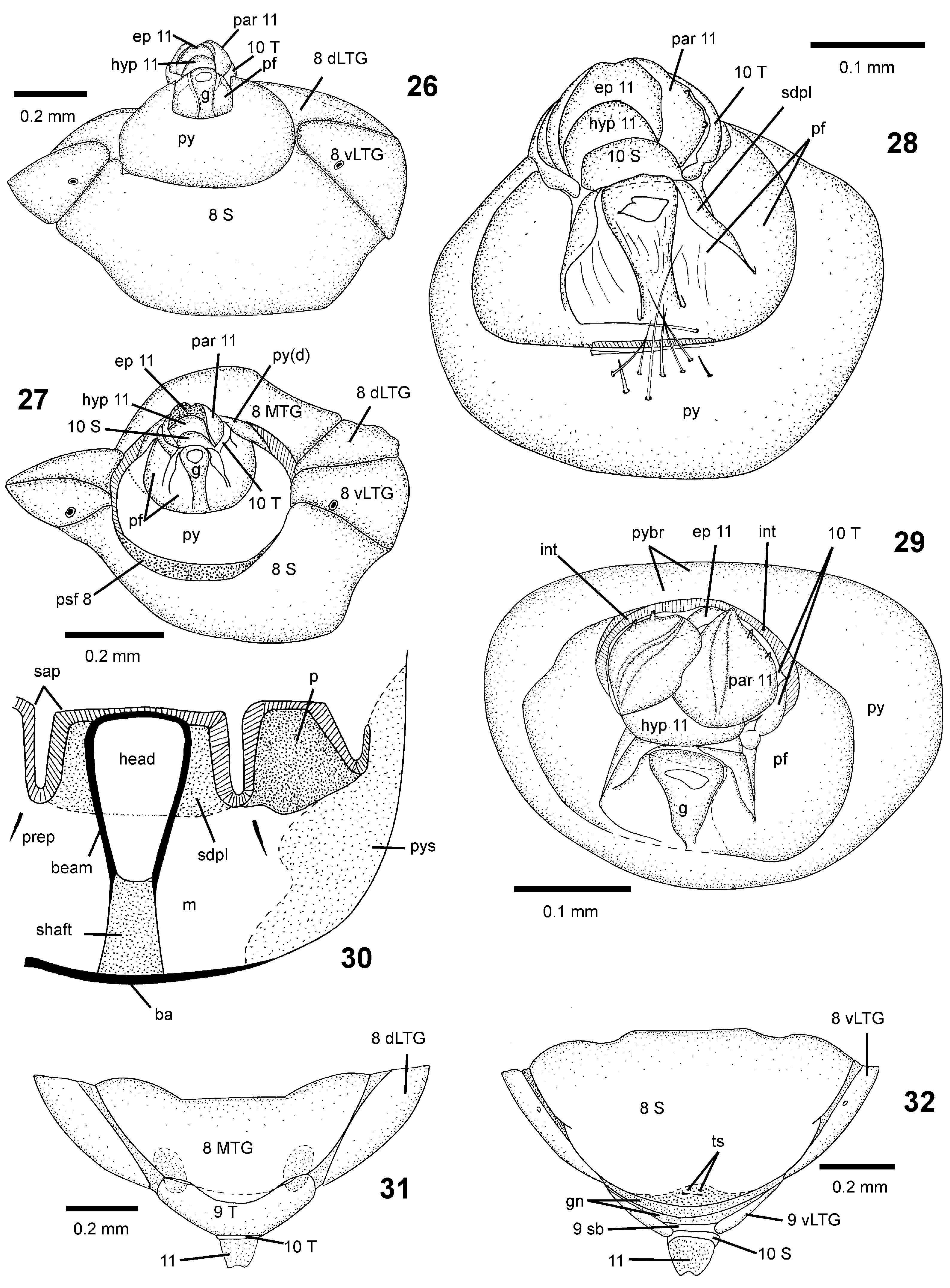
Abdominal segments 8–11 in males ( Figs. 17–20 View FIGURES 13 – 20 , 26–30 View FIGURES 26 – 32 ): Ve n t r i t e 8 represented by (a) strongly sclerotized sternum forming male subgenital plate; its anterior margin nearly straight and posterior margin moderately concave (the shape in situ strongly dependent on slightest tilting of the longitudinal axis; the posterior margin of subgenital plate may even appear convex in detached terminalia); (b) ventral laterotergites as in pregenital segments, triangular. Ventral laterotergites separated from dorsal laterotergites by non-sclerotized strips. (c) The anterior part of intersegmental membrane 8–9 semi-sclerotized, forming a narrow posterosternal fringe along the posterior margin of subgenital plate, while the infolded posterior part of this membrane allows for partial telescopation of the pygophore. Dorsum 8 with posterior margin slightly bisinuate; formed by (a) transverse mediotergite 8 (anterior margin straight, posterior vaguely delimited; paratergal lateral parts less sclerotized), (b) narrow intratergal membranes separating the mediotergite from (c) triangular and fully sclerotized dorsal laterotergites 8 with well expressed membranous connexival line.
Medio-dorsal part of the intersegmental membrane 8–9 (lateral parts not visible) protruding and forming (d) a semisclerotized posterotergal fringe 8–9 equivalent to its ventral counterpart.
Pygophore 1 (segment 9) strongly sclerotized, short, ring-shaped, transverse, basal ventral part telescoped under subgenital plate. Ventral view: anterior and posterior margins moderately concave, lateral margins distinctly concave, ventral (posterior) margin of the posterior foramen strengthened by a prominent linear basal apodeme. The paired ventral areas of posterior foramen (lateral to the guide, ventral to paramere and mesal to posterolateral margin of pygophore) closed by a membrane turning laterad to vaguely delimited pygophoral sclerites associated with posterolateral walls of the pygophore. Lateral view: anterior and posterior margins of the pygophore parallel-sided, straight. Dorsal view: anterior margin deeply emarginate, bisinuate, strengthened by marginal apodeme; medial sector convex; pygophoral bridge broad, its posterior margin deeply concave, somewhat diffuse.
1. It must be stressed that some aspects of the SEM photographs ( Figs. 17–20 View FIGURES 13 – 20 ) of male terminalia (as well as derived interpretative drawings, Figs. 26–29 View FIGURES 26 – 32 ) are misleading. They cannot distinguish between sclerites and membranes, and, consequently, the shapes and sizes of genital structures situated within the posterior foramen (supradistal plate, parameres and pygophoral sclerites; frame of the guide) are simplified, distorted and their shapes and mutual relationships are inaccurate. For the sake of clarity, the description is therefore supplemented by a hand-drawn scheme ( Fig. 30 View FIGURES 26 – 32 ) based on cleared detached terminalia in alcohol.
Guide directed posterodorsad, racquet-shaped, with strongly sclerotized parallel-sided shaft and much broader, sharply defined subtrapezoid head with a rounded apex exceeding the supradistal plate in ventral view; the beam thin-walled. Supradistal plate represented by a sclerotized arcuate arch situated anterodorsal to the guide frame and mesad to plate-shaped parameres; lateral sectors of the arch apodeme-like. Each paramere inversely U-shaped, mesal and dorsal sectors narrow, strongly sclerotized and apodeme-like, lateral sector broad; the mesal part close to a needle-like, diagonal, more proximal preparameral apodeme. (Basis of the lateral sector of the supradistal arch merging with the mesal basis of the paramere. When superficially examined, illusory existence of a pair of isolated loop-like sclerites situated laterad to frame of the guide might be assumed).
Segment 10 with a short, semi-ring-shaped, little sclerotized, inversely U-shaped tergum situated behind the pygophoral bridge and laterad to parandria; the plate-shaped sternum only visible in strictly posterior view, provided with macrotrichia, and situated between the supradistal plate and hypandrium. Segment 11 long, prominent, protruding out of segment 10 (in alcohol-preserved individuals), formed by four semisclerotized flaps, namely a small dorsal epandrium with an emarginate apex, large ventral hypandrium and a pair of large lateral parandria; the latter provided with a percurrent keel and a pair of lateral finger-shaped tubercles each.
Abdominal segments 8–11 in females ( Figs. 31–32 View FIGURES 26 – 32 ): Sternum 8 strongly sclerotized, forming female subgenital plate; its anterior margin, irregularly straight with lateral sectors receding distad; lateral margins moderately converging distad, in their mid-lengths distinctly notched; posterior margin convex, its most caudal point provided with a minute, transverse, black terminal structure. (This structure as well as a complex, narrow, desclerotized genital area between the subgenital plate and ventral bridge 9 to be studied later.) Ventral laterotergites 8 triangular, strongly sclerotized, separated from dorsal laterotergites by a sharp connexival line. Dorsal laterotergites 8 sclerotized excepting narrow lateral strips, their anterior regions separated from mediotergite 8 by a sulcus, posterior regions fused with the mediotergite 8. The latter sclerotized except for a pair of posterolateral irregular spots; anterior margin with a broad and short rectangular excision, posterior margin straight, nearly adjoining tergum 9. Both ventral and dorsal laterotergites 8 distally pointed, exceeding subgenital plate and tergum 8, respectively. Tergum 9 shaped as a short half-circle, posterior margin excised to accommodate segment 10, lateral regions less sclerotized (in series with dorsal laterotergites of preceding segments). Venter 9 represented by mutually approached laterotergites 9 connected by and fused with a narrow, arcuate medial sternal bridge 9; posterior margin of the latter with a rounded excision filled up by a ring-shaped, short segment 10 out of which the segment 11 is protruding (architecture of the latter same as in M, both 10 and 11 visible in dorsal and ventral views).
Abdominal spiracles: Spiracles 1 large, nearly dorsally situated on ventral (lateral) side of laterotergite 1; spiracles 2 on ventral margins of ventral laterotergites 2, in more anterior position than spiracles on the more posterior segments. Spiracles 3–7 minute, situated submarginally in the middle of ventral sides of the respective laterotergites (spiracles larger and their rims more sclerotized in females). Spiracles 8 larger than the preceding ones, located at ventral margins of ventral laterotergites 8 (males) or close to these margins (females).
Etymology: inexpectatus , lat. = unexpected.
Comparative notes: The fauna of enicocephalomorphans of Borneo is poorly known, but rich in undescribed genera and species. All Oncylocotis species from Borneo that we have seen are undescribed endemics. Oncylocotis inexpectatus n. sp. differs from all the other undescribed Oncylocotis species from Borneo by its unusually long and slender labial segment III and numerous blackish granules on hindlobe of pronotum. O. inexpectatus n. sp. also differs from all the described Oriental species ( Jeannel 1942); it is similar to O. lombocensis (Breddin, 1899) , but the posterior lobe of the head of the latter is not transverse. In Villiers´s (1969) key to Afrotropical and Madagascan species (taken into account as well owing to uncertain geographical origin of the host ant species), O. inexpectatus would run into the group with unicolorous legs and forewings, slender forelegs, transverse posterior lobe of head, and would key under Oncylocotis usingeri (Villiers, 1962) from RSA (Cape Province), a species with remarkably different forewing venation (cf. Villiers 1969: Fig. 101).
Ethology and field ecology of Oncylocotis inexpectatus n. sp.: Oncylocotis inexpectatus n. sp. was comparatively rarely seen during our (JD) sampling. In total twenty adult individuals (mentioned under Material, 14 M, 4 F, 2 unsexed) were found after repetitive sampling from two A. gracilipes supercolonies (ca. 100.000 ants sampled). O. inexpectatus was never observed to be attacked by A. gracilipes and it was able to live in a laboratory colony for at least two months (JD pers. obs). All O. inexpectatus specimens were found within the bamboo and drainage pipes that A. gracilipes had moved into; consequently, we assume that O. inexpectatus n. sp. also occurs within the underground nests of Anoplolepis gracilipes . The two colonies that contained O. inexpectatus n. sp. were in a fruit plantation of rambutan, mango and durian, and an ethnobotanical garden with various herbs, banana and some larger trees, respectively.
The insect species found with Anoplolepis gracilipes in Sabah: During the survey of A. gracilipes in Sabah several guest insect species were found (JD):
(a) Myrmecophilus leei Kistner & Chong 2007 ( Kistner et al. 2007) ( Orthoptera : Gryllomorpha: Myrmecophilidae ); originally described from Malaysia: Penang as associated with A. gracilipes . The Myrmecophilidae include kleptoparasitic inquilines of ants inducing their hosts to feed them by regurgitated liquid food.
(b) Oncylocotis inexpectatus n. sp. ( Heteroptera : Enicocephalidae )
(c) Oncylocotis n. sp. ("Sabah 2") ( Heteroptera : Enicocephalidae )—one male. Collected with O. inexpectatus n. sp., much larger, with strikingly bicolorous legs. To be described in a revision of Oncylocotis of Sabah (Baňař & Štys, in prep.).
(d) Trochoideus desjardinsi Guérin-Meneville, 1838 ( Coleoptera : Endomychidae )—the ‘handsome fungus beetle’ a mycetophagous species normally found in termite nests as other species of the genus ( Skelley & Burgess 1995).
(e) Aphanocephalus sp., two species ( Coleoptera : Discolomatidae )—‘tropical log beetles’ feeding on fruit bodies of fungi.


No known copyright restrictions apply. See Agosti, D., Egloff, W., 2009. Taxonomic information exchange and copyright: the Plazi approach. BMC Research Notes 2009, 2:53 for further explanation.