
Pandalopsis spinosior Hanamura, Kohno & Sakaji, 2000
|
publication ID |
https://doi.org/ 10.11646/zootaxa.4040.2.6 |
|
publication LSID |
lsid:zoobank.org:pub:15FCF489-080E-40FD-9840-21201588C312 |
|
DOI |
https://doi.org/10.5281/zenodo.5696416 |
|
persistent identifier |
https://treatment.plazi.org/id/03C8056E-FF96-9606-FF00-5703FC686F33 |
|
treatment provided by |
Plazi |
|
scientific name |
Pandalopsis spinosior Hanamura, Kohno & Sakaji, 2000 |
| status |
|
Pandalopsis spinosior Hanamura, Kohno & Sakaji, 2000 View in CoL
[New Japanese name: Rausu-budou-ebi]
( Figs. 1–3 View FIGURE 1 View FIGURE 2 View FIGURE 3 , 4A–C View FIGURE 4. A – C , 5A View FIGURE 5. A )
Pandalopsis spinosior Hanamura, Kohno & Sakaji, 2000: 27 View in CoL , figs. 1–4. [type locality: Urup (or Etorofu) Strait, South Kurile Islands, about 400 m]. –– De Grave & Fransen 2011: 445.
Material examined. Off Rausu, Shiretoko Peninsula, Hokkaido, Sea of Okhotsk, ca. 400 m, June 2003, coll. Yoji Matsuzawa, 2 ovigerous female (cl 43.4, 47.1 mm), CBM-ZC 7483; off Rausu, 500–800 m, reared in aquarium, 2 males (cl 30.6, 32.4 mm), 1 female (cl 40.0 mm), 1 ovigerous female (cl 42.6 mm), AMF-ZC00003. Off Iturup (Etorofu) Island, South Kurile Islands, 400–450 m, May 2007, trawl, 1 female (cl 42.0 mm), 7 ovigerous females (cl 38.0– 41.8 mm), CBM-ZC 9407.
Description. Body ( Fig. 1 View FIGURE 1 ) moderately robust; integument glabrous.
Rostrum ( Fig. 2 View FIGURE 2 A, B) distinctly overreaching distal margin of antennal scale, 1.1–1.3 times as long as carapace; dorsal margin armed with 13–15 movable spines, including 6 or 7 on rostrum proper and 6–8 on carapace posterior to orbital margin, and 1 or 2 small fixed teeth near apex of rostrum, subdistal half leaving unarmed, posteriormost (first) spine arising at about midlength of carapace (0.48–0.49 of carapace length); ventral margin armed with 8–11 fixed teeth over entire length, posteriormost tooth slightly smaller than preceding one; lateral carina well delimited throughout entire length.
Carapace ( Fig. 2 View FIGURE 2 A) with postrostral carina moderately high, but not crested, extending to posterior two-fifths, dorsal margin gently convex in lateral view; antennal tooth moderately strong; branchiostegal tooth small; pterygostomial margin broadly rounded; no conspicuous ridges or carinae on lateral surface.
Pleon ( Fig. 2 View FIGURE 2 C) dorsally smooth. Third pleomere without middorsal protuberance or posterodorsal median tooth. Fourth and fifth pleura each with sharp posteroventral tooth. Sixth pleomere about 0.5–0.6 times as long as carapace and 2.0–2.4 times longer than high; posterolateral process terminating in acute tooth.
Telson ( Fig. 2 View FIGURE 2 F) 1.4–1.6 times as long as sixth pleomere, armed with 7–9 dorsolateral spines on either side; posterior margin terminating in small blunt triangular projection, armed with 3 pairs of unequal spines. Eye ( Fig. 2 View FIGURE 2 D) broadly subpyriform, maximal diameter of cornea 0.2 of carapace length; ocellar sinous present, but ocellus absent.
Antennular peduncle ( Fig. 2 View FIGURE 2 D) reaching midlength of antennal scale. First segment 1.1–1.3 times as long as length of second and third segments combined; stylocerite rounded. Second segment with few spinules on dorsodistal margin. Aesthetasc-bearing portion of outer flagellum distinctly shorter than carapace. Antenna with stout basicerite bearing moderately strong ventrolateral tooth. Carpocerite moderately stout, not reaching midlength of antennal scale. Antennal scale 0.7–0.8 times as long as carapace and 3.3–3.6 times longer than broad, lateral margin nearly straight; distolateral tooth ( Fig. 2 View FIGURE 2 E) slightly reaching beyond distal margin of rounded lamella.
Third maxilliped ( Fig. 3 View FIGURE 3 A) not reaching distal margin of antennal scale. Ultimate segment subequal in length to penultimate segment (= carpus), terminating in small corneous spine, with numerous tufts of short transverse or obliquely transverse rows of long stiff setae on dorsal to lateral surfaces; terminus with some spinules proximal to base of apical spine ( Fig. 3 View FIGURE 3 B); mesial face with numerous transverse tracts of stiff setae. Carpus also with numerous tufts of stiff setae on lateral surface and transverse tracts of dense stiff setae on mesial surface. Antepenultimate segment (merus-ischium-basis fused segment) longer than distal 2 segments combined, with small rounded tubercle basally on dorsal surface; dorsal surface elevated at midlength; ventral margin forming blunt edge fringed with row of numerous setae; exopod absent.
First pereopod ( Fig. 3 View FIGURE 3 C) minutely chelate, reaching midlength of antennal scale. Propodus about 0.8–0.9 times as long as carpus, tapering distally, with field of numerous stiff setae, arranged in transverse or obliquely tracts, in proximal three-fourths of mesial surface. Ischium ventrally with broad laminar expansion, fringed with row of numerous short setae.
Second pereopods ( Fig. 3 View FIGURE 3 D) subequal, extending beyond antennal scale by length of fingers to length chela. Chela small, dactylus slightly shorter than palm. Carpus divided in 14–19 articles. Merus without annulation. Ischium with ventral margin slightly expanded in proximal half to accommodate chela.
Third to fifth pereopods generally similar, decreasing in length posteriorly. Third pereopod ( Fig. 3 View FIGURE 3 E) overreaching antennal scale by length of dactylus and slightly more than distal one-fourth of propodus; dactylus ( Figs 3 View FIGURE 3 F; 4A) 0.22–0.26 times as long as propodus, slightly curved, terminating in acute unguis not clearly demarcated basally, flexor margin with 5–7 accessory spinules in proximal 0.5–0.7, becoming longer and more widely spaced distally, and 1 subterminal slender spinule appressed to unguis; propodus becoming slightly wider distally, dorsal surface with tufts of short stiff setae (setae becoming longer distally), lateral surface with tufts of short setae and scattered spinules particularly numerous on ventral half, mesial surface ( Fig. 4B View FIGURE 4. A – C ) with longitudinal row of stiff setae and scattered spinules on dorsal half and single longitudinal row of longer spinules and covering of minute spinules on ventral half and midline unarmed, ventral (flexor) surface with slender spinules arranged in 3 irregular longitudinal rows; carpus 0.5–0.6 times as long as propodus, lateral surface with sparse tufts of short setae and some spinules dorsally and 2 or 3 spines ventrally, mesial surface with dense covering of minute spinules mixed with few tufts of short setae ( Fig. 4C View FIGURE 4. A – C ); merus 0.6–0.9 times as long as carapace, armed with 7–11 lateral and 6–8 ventral spines; ischium with 1 ventrolateral spine. Fourth pereopod ( Fig. 3 View FIGURE 3 G, H) overreaching antennal scale by length of dactylus; dactylus ( Fig. 3 View FIGURE 3 I) armed with 5–8 spinules in proximal 0.5–0.7 of flexor margin in addition to subapical spinule; carpus 0.5 times as long as propodus ( Fig. 3 View FIGURE 3 H), with 2–4 lateral spine; merus 0.6–0.8 times as long as carapace, with 7–10 lateral and 5–8 ventral spines; ischium with 1 ventral spine. Fifth pereopod ( Fig. 3 View FIGURE 3 J) reaching as far as distal margin of antennal scale; dactylus ( Fig. 3 View FIGURE 3 K, L) 0.14–0.18 times as long as propodus, flexor surface forming narrow facet, armed with 5–9 spinules in proximal 0.7 of flexor margin, and subapical spinule; propodus becoming wider distally, bearing numerous tufts of short stiff setae (becoming more dense distally and forming grooming apparatus), mesial surface with single row of widely spaced slender spinules and sparse tufts of short setae, ventral (flexor) surface with slender, widely spaced spinules arranged in 3 rows, distal margin with 3 strong spinules reaching about midlength of dactylus; carpus about 0.4–0.6 times as long as propodus, with 2 or 3 lateral spines, and with sparse tufts of short setae on dorsal margin, mesial face nearly glabrous; merus 0.6–0.7 times as long as carapace, with 6–8 lateral and 1–4 ventral spines; ischium with 1 ventral spine.
Endopod of first pleopod in male ( Fig. 2 View FIGURE 2 G) bilobed distally, distomesial lobule (appendix interna) long and narrow. Male second pleopod with appendix masculina ( Fig. 2 View FIGURE 2 H) as long as appendix interna, distal margin with row of numerous spiniform setae. First and second pleopods in females as described in Hanamura et al. (2000).
Uropod with rami subequal in length, reaching nearly tip of telson.
Eggs large, 3.7–4.6× 2.9–3.4 mm (eyed stage).
Colour in life. Carapace and pleon mottled with reddish violet; rostrum with white ring subterminally. Antennular and antennal flagella alternated with reddish violet and white. Appendages generally red, third to fifth pereopods sometimes whitish. Eggs yellowish in eyed stage. See Fig. 5 View FIGURE 5. A .
Distribution. Urup Strait to Nemuro Strait, South Kurile Islands, 400– 800 m.
Remarks. In their original description, Hanamura et al. (2000) compared Pandalopsis spinosior only with P. miyakei . This was based primarily on the similarity in the armature of the dactyli of the third and fourth pereopods. During this study, we found that P. spinosior is also strikingly similar to P. coccinata in many morphological aspects and particularly in the live colouration. The extension of the row of accessory spinules on the dactyli of the third and fourth pereopods is more variable than described by Hanamura et al. (2000). There are no clear differences in the colouration between the two species (see Fig. 6), although colouration has been employed in differentiating similar species in pandalids (e.g., Chan & Yu 1991; Chan & Crosnier 1991, 1997; Komai 1999; Chan 2004). Actually, the shrimp fished at Rausu has been believed to represent P. coccinata .
Differentiating characters between P. spinosior and P. coccinata are summarized in Table 1 View TABLE 1 . Pandalopsis spinosior is distinguished from P. c o cc i na t a by the proportionally shorter and stouter dactylus of the third pereopod with accessory spinules distributed on the proximal 0.5–0.7 length of the flexor margin ( Fig. 4A View FIGURE 4. A – C ). In P. coccinata , minute accessory spinules are restricted to the proximal 0.3–0.4 of the flexor margin ( Fig. 4 View FIGURE 4. A – C D). Furthermore, the setation and armature of the propodus and carpus of the third pereopod (and also those of the fourth pereopod, but the third pereopod is used as a representative) are different between the two species, although little attention has been paid for these characters in previous taxonomic literature on Pandalopsis (e.g., Komai 1994; Hanamura 2000). In P. spinosior , the propodus of the third pereopod bears numerous tufts of short stiff setae on the lateral surfaces [ Hanamura et al. (2000) did illustrate the setation but did not properly describe it] and numerous scattered minute spinules on the mesial surface ( Fig. 3 View FIGURE 3 F, H; Fig. 4B View FIGURE 4. A – C ), but these setation and armature are much fewer in P. coccinata ( Fig. 4 View FIGURE 4. A – C E). Similar difference is also seen in the mesial armature of the carpus. In P. spinosior , the mesial face is covered with minute scattered spinules ( Fig. 4C View FIGURE 4. A – C ), but these spinules are fewer and much reduced in the size in P. coccinata ( Fig. 4 View FIGURE 4. A – C F). The third to fifth pereopods are proportionally shorter in P. s p i no s i o r than in P. coccinata . For example, the merus of the third pereopod is 0.6–0.9 times as long as the carapace in P. s p i no s i o r, rather than 0.9–1.0 times as long in P. coccinata . The telson can have more numerous dorsolateral spines in P. spinosior than in P. coccinata (seven to nine pairs versus five to seven pairs), although the variation ranges partially overlap.
Distinction between P. spinosior and P. zarenkovi has remained unclear, because Ivanov & Sokolov (2001) did not compare their new species with P. spinosior . In fact, these two species are similar to one another in many diagnostic aspects. Hayashi (2007) suggested that the two taxa might be conspecific. Nevertheless, P. z a re nk o v i seems to differ from P. s p i no s i o r in the relatively longer rostrum (1.4–1.5 times as long as the carapace versus 1.1– 1.3 times as long) and the more strongly produced dorsodistal tooth of the antennal scale. Furthermore, the figures given by Ivanov & Sokolov (2001: Figs. 1 View FIGURE 1 a, 3c) show sparse setation on the propodi of the third to fifth pereopods, like in P. coccinata . As described above, P. spinosior exhibits a characteristic setation of the propodi of the third to fifth pereopods. Furthermore, live colouration seems to be also different between P. z a re nk o v i and P. spinosior . Ivanov & Sokolov (2001: 164) stated “Pink with violet tint on upper side of cephalothorax and abdomen; rostrum with white band subapically; anterior 5 abdominal somites with obscure white bands”. On the other hand, in P. spinosior , the body is mottled violet. Consequently, we maintain P. zarenvkovi as a valid species for the time being.
Komai (1994) briefly reviewed species of Pandalopsis with an identification key to 14 species known at the time. Since Komai (1994), four new species, including P. spinosior , have been described in Pandalopsis ( Jensen 1998; Hanamura et al. 2000; Ivanov & Sokolov 2000; Komai & Takeda 2002). Hayashi (2007, 2008) reported on nine species of the genus known from Japan: P. coccinata , P. gibba Komai & Takeda, 2004 , P. glabra Kobjakova, 1936 , P. japonica Balss, 1914 , P. longipes Komai, 1994 , P. miyakei , P. ochotensis Kobjakova, 1936 , P. pacifica Doflein, 1902 and P. r u br a Komai, 1994. In addition to these nine species, Komai (1994) recorded P. longirostris Rathbun, 1902 from off eastern Hokkaido, although the identification remains provisional. This study confirms the presence of P. spinosior in Japanese waters to be a 11th of the genus from the country. The occurrence of P. spinosior is quite scarce (presently known only from the Urup Strait in the Kurile Islands and the Nemuro Strait), although the species represents a commercially important fishery recourse in eastern Hokkaido. It is likely that P. spinosior prefers steep slopes or rather uneven sea bottoms where trawl operation is not easy. On the other hand, P. coccinata , also a representative of commercially important shrimp species in northern Japan, inhabits flat muddy bottoms ( Komai 1994; Komai & Komatsu 2009) and is commercially fished by off shore trawlers in Hokkaido through Chiba Prefecture ( Urita 1941; T. Komai, unpublished data).


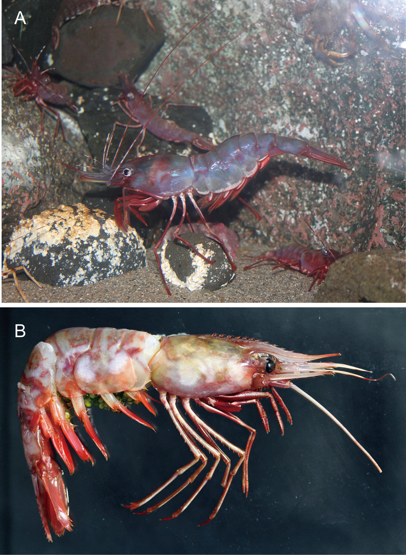
TABLE 1. Summary of differentiating characters among Pandalopsis spinosior Hanamura, Kohno & Sakaji, 2000, P. coccinata Urita, 1941 and P. zarencov i Ivanov & Sokolov, 2001. Average values, if available, are given in parentheses.
| Characters/species | P. spinosior (n = 13; cl 30.6–47.1 mm) | P. coccinata (n 10; cl 33.1–40.1 mm) | P. zarenkovi Ivanov & Sokolov (2001) |
|---|---|---|---|
| Rostrum | |||
| Rostrum length/cl | 1.1–1.3 (1.2; n = 8) | 1.3-1.5 (1.4) | 1.41–1.50 |
| No. of postrostral spines | 6 7 7 | 5–7 6 | 8 |
| No. of ventral teeth | 8 11 10; n = 8 | 9–12 10 | 10–14 |
| Telson | |||
| No. of dorsolateral spines | 7 9 on either side 8 | 5–7 on either side 7 | 8 on either side |
| Antennal scale | |||
| Distolateral tooth | slightly reaching beyond lamella | slightly reaching beyond lamella | distinctly overeraching lamella |
| Third pereopod | |||
| Flexor spinules of dactylus | in proximal 0.5–0.8 | in proximal 0.3–0.4 | in proximal 0.7–0.8 |
| Dactylus length/propodus length | 0.22–0.26 0.24 | 0.31–0.36 0.32 | 0.2 |
No known copyright restrictions apply. See Agosti, D., Egloff, W., 2009. Taxonomic information exchange and copyright: the Plazi approach. BMC Research Notes 2009, 2:53 for further explanation.
|
Kingdom |
|
|
Phylum |
|
|
Class |
|
|
Order |
|
|
Family |
|
|
Genus |
Pandalopsis spinosior Hanamura, Kohno & Sakaji, 2000
| Hibino, Mai, Matsuzaki, Koji & Komai, Tomoyuki 2015 |
Pandalopsis spinosior
| De 2011: 445 |
| Hanamura 2000: 27 |