
Parapharyngodon thulini, Rahimian, Hassan, Pazoki, Samaneh & Habashi, Sima Abbasi, 2014
|
publication ID |
https://doi.org/ 10.11646/zootaxa.3852.1.2 |
|
publication LSID |
lsid:zoobank.org:pub:CE7E8E7A-073D-442A-B1D8-4CD661B59205 |
|
DOI |
https://doi.org/10.5281/zenodo.6129708 |
|
persistent identifier |
https://treatment.plazi.org/id/03AB505F-785F-A374-1FC9-FF37A9E300BC |
|
treatment provided by |
Plazi |
|
scientific name |
Parapharyngodon thulini |
| status |
sp. nov. |
Parapharyngodon thulini sp. nov.
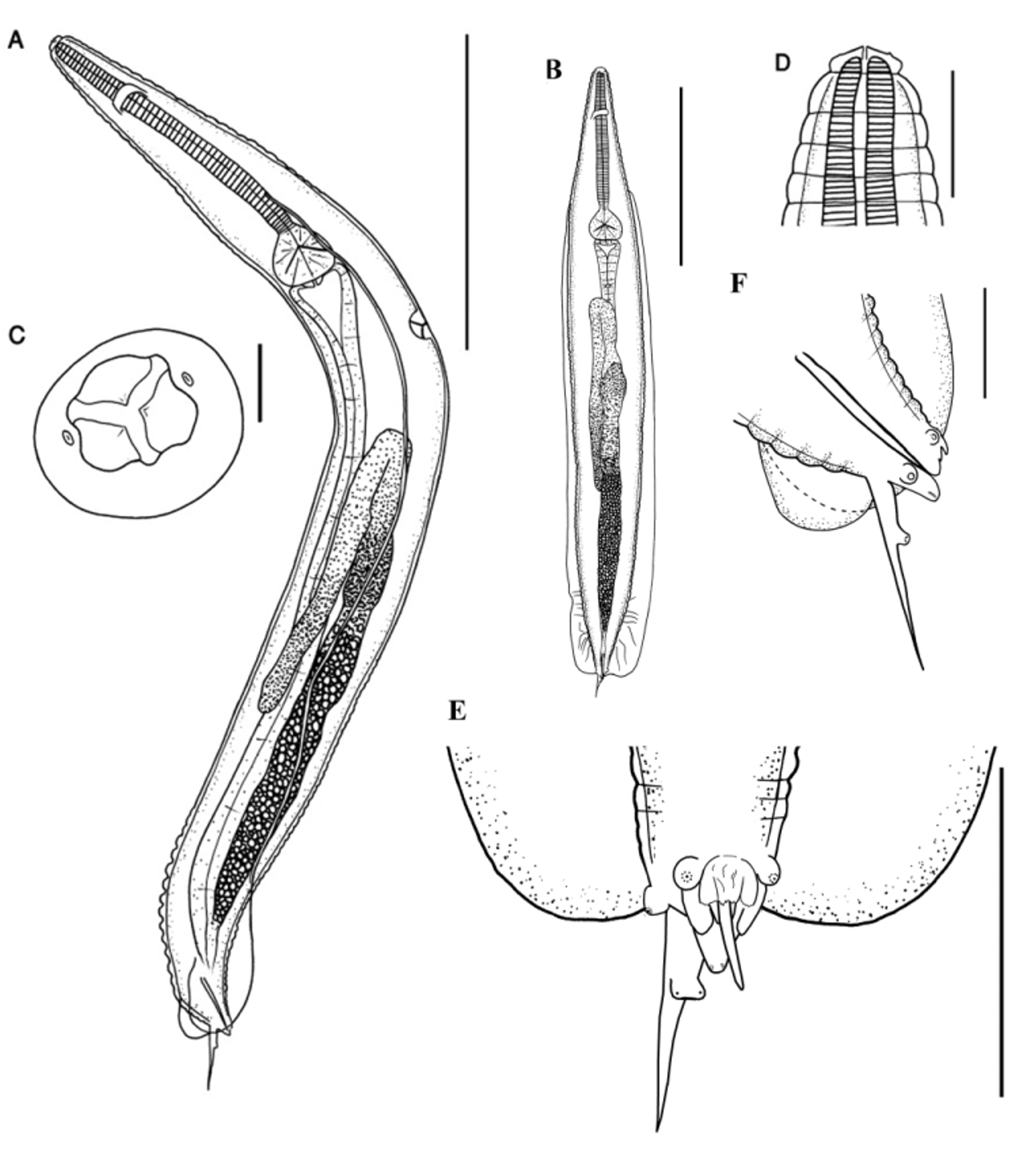
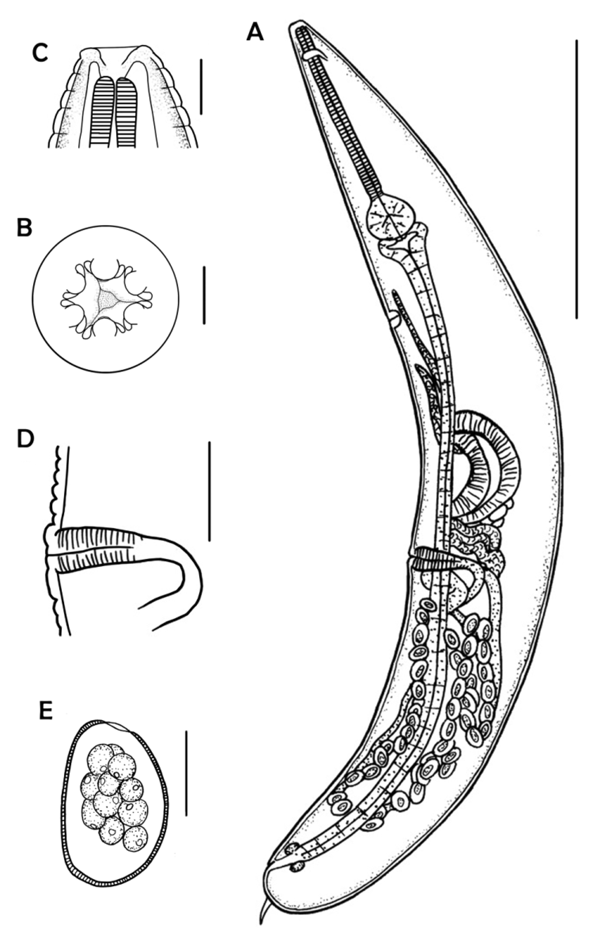
Figs. 2–4 View FIGURE 2 View FIGURE 3 View FIGURE 4
General: Very robust, white nematodes, with prominent annulations from cephalic extremity to posterior end. Excretory pore posterior to bulb.
Male (based on holotype and 9 paratypes; mean ± SD [range]): Body 2.17 ± 0.42 mm (1.87–3.31 mm) long, 225 ± 30 (178–271) wide at level of excretory pore. Head well marked-off from body proper; cephalic extremity with three cuticular membranes; two amphids immediately next to the mouth opening ( Fig. 4 View FIGURE 4 A). Cuticular annulations approximately 15 µm wide, more prominent in both anterior and posterior regions of body. Oesophageal corpus 504 ± 61 (371–578) long; isthmus 26 ± 5 (19–35); bulb 91 ± 6 (83–103) long, 105 ± 10 (85–117) wide. Nerve ring and excretory pore 144 ± 15 (124–178) and 776 ± 78 (654–865) from anterior end, respectively. Lateral alae originate 473 ± 42 (395–529) from anterior end, at level of isthmus, rounded at level of genital cone, terminate approximately 96 ± 27 (56–144) from tip of genital cone, narrow anteriorly, enlarged to form broad vanes in cloacal region; maximum width 106 ± 22 (65–133). Three pairs of caudal papillae: precloacal ( Fig. 4 View FIGURE 4 D), postcloacal ( Fig. 4 View FIGURE 4 C) and the last pair fusing to form common base with two prominent sessile organs at tip; 47 ± 8 (38–59) from tip of tail filament ( Fig. 4 View FIGURE 4 B). Two subsurface structures at tip of well-developed genital cone ( Fig. 4 View FIGURE 4 E). Caudal alae absent. Anterior cloacal lip well developed, extends between 2 precloacal papillae, comprising 2 symmetrical pairs of cuticular lips in tandem covering genital cone anteriorly; anterior pair overlies base of posterior pair; tip of anterior pair serrate; posterior pair smooth; central, cuticular and serrate process located anterior to bilobed posterior cloacal lip. Posterior cloacal lip, opening to spicular pouch, bilobed. Spicule well sclerotized, 89 ± 9 (71–101) long, straight, pointed. Tail filament (caudal appendage) 81 ± 9 (68–94) long.
Female (based on allotype and 9 paratypes; mean ± SD [range]): Body 4.01 ± 0.43 mm (3.53–4.54 mm) long, 469 ± 114 (325–617) wide at level of vulva. Cephalic extremity flattened in lateral view; mouthsubtriangular surrounded by 6 prominent spherical processes, with circular ridge around periphery of each ( Fig. 4 View FIGURE 4 F, G). Cuticle with annulations approximately 27 µm wide. Oesophageal corpus 791 ± 118 (546–920); isthmus 40 ± 5 (33–48); bulb 152 ± 15 (130–179) long, 185 ± 23 (160–229) wide. Nerve ring, excretory pore, and vulva 150 ± 25 (96–186), 1.22 ± 0.12 mm (1.06–1.36 mm), and 2.21 ± 0.23 mm (1.87–2.52 mm) from anterior end of body, respectively. Ovaries reach level of oesophageal bulb, do not form coils around oesophagus. Vulva in middle of body; pre- and postvulvar cuticle protrudes to varying degrees in different specimens. Body terminates in tail spike, 115 ± 23 (66–146) long. Eggs oval, 107 ± 11 (91–121) long, 65 ± 5 (57–71) wide, slightly flattened on one side, possessing subterminal operculum.

No known copyright restrictions apply. See Agosti, D., Egloff, W., 2009. Taxonomic information exchange and copyright: the Plazi approach. BMC Research Notes 2009, 2:53 for further explanation.