
Pestalotiopsis licualacola K. Geng, Y.
|
publication ID |
https://doi.org/ 10.11646/phytotaxa.88.3.2 |
|
DOI |
https://doi.org/10.5281/zenodo.5074090 |
|
persistent identifier |
https://treatment.plazi.org/id/03A6A121-564D-FF9D-1FEA-F9920C40FEF8 |
|
treatment provided by |
Felipe |
|
scientific name |
Pestalotiopsis licualacola K. Geng, Y. |
| status |
|
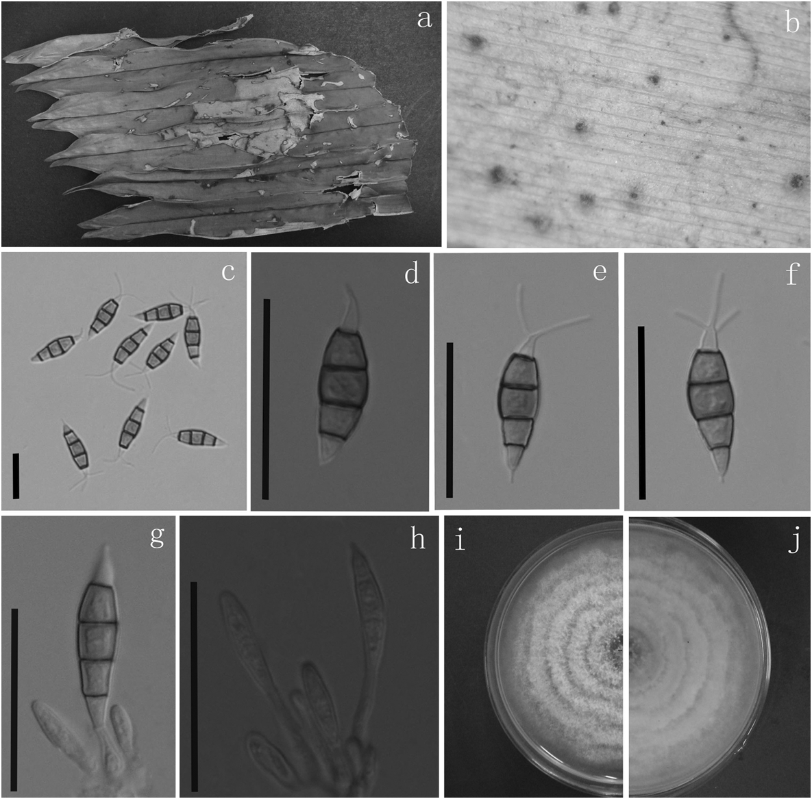
Pestalotiopsis licualacola K. Geng, Y. Song, K.D. Hyde & Yong Wang bis, sp. nov. ( Fig. 2 View FIGURE 2 ) MycoBank MB 803183
Type: — CHINA. Hainan Province: Xinglong County, Tropical Botanical Garden , living leaves of Licuala grandis , 8 March 2012, HGUP 4057, K. Geng, HGUPd4057, holotype !
Differs from related Pestalotiopsis and Pestalosphaeria species mainly by its noticeably narrower, fusiform conidia with mostly a single apical appendage.
Colonies on PDA attaining 7 cm diam. after 7 days at 25° C, with edge undulate, whitish, aerial mycelium on surface, fruiting bodies black, concentric; reverse of culture yellow to pale brown. Conidiophores most often indistinct. Conidiogenous cells discrete, hyaline, simple, filiform, 4–10 µm long. Conidia 16–20 × 3–5 µm (x = 17.4 × 3.9 µm), fusiform, straight to slightly curved, 4-septate, smooth, greyish brown; basal cell conical, hyaline, thin-walled, 2–4 µm long (x = 2.4 µm); with three median cells, dark brown, concolorous, septa and periclinal walls darker than the rest of the cell, together 9.5–12 µm long (x = 11 µm); second cell from base 2.7–4.2 µm (x = 3.6 µm); third cell 2.4–4 µm (x = 3.3 µm); fourth cell 2.5–3.8 µm (x = 3.2 µm); apical cell hyaline, conic to subcylindrical, 1.8–3.6 µm (x = 2.4 µm); with 1–3 tubular apical appendages (mostly 1) without knobs, arising from the apex of the apical cell, 4–9.5 µm long (x = 6.6 µm); basal appendage filiform, short.
Etymology: —In reference to the host, Licuala grandis , from which this fungus was first isolated.
| K |
Royal Botanic Gardens |
| PDA |
Royal Botanic Gardens |
| C |
University of Copenhagen |
No known copyright restrictions apply. See Agosti, D., Egloff, W., 2009. Taxonomic information exchange and copyright: the Plazi approach. BMC Research Notes 2009, 2:53 for further explanation.