
Philactinoposthia coneyi, Hooge, Matthew D. & Rocha, Carlos E. F., 2006
|
publication ID |
https://doi.org/ 10.5281/zenodo.174287 |
|
DOI |
https://doi.org/10.5281/zenodo.6263413 |
|
persistent identifier |
https://treatment.plazi.org/id/03DE87D4-E62E-FFB4-FE84-126AFD35FE1B |
|
treatment provided by |
Plazi |
|
scientific name |
Philactinoposthia coneyi |
| status |
sp. nov. |
Philactinoposthia coneyi sp. nov.
( Figs. 1–3 View FIGURE 1 View FIGURE 2 View FIGURE 3 )
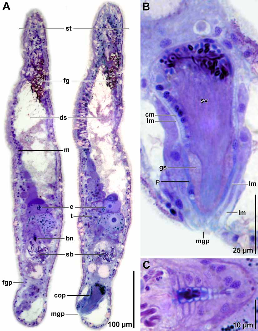
Type material. Holotype. MZUSP PL. 179, one set of 2-µm-thick serial sagittal sections of epoxy-embedded specimen stained with toluidine blue. Paratype. MZUSP PL. 180, epoxy-embedded whole mount.
Type locality. Praia de Feiticeira, Ilhabela, São Paulo, Brazil, from subtidal finegrained sand (23°47’16.0”S, 45°42’31.2”W).
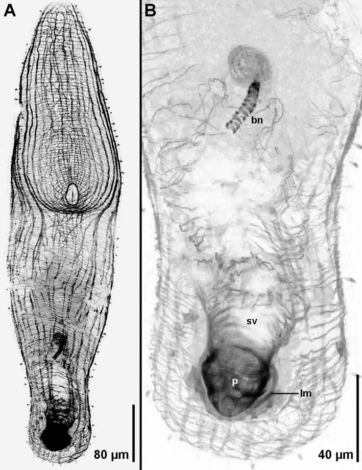
Other material examined. Whole mounts for fluorescence imaging of musculature; photographs of living specimens in squeeze preparations; two sets of 2-µm-thick serial sections of epoxy-embedded specimen.
Etymology. Species name in honor of Mr. Jon Coney of Portland, Oregon, USA.
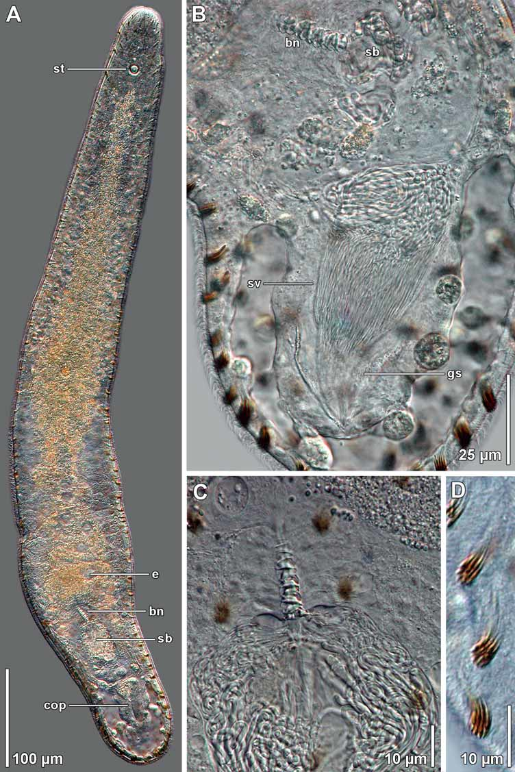
Description. Living specimens ~700 µm long and ~85 µm wide ( Fig. 1 View FIGURE 1 A). Body cylindrical, elongate. Anterior and posterior ends rounded. Body mostly without color by transmitted light, except for some yellow coloration of digestive syncytium ( Fig. 1 View FIGURE 1 A). Epidermis completely ciliated. Large, red-colored rhabdoid glands scattered across body ( Fig. 1 View FIGURE 1 D). Frontal organ well developed; cell bodies of frontal glands positioned approximately one-third of body-length behind frontal pore ( Fig. 2 View FIGURE 2 A). Mouth opening on ventral surface, middle of body. Digestive central syncytium extends from level anterior to position of mouth posteriorly to level of seminal bursa ( Fig. 2 View FIGURE 2 A).
Body-wall musculature with circular muscles that encircle the body along entire length of animal; straight longitudinal muscles present between frontal pore and anterior edge of mouth; longitudinal-cross-over muscles (fibers with a longitudinal orientation anteriorly, but bend medially to cross diagonally) present in both dorsal and ventral body wall; longitudinal muscles in anterior half of body that wrap around posterior rim of mouth (U-shaped muscles) present in ventral body wall; anterior end with ventral diagonal muscles positioned between outer circular and inner longitudinal muscles ( Fig. 3 View FIGURE 3 A).
Ovary unpaired, ventral; extends from level of mouth posteriorly to bursal nozzle ( Figs. 1 View FIGURE 1 A, 2A). Testes paired, lateral to ovary, compact. Testes extend anteriorly to level of mouth and posteriorly to male copulatory organ.
Female gonopore at level slightly anterior to seminal vesicle; connection to bursa poorly defined ( Fig. 2 View FIGURE 2 A). Bursa with wall; connects anteriorly to well-defined bursal nozzle with appearance like a stack of hats ( Figs. 1 View FIGURE 1 A–C, 2A, C).
Male gonopore positioned subterminally on ventral side. Opens immediately to muscular, glandular male copulatory organ with sclerotized penis ( Figs. 1 View FIGURE 1 B, 2A, B, 3B). Seminal vesicle composed of outer longitudinal and inner circular muscles and packed with wellaligned sperm ( Figs. 1 View FIGURE 1 B, 2B, 3B). Penis not invaginated into seminal vesicle; positioned between seminal vesicle and male gonopore; composed of a cone of longitudinal muscles, some of which are continuous with longitudinal fibers of seminal vesicle. Inside longitudinal fibers is a cone-shaped sclerotized penis, which stains blue with toluidine blue in histological sections ( Fig. 2 View FIGURE 2 B), and stains with Alexa-488 labeled phalloidin in specimens viewed with confocal microscopy ( Fig. 3 View FIGURE 3 B). Penis surrounds a layer of glandular secretions that surround the sperm and stain metachromatically for glycoseaminoglycans ( Figs. 1 View FIGURE 1 B, 2B)
Remarks. The family Actinoposthiidae was erected by Hooge (2001) as means of separating the ~30 species with typical body-wall muscle layering from species of Childia (Childiidae) , which have reversed body-wall muscle layering (longitudinal fibers positioned outside of circular fibers). The Actinoposthiidae includes species that have either a sclerotized or muscular penis that is not invaginated into a seminal vesicle. Unfortunately, the family and several of its genera appear to be polyphyletic and the distinctive characters have yet to be identified. The genus Philactinoposthia is particularly problematic because it contains some species with sclerotized penes and others with muscular penes, despite the unlikelihood that these structures are homologous. We place our new species in the genus Philactinoposthia because of its strong similarity to P. viridorhabditis Dörjes, 1968 , a species that shares with P. coneyi an unpaired ovary, a male copulatory organ with a muscular seminal vesicle and an uninvaginated cone-shaped penis. P. coneyi differs from P. viridorhabditis in having a more elongate body, a shorter, less-irregularly shaped penis surrounded by longitudinal muscle fibers, and a distinctive stack-of-hats shaped bursal nozzle.
| MZUSP |
Museu de Zoologia da Universidade de Sao Paulo |
No known copyright restrictions apply. See Agosti, D., Egloff, W., 2009. Taxonomic information exchange and copyright: the Plazi approach. BMC Research Notes 2009, 2:53 for further explanation.