
Philocelis robrochai, Hooge, Matthew D. & Rocha, Carlos E. F., 2006
|
publication ID |
https://doi.org/ 10.5281/zenodo.174287 |
|
DOI |
https://doi.org/10.5281/zenodo.6263461 |
|
persistent identifier |
https://treatment.plazi.org/id/03DE87D4-E605-FF90-FE84-117FFEC9FB2B |
|
treatment provided by |
Plazi |
|
scientific name |
Philocelis robrochai |
| status |
sp. nov. |
Philocelis robrochai sp. nov.
( Figs. 27–29 View FIGURE 27 View FIGURE 28 View FIGURE 29 )
Type Material. Holotype. MZUSP PL. 196, one set of 2-µm-thick serial sagittal sections of epoxy-embedded specimen stained with toluidine blue. Paratype. MZUSP PL. 197, one set of 2-µm-thick oblique sections of epoxy-embedded specimen stained with toluidine blue.
Type locality. Praia da Vila, Ilhabela, São Paulo, Brazil, from subtidal coarse-grained sand (23°46’44.6”S, 45°21’32.6”W).
Other material examined. Living specimens in squeeze preparations from Praia de Pequeá, Ilhabela; three sets of 2-µm-thick serial oblique sections of epoxy-embedded specimens stained with toluidine blue; whole mounts for fluorescence imaging of musculature; photographs of living specimens in squeeze preparations.
Etymology. Species name in honor of Mr. Roberto Rocha of San Jose, CA.
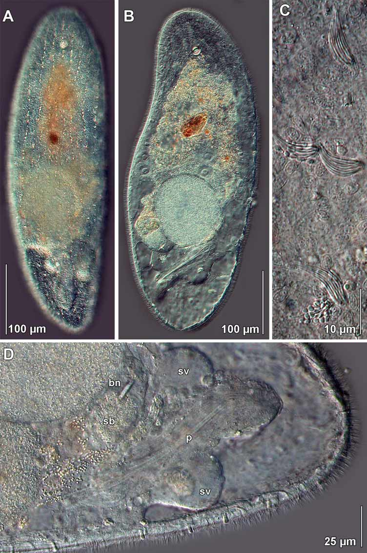
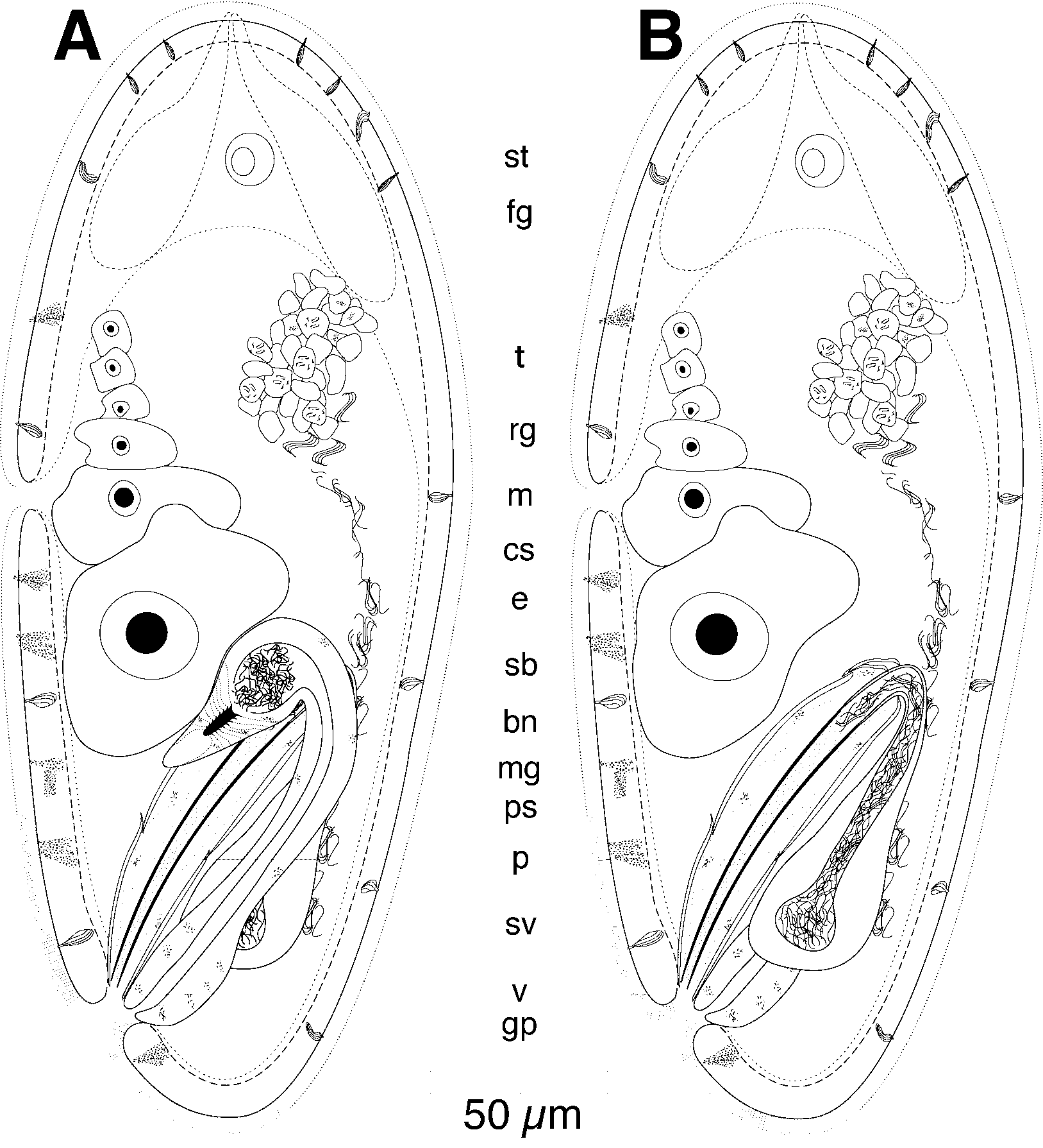
Description. Living specimens ~500 µm long and ~140 µm wide ( Figs. 27 View FIGURE 27 A, B). Anterior and posterior ends rounded. Body cylindrical. Epidermis completely ciliated. Rhabdoid glands in distinct rows ( Fig. 27 View FIGURE 27 A, C). Mucoid glands present mostly in ventral body wall ( Fig. 28 View FIGURE 28 A). Body colorless in transmitted light, but digestive syncytium has orange coloration. Frontal organ well developed. Cell bodies of frontal glands positioned 70 µm behind frontal pore ( Figs. 27 View FIGURE 27 B, 28A). Mouth opening on ventral surface, middle of body. Digestive central syncytium extends from frontal glands posteriorly to male copulatory apparatus.
Body-wall musculature with circular muscles that encircle the body along entire length of animal; straight longitudinal muscles present between frontal organ and anterior edge of mouth; longitudinal muscles with a longitudinal orientation anteriorly that bend medially to cross diagonally over the body (longitudinal-cross-over fibers) present in dorsal and ventral body walls; longitudinal muscles in the anterior half of the body that wrap around the posterior rim of the mouth (U-shaped muscles) present in ventral body wall.
Ovaries paired, ventral; extend from frontal glands posteriorly to bursal nozzle ( Figs. 27 View FIGURE 27 B, D, 28A). Testes paired, dorsal; separate from ovary. Testes extend from frontal gland posteriorly to seminal vesicles ( Fig. 28 View FIGURE 28 B).
Common gonopore positioned ventrally at posterior end of body; opens directly to male copulatory organ anteriorly, and to vagina caudally ( Figs. 28 View FIGURE 28 A, B).
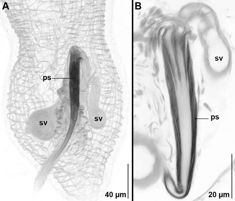
Male copulatory organ composed of a long (~120 µm), curved, conical sclerotized stylet surrounded by a weakly muscular penis sac ( Figs. 27 View FIGURE 27 B, D, 28B, 29A, B). Penis flanked laterally by muscular seminal vesicles with long muscular ducts that connect to proximal end of penis.
Vagina with thick wall; positioned dorsal to male copulatory organ; extends anteriorly to well-developed seminal bursa ( Figs. 27 View FIGURE 27 D, 28A). Bursa with bursal nozzle directed ventrally and slightly posteriorly ( Figs. 27 View FIGURE 27 D, 28A).
Remarks. Species of the genus Philocelis are united in having the vagina positioned posterior to the male copulatory organ, a seminal bursa with attached bursal nozzle, and a male copulatory organ composed of a muscular penis-sac surrounding sclerotized stylets. Although the penis stylet of P. robrochai is more compact than that of P. brueggemanni Hooge and Tyler, 2003 , P. cellata Dörjes, 1968 , and P. karlingi ( Westblad, 1946) , which have widely spaced stylet needles, the overall organization of the male copulatory organ appears to be homologous. Preliminary analysis of both cytochrome oxidase I and 18S rDNA molecular sequence data support a strong relationship between P. robrochai and P. brueggemanni (unpublished data). Interestingly, 18S sequence data also supports a close relationship between Philocelis species and Philactinoposthia saliens (Graff, 1882) (Actinoposthiidae) —a relationship that is further supported by similarities in sperm ultrastructure ( Petrov et al. 2004). Several species of Philactinoposthia appear to have male copulatory organs with a similar composition to that of Philocelis , and given that the Otocelididae has been shown to be non-monophyletic ( Hooge & Tyler 2005), any future revision of the Otocelididae should include an examination of the relationship of these two genera.
| MZUSP |
Museu de Zoologia da Universidade de Sao Paulo |
No known copyright restrictions apply. See Agosti, D., Egloff, W., 2009. Taxonomic information exchange and copyright: the Plazi approach. BMC Research Notes 2009, 2:53 for further explanation.