
Pliocaloca kleithria, Shackleton, Michael, 2010
|
publication ID |
https://doi.org/ 10.5281/zenodo.195305 |
|
DOI |
https://doi.org/10.5281/zenodo.6196832 |
|
persistent identifier |
https://treatment.plazi.org/id/03C887F3-F374-DC61-FF40-FA49A4EFFE47 |
|
treatment provided by |
Plazi |
|
scientific name |
Pliocaloca kleithria |
| status |
sp. nov. |
Pliocaloca kleithria sp.nov.
Figs 5–8 View FIGURES 1 – 8 , 11–23 View FIGURES 9 – 12 View FIGURES 13 – 16 View FIGURES 17 – 23
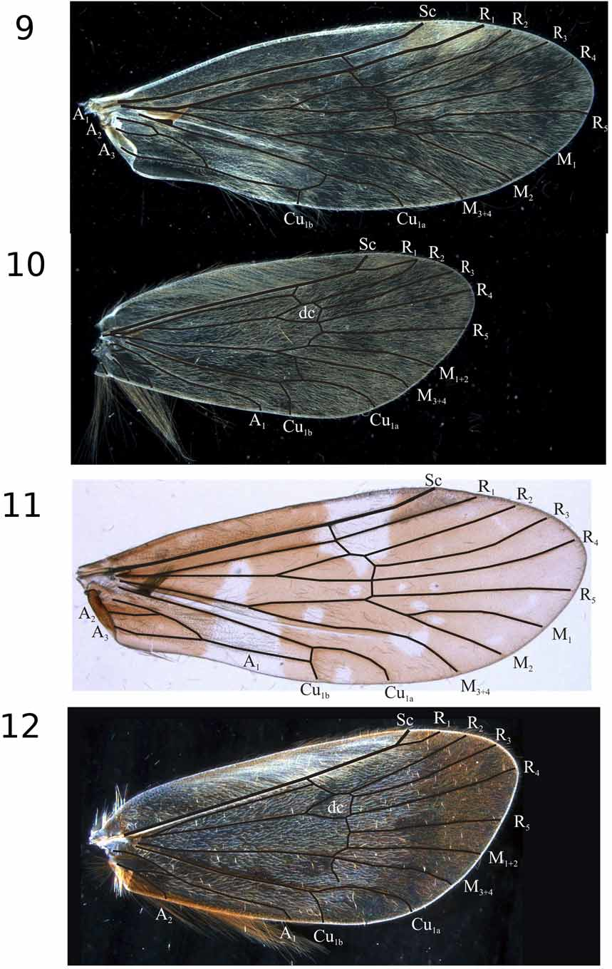
Diagnosis. As stated above, P. kleithria and P. fidesria possess many similar characteristics, suggesting a close alliance between the two. Of the previously described species, P. d a s o d e s ( Neboiss 1984) appears to be most similar to P. kleithria . In both species the forewings ( Fig. 11 View FIGURES 9 – 12 ) lack a thyridial cell and possess a setose, sclerotised knob on the underside of vein Cu1. Morphology of the genitalia is similar in both species and a slender process rises, medially, from the base of the inferior appendage ( Fig. 6 View FIGURES 1 – 8 ).
The following characters separate Pliocaloca kleithria from P. dasodes : the preanal appendages ( Fig. 5 View FIGURES 1 – 8 ) and the slender processes of the inferior appendages ( Fig. 6 View FIGURES 1 – 8 ) are much longer, segment X ( Fig 5 View FIGURES 1 – 8 ) is not as tapered and has a much wider apical incision, and the sclerotised knob on vein Cu1 of the forewing ( Fig. 11 View FIGURES 9 – 12 ) is located more basally. The shape of the apices of segment X also differs between these species. In P. dasodes , the lateral margins of this segment gently taper along its length ( Neboiss 1984). The segment is deeply and narrowly incised apically. In P. kleithria , the lateral margins are curved inwards at about 2/3rds their length ( Fig. 5 View FIGURES 1 – 8 ). The medial incision of the segment is wider but not as deep as in P. d a s o d e s and the apices are somewhat rounded.
The shapes of the genital features distinguish Pliocaloca kleithria from P. fidesria . The slender process arising from the inferior appendage ( Fig. 6 View FIGURES 1 – 8 ) is gently curved outwards in P. kleithria , while in P. f i d e s r i a it is abruptly curved outwards at about 1/3rd the length of the segment ( Fig 2 View FIGURES 1 – 8 ). In P. kleithria , segment X ( Fig 5 View FIGURES 1 – 8 ) bears a pair of strong dorsal ridges that diverge apically. The apices of the segment are somewhat rounded, with the lateral margins curved outwards. In P. fidesria , the dorsal ridges of segment X ( Fig. 1 View FIGURES 1 – 8 ) are parallel to each other and the lateral margins of the posterior 1/3rd are relatively straight and tapered inwards.
Description. Male. Length of fore wing 5.7–6.7 mm. Wings brown. Fore wing with distinct white patches, 1 large patch adjacent to costal margin basal of pterostigma, 1 large patch on anal region, scattered smaller patches. Forewing ( Fig. 11 View FIGURES 9 – 12 ): thyridial cell absent; vein A1 joins Cu1b at arculus just behind position where Cu1b separates from Cu1a; base of Cu1 with oval sclerotised knob bearing tuft of dark, long, setae on underside of wing; membrane between veins A1 and Cu1 with long setae. Hind wing ( Fig. 12 View FIGURES 9 – 12 ): crossvein m–cu not quite joining with Cu1a. Genitalia ( Figs 5–7 View FIGURES 1 – 8 ): Segment X long and narrow, strongly incised apically, 2 strong dorsal ridges extending along most of length of segment and diverging apically; preanal appendages slender, rounded apically, strongly curved medially, enlarged sub-apically, extending to approximately half length of segment X; inferior appendages relatively narrow, curved inwards, apices pointed and projecting dorsad, each with slender process arising medially from base; slender process extending almost to apex of inferior appendage, gently curved, tapered to point. Phallus ( Fig. 8 View FIGURES 1 – 8 ): with a pair of rounded lobes within distal1/2, projecting from the lateral margin.
Female unknown.
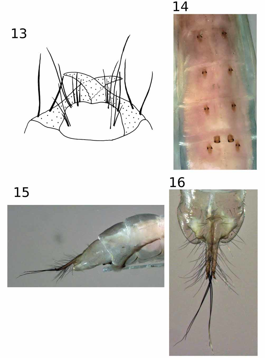
Pupa. Head: antennae as long as body; labrum ( Fig. 13 View FIGURES 13 – 16 ) with anterior margin straight or slightly concave, 3 dark setae towards each posterolateral corner, each anterolateral corner with 3 dark sub-apical setae and 2 transparent apical setae; mandibles ( Fig. 13 View FIGURES 13 – 16 ) with small serrations along inner margins, each with 2 setae on basal 1/4th of outer margin. Middle legs: each tibia with dorsal fringe of setae. Abdomen ( Fig. 14 View FIGURES 13 – 16 ): lateral fringes present; anterior hookplates on tergites 3–6, posterior hookplates on segment 5 only; all hookplates with 2 hooks; terminal processes ( Figs 15, 16 View FIGURES 13 – 16 ) narrow, apically acute and upturned, each with 2 long black setae subapically, rows of finer setae on ventral and dorsolateral surfaces.
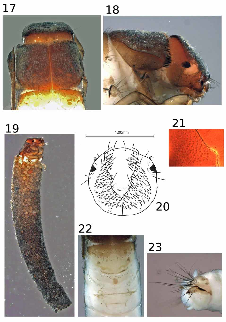
Larva. Length: 8 mm. Head ( Figs 20, 21 View FIGURES 17 – 23 ): brown, dorsum covered by spinules, slight depression in posterior region of frontoclypeus; strong band of pale secondary setae mostly outside frontoclypeus, those inside frontoclypeus longer and directed mesad; frontoclypeus with 2 pairs of primary setae on anterior margin; antennae small, situated about halfway between anterior margin of head capsule and eye; carina not present. Pronotum ( Figs 17, 18 View FIGURES 17 – 23 ): dorsum covered by spinules; each sclerite with anterior margin curved forward between suture line and lateral margin; lateral carina present, almost reaching posterior margin, not extending onto dorsum of segment; anterior margin with dense row of short blunt setae; foretrochantin not fused to propleuron, apically acute. Mesonotum ( Fig. 22 View FIGURES 17 – 23 ): lightly sclerotised, pigmentation predominantly in anterior half. Metanotum ( Fig. 22 View FIGURES 17 – 23 ): with single, round, medial sclerite; setal area 1 with about 6 setae, setal area 2 with 1 seta, setal area 3 with about 8 setae. Abdomen ( Fig. 23 View FIGURES 17 – 23 ): segment I lateral humps without sclerites; abdominal gills absent; tergite 9 with 5 pairs of hairlike setae; anal lateral sclerites with many setae, 2 setae darker, thicker, and slightly longer than rest; abdominal prolegs each with 1 accessory tooth. Legs ( Fig. 19 View FIGURES 17 – 23 ): hind legs almost twice as long as forelegs. Case ( Fig. 19 View FIGURES 17 – 23 ): curved cylinder of sand grains, posterior aperture round.
Holotype male: New South Wales, Wilson River Res nr. Bellangry, 5 Dec 1988, G. Theischinger, (MV TRI–26295).
Paratype (specimen figured): 1 male, New South Wales, Terania Creek N of Lismore 28°25 S, 153°18 E, 21 Jan 1986, G. Theischinger, (MV TRI–26285).
Other material examined: New South Wales. Tributary of Wilson River, 70 meters along Falls Walk Track 13°12’ S, 152°29’ E, 4 Dec 2007, A. Glaister, J. Dean, and R. St Clair 1 male pupa, 3 female pupae, 5 larvae, (AM); 4 male pupae, 2 female pupae, 3 larvae (MV).
Etymology. From the Greek kleithria , meaning “keyhole,” pertaining to the keyhole shape formed by the apical incision in segment X.
Remarks. Jackson (1998, figs 1.12–1.17) has previously illustrated the larvae of what she called Pliocaloca sp. AV1, with a distribution in southern Queensland and northern New South Wales. She makes reference to a pupal specimen associating this larva with an undescribed adult of Pliocaloca . Unfortunately, the specimens Jackson used to associate this larva are missing from the Australian voucher collection. The larva of P. kleithria is similar to the illustrations given by Jackson (1998). However, it is not known if the larva Jackson illustrated represents P. kleithria or P. fidesria , or if the larvae of these species can be distinguished based on morphology.

No known copyright restrictions apply. See Agosti, D., Egloff, W., 2009. Taxonomic information exchange and copyright: the Plazi approach. BMC Research Notes 2009, 2:53 for further explanation.