
Pseudorhabdosynochus marcellus, Justine, Jean-Lou & Sigura, Aude, 2007
|
publication ID |
https://doi.org/ 10.5281/zenodo.177917 |
|
DOI |
https://doi.org/10.5281/zenodo.6238102 |
|
persistent identifier |
https://treatment.plazi.org/id/039C879B-4B3E-422E-EEC9-FB6CE09234A1 |
|
treatment provided by |
Plazi |
|
scientific name |
Pseudorhabdosynochus marcellus |
| status |
sp. nov. |
Pseudorhabdosynochus marcellus View in CoL n. sp.
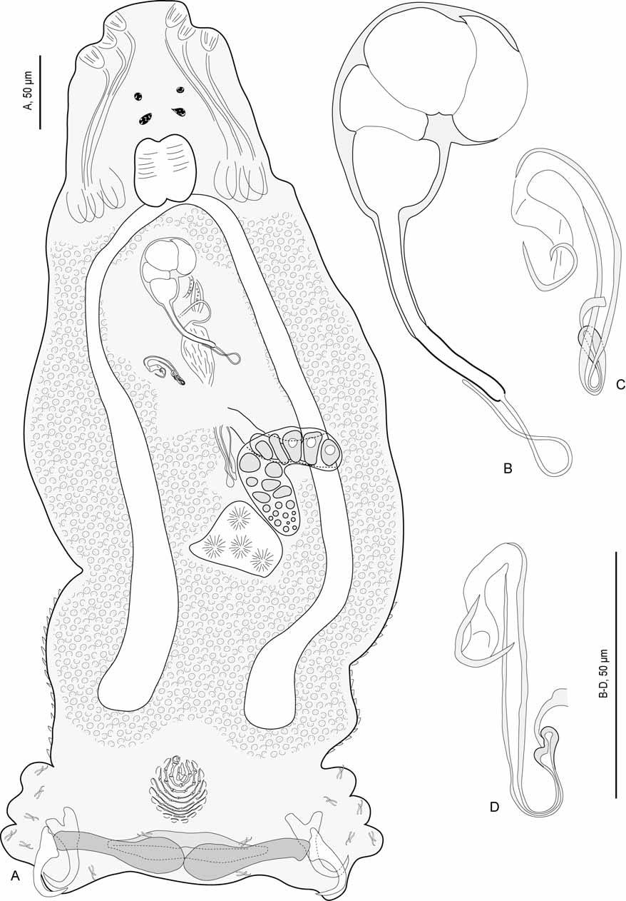
( Figs 15–16 View FIGURE 15 View FIGURE 16 )
Type host: Epinephelus malabaricus Bloch & Schneider (Serranidae) .
Type locality: Lagoon off Nouméa, New Caledonia.
Site: Between secondary gill lamellae.
Type specimens: Holotype, JNC 1536A104, off Ouen Toro, Nouméa, New Caledonia (22°19'S, 166°27'E, 18.v.2005).
Material examined: 2 specimens in carmine.
Material deposited: Holotype (c) and 1 paratype, MNHN.
Prevalence: 50% (1/2).
Intensity: See Table 1. A very rare species (1 % of the diplectanids).
Etymology: refers to a character of a well-known Caledonian cartoon, Tonton Marcel.
Description. [Measurements given for holotype (h) and paratype (c)]. Body elongate; length h 590, c 580, width h 230, c 280. Tegument scaly; posterior region with scales on ventral and dorsal faces from squamodiscs to level of ovary and testis. Anterior region with 3 pairs of head organs and 2 pairs of eye-spots; distance between outer margins of anterior eye-spot pair h 36, c 37, of posterior eye-spot pair h 33, c 40.
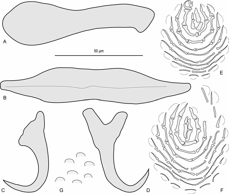
Haptor differentiated from rest of body, as wide as body, width h 250, c 290, provided with 2 similar squamodiscs, 2 pairs of lateral hamuli, 3 bars and 14 marginal hooklets. Squamodiscs observed only in holotype; round in shape, made up of rows of rodlets; central rows forming almost closed ovals; rodlets progressively thinner from centre to periphery; rodlets adjacent in central rows, separated in peripheral rows; last row with very thin, separated rodlets; ventral and dorsal squamodiscs similar, but ventral slightly larger; ventral squamodisc length h 44, width h 50, with 9 rows of rodlets and 1 closed oval, total number of rodlets h 59; dorsal squamodisc, length h 44, width 36, with 10 rows of rodlets and 1 closed oval, total number of rodlets h 46. Ventral hamulus with distinct handle and guard, outer length h 54, c 54, inner length h 46, c 46. Dorsal hamulus with indistinct guard, outer length h 47, c 47, inner length h 28, c 29. Dorsal (lateral) bars straight, with flattened medial extremity and thick cylindrical lateral extremity, length h 86, c 90, maximum width h 25, c 24. Ventral bar flat, with slightly constricted median portion and blunt extremities, length h 106, c 111, maximum width h 20, c 22; groove visible on its ventral side.
Pharynx subspherical, length h 50, c 47, width h 43, c 40. Oesophagus apparently absent, such that intestinal bifurcation immediately follows pharynx. Caeca simple, terminate blindly at level of posterior margin of vitelline field.
Testis subspherical, intercaecal, length h 70, c 55, width h 55, c 45. Vas deferens inconspicuous; seminal vesicle in middle region of body, transforms into duct. Very small prostatic reservoir and inconspicuous prostatic duct both connect with quadriloculate organ. Quadriloculate organ with fourth (posterior) chamber as sclerotised as 3 anterior chambers; fourth chamber ends in very elongate sclerotised cone, prolonged by sclerotised tube; end of tube prolonged by thin unsclerotised filament of variable length. Inner length of quadriloculate organ h 42, c 42; cone length h 25, c 25; tube length h 21, c 20; tube diameter h 3, c 3; filament length h 49, c 3.
Ovary subequatorial, intercaecal, pre-testicular, encircles right caecum. Ovary width h 75, c 80. Oviduct passes medially to form oötype, surrounded by Mehlis’ gland; oötype short, opens into uterus. Uterus dextral. Unsclerotised vagina inconspicuous. Duct from sclerotised vagina to oötype inconspicuous. Vitelline fields extend posteriorly from posterior to pharyngeal level in 2 lateral bands, confluent in post-testicular region and terminate anterior to peduncle. Bilateral connections from vitelline fields to oötype inconspicuous. Egg unknown.
Sclerotised vagina (nomenclature of parts according to Justine 2007a; see Figure 20 View FIGURE 20 ). Sinistral, a complex sclerotised structure. Sclerotised vagina comprises anterior trumpet, followed by primary canal, primary chamber and secondary chamber; primary canal cylindrical, bent once in its anterior part and once in its posterior part; anterior part (before first bend) cylindrical, with very thin wall; median part (between bends), cylindrical with sclerotised wall; posterior part (forming posterior bend) with small diameter and thin wall; primary chamber in continuity with primary canal; primary and secondary chamber embedded in same distal heavily sclerotised part, thus secondary canal absent; accessory structure connects to distal sclerotised part. Total length of sclerotised vagina (measured from anterior extremity of first bend of primary canal to posterior extremity of second bend of primary canal, i.e. not taking in account curved length along bend and coil of canal) h 32, c 35. Orientation of sclerotised vagina: first bend of primary canal anterior.
Differential diagnosis. Two species have a sclerotised vagina with a long primary canal with a bend in the anterior part and a second bend in the posterior part, and the chambers small and apparently united in a common sclerotised part.
A species of Pseudorhabdosynochus from Cephalopholis argus off New Caledonia and Australia ( Justine 2007b) has a vagina with structure and measurements similar to P. m a rc e l l u s, but can be distinguished by more numerous squamodisc rodlets (80–90 vs 46–59), and a massive and shorter ventral bar (73 vs 109).
P. l a n t a u e n s i s ( Beverley-Burton & Suriano, 1981) from E. bruneus Bloch and E. longispinis (Kner) has a vagina (see redescription in Justine, 2005a) which shares with P. marcellus a similar structure, a quadriloculate organ with a very long cone and squamodiscs with small numbers of rows (9–10) and rodlets (60); however, the anterior bend of its primary canal is double, not simple. Clearly, these species are closely related but can be differentiated on the basis of vaginal structure.
P. marcellus can be differentiated from all other species of Pseudorhabdosynochus by its vaginal structure.

| MNHN |
Museum National d'Histoire Naturelle |
No known copyright restrictions apply. See Agosti, D., Egloff, W., 2009. Taxonomic information exchange and copyright: the Plazi approach. BMC Research Notes 2009, 2:53 for further explanation.
|
Kingdom |
|
|
Phylum |
|
|
Class |
|
|
Order |
|
|
Family |
|
|
Genus |