
Pseudorhabdosynochus cf. shenzhenensis Yang, Zeng & Gibson, 2005
|
publication ID |
https://doi.org/ 10.5281/zenodo.177917 |
|
DOI |
https://doi.org/10.5281/zenodo.6238114 |
|
persistent identifier |
https://treatment.plazi.org/id/039C879B-4B38-4214-EEC9-FA86E25E3731 |
|
treatment provided by |
Plazi |
|
scientific name |
Pseudorhabdosynochus cf. shenzhenensis Yang, Zeng & Gibson, 2005 |
| status |
|
Pseudorhabdosynochus cf. shenzhenensis Yang, Zeng & Gibson, 2005 View in CoL
( Figure 19 View FIGURE 19 )
Host: Epinephelus malabaricus Bloch & Schneider (Serranidae) .
Type Host: Epinephelus coioides (Hamilton)
Locality: Lagoon of New Caledonia.
Site: Between secondary gill lamellae.
Material examined: 2 specimens in picrate, MNHN JNC 2130A5, JNC 2130A6, Baie Maa , New Caledonia (22°12’780S, 166°19’933E, 9.iii.2007)
Prevalence: 50% (1/2).
Intensity: See Table 1. A rare species (1.5% of the diplectanids).
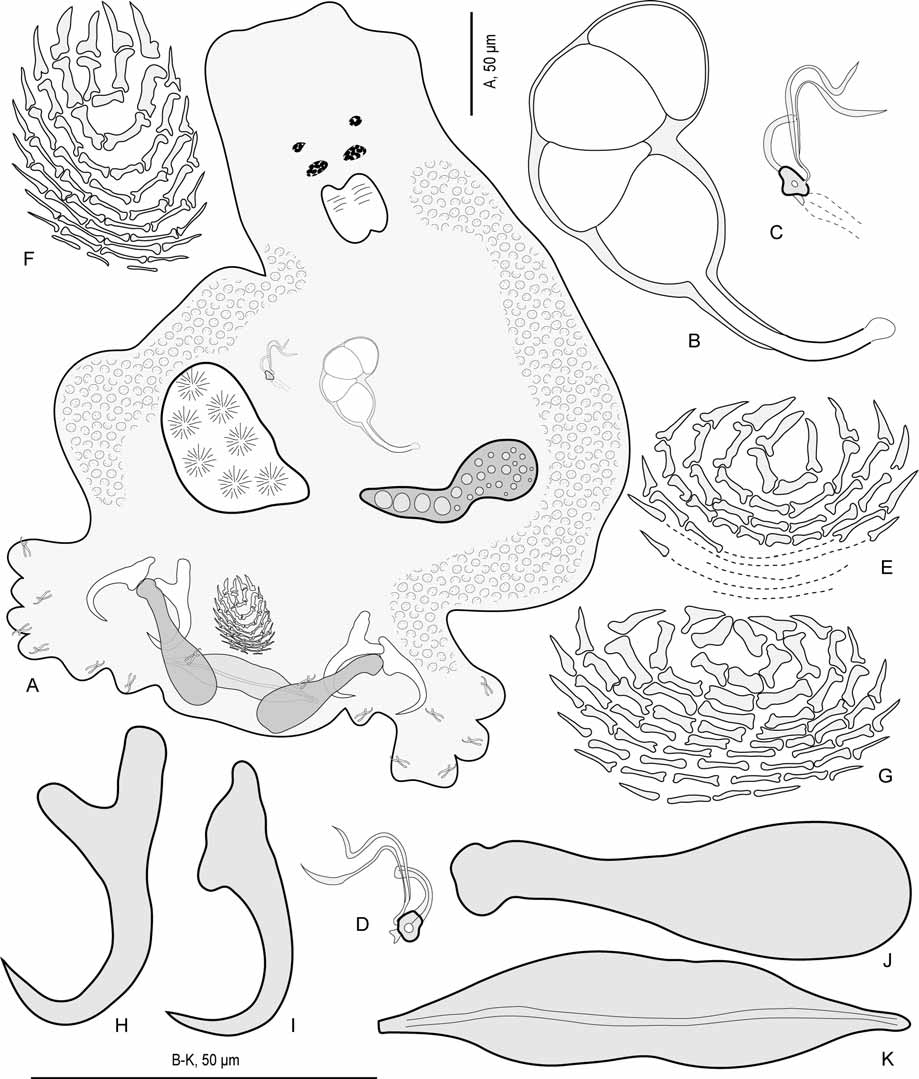
Description. Body length 370–420, width 220–250. Tegumental scales apparently absent. Anterior region with 2 pairs of eye-spots; distance between outer margins of anterior eye-spot pair 36, of posterior eye-spot pair 30–32.
Haptor differentiated from rest of body, width 170–230, provided with 2 similar squamodiscs, 2 pairs of lateral hamuli, 3 bars and 14 marginal hooklets. Squamodiscs deformed in available specimens, made up of rows of rodlets; central rows forming almost closed ovals; rodlets progressively thinner from centre to periphery; rodlets adjacent in central rows, separated in peripheral rows; last row with very thin, separated rodlets; ventral and dorsal squamodiscs similar; ventral squamodisc length 28–29, width 40–50 with 9 rows of rodlets and 1 closed oval, total number of rodlets 64; dorsal squamodisc, length 38, width 26, with 10 rows of rodlets and 1 closed oval, total number of rodlets 55. Ventral hamulus with distinct handle and guard, outer length 45, inner length 35. Dorsal hamulus with indistinct guard, outer length 38, inner length 22. Dorsal (lateral) bars straight, with flattened medial extremity and cylindrical lateral extremity, length 63–66, maximum width 19. Ventral bar flat, with slightly constricted median portion and pointed extremities, length 68–75, maximum width 15–16; groove visible on its ventral side.
Pharynx subspherical, length 30–35, width 26–29. Caeca not seen in picrate specimens.
Testis subspherical, intercaecal, length 70, width 45. Vas deferens, seminal vesicle, prostatic reservoir not seen. Quadriloculate organ with fourth (posterior) chamber as sclerotised as 3 anterior chambers; fourth chamber ends in elongate sclerotised cone, prolonged by sclerotised tube; end of tube prolonged by thin unsclerotised filament of variable length. Inner length of quadriloculate organ 40–42; cone length 15–16; tube length 12–13; tube diameter 2; filament length 4–10.
Ovary subequatorial, intercaecal, pre-testicular. Ovary width 80. Uterus dextral. Unsclerotised vagina inconspicuous. Duct from sclerotised vagina to oötype inconspicuous. Vitelline fields extend posteriorly from posterior to pharyngeal level in 2 lateral bands and terminate anterior to peduncle. Egg unknown.
Sclerotised vagina (nomenclature of parts according to Justine 2007a, see Figure 20 View FIGURE 20 ). Sinistral, a complex sclerotised structure. Sclerotised vagina comprises anterior trumpet, followed by primary canal, and distal heavily sclerotised part; primary canal conical, bent once in its anterior part and once in its posterior part; wall of primary canal progressively thinner from trumpet to posterior extremity; primary chamber embedded in distal heavily sclerotised part; secondary chamber and secondary canal not seen; accessory structure weakly sclerotised, connects to distal sclerotised part. Total length of sclerotised vagina (measured from anterior to posterior extremity, i.e. not taking in account curved length along bend and coil of canal) 20–22. Orientation of sclerotised vagina: trumpet anterior.
Remarks. Only two specimens of this species could be examined, and they were both picrate slides. We are not completely convinced that these specimens are conspecific with P. shenzhenensis Yang, Zeng & Gibson, 2005 , described from E. coioides off South China, and identification as P. cf shenzhenensis is only provisional. Observations of more numerous specimens, and particularly of carmine preparations, could reveal that this is a new species. The structure of the vagina, with conical bent primary canal and small chambers, is similar to that of P. shenzhenensis , but not exactly the same: the primary chamber seems more elongate in P. s h e n - zhenensis than in this material.

| MNHN |
Museum National d'Histoire Naturelle |
No known copyright restrictions apply. See Agosti, D., Egloff, W., 2009. Taxonomic information exchange and copyright: the Plazi approach. BMC Research Notes 2009, 2:53 for further explanation.
|
Kingdom |
|
|
Phylum |
|
|
Class |
|
|
Order |
|
|
Family |
|
|
Genus |