
Retevirgula triangulata ( Silén, 1941 )
|
publication ID |
https://doi.org/ 10.11646/zootaxa.5379.1.1 |
|
publication LSID |
lsid:zoobank.org:pub:430102D2-4EAA-41B3-B57F-CC532F929DA3 |
|
DOI |
https://doi.org/10.5281/zenodo.10248885 |
|
persistent identifier |
https://treatment.plazi.org/id/4B6E902E-FFBC-FF8D-FF46-F9301DB7FC22 |
|
treatment provided by |
Plazi |
|
scientific name |
Retevirgula triangulata ( Silén, 1941 ) |
| status |
|
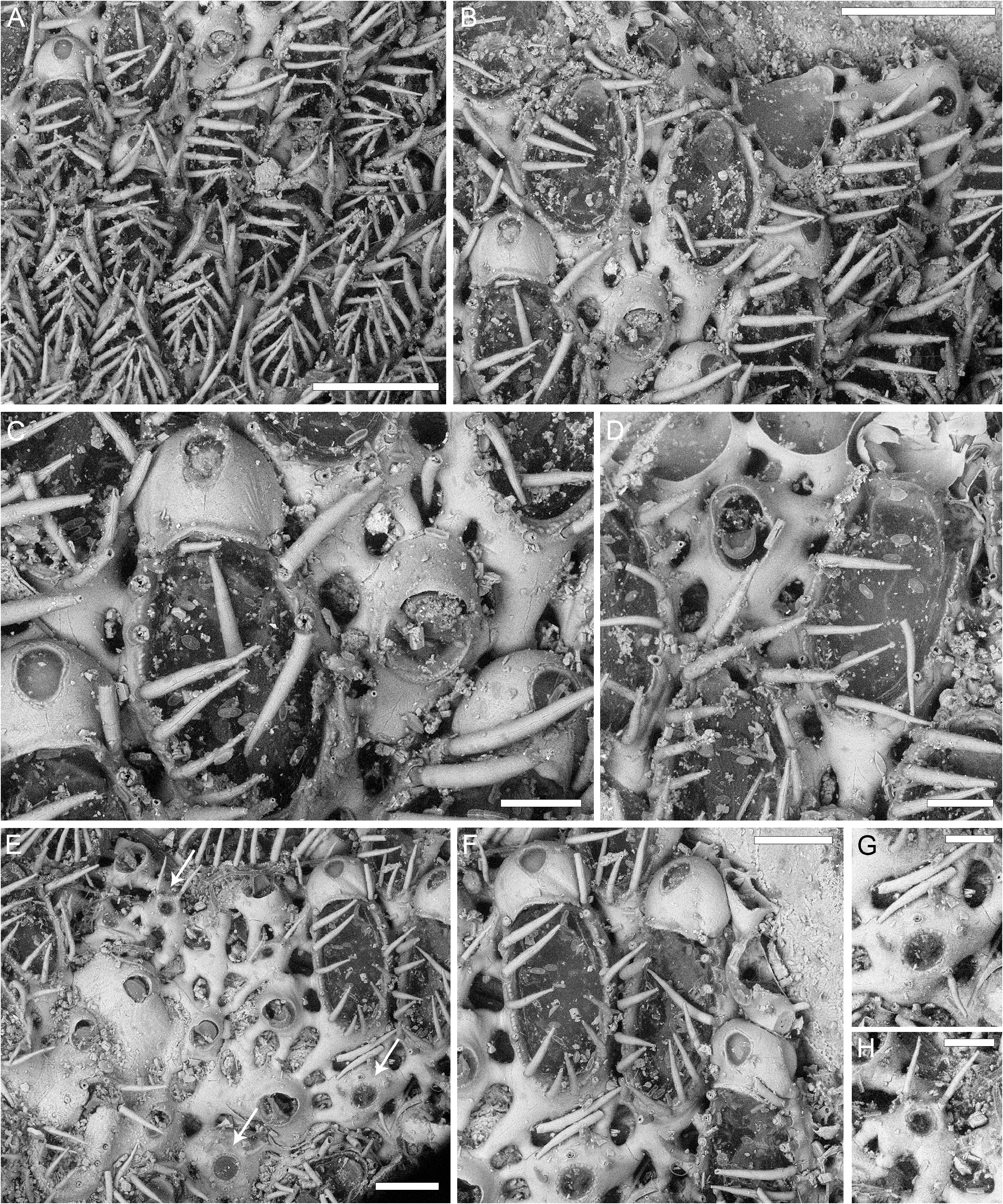
Retevirgula triangulata ( Silén, 1941)
( Fig. 9 View FIGURE 9 ; Table 10)
Pyrulella tubulata var. triangulata Silén, 1941: 28 , figs 25, 26.
Retevirgula tubulata var. triangularis [sic]: Mawatari & Mawatari, 1980: 89, fig. 31.
Retevirgula triangulata : Min et al., 2017: 475.
Material examined. Holotype by original designation UPSZTY 2471 , Bonin Islands (Ogasawara), Takino Ura, Japan; depth 45–60 m. Leg. Prof. S. Bock 1914.
Description. Colony encrusting, multiserial, unilaminar ( Fig. 9A, B View FIGURE 9 ).
Autozooids oval to almost parallel-sided elliptical ( Fig. 9B, F View FIGURE 9 ), twice as long as wide (mean L/ W 1.96), quincuncially or irregularly arranged, disjunct with 8–14 tubular connections (30–90 µm wide) to adjacent autozooids and heterozooids (both avicularia and kenozooids); tubular connections more visible in certain portions of the colony, leaving large (70–225 × 40–110 µm), irregularly elliptical lacunae between them ( Fig. 9B, E View FIGURE 9 ). Gymnocyst smooth, more extensive proximally (80–90 µm), sloping steeply laterally; cryptocyst forming a very narrow (10–15 µm) coarsely beaded rim around the opesia, indented by periopesial spines ( Fig. 9B, C, F View FIGURE 9 ).
Opesia oval or elliptical, somewhat mirroring autozooidal shape, occupying most of the frontal surface (mean OpL/ZL 0.77), encircled by 14–18 articulated spines, 20–30 µm in diameter, 140–230 µm long, the two distalmost pairs more erect, the remaining pairs overarching the frontal membrane ( Fig. 9A View FIGURE 9 ).
Avicularia and kenozooids interzooidal, sparse ( Fig. 9A–D View FIGURE 9 ) or forming clusters ( Fig. 9E View FIGURE 9 ), the shape, size and convexity of the cystid/zooid variable. Avicularia elliptical, slightly constricted at about mid-length, an extremely narrow rim of beaded cryptocyst outlining its proximal half ( Fig. 9C, D View FIGURE 9 ); mandible and rostrum semicircular, the latter raised and directed distally or distolaterally to either side; pivotal bars or condyles absent. Kenozooids with circular openings surrounded by a rim of beaded cryptocyst (10–15 µm wide) and 5–6 erect spines, 10–20 µm in diameter, 100–150 µm long, not indenting the cryptocyst but placed at a short distance (10–25 µm) from it ( Fig. 9E–H View FIGURE 9 , see arrows).
Ovicells globular, convex; ooecium smooth, ectooecium partially calcified, with a plectrum-shaped or teardrop-shaped fenestra, placed medially ( Fig. 9A, C, F View FIGURE 9 ).
Remarks. Min et al. (2017) pointed out that in the description of this species neither Mawatari & Mawatari (1980) nor Silén (1941) mentioned the presence of spinose interzooidal kenozooids, which were indeed observed in the holotype (see Fig. 9E–H View FIGURE 9 ).
No known copyright restrictions apply. See Agosti, D., Egloff, W., 2009. Taxonomic information exchange and copyright: the Plazi approach. BMC Research Notes 2009, 2:53 for further explanation.
|
Kingdom |
|
|
Phylum |
|
|
Class |
|
|
Order |
|
|
Family |
|
|
Genus |
Retevirgula triangulata ( Silén, 1941 )
| Martino, Emanuela Di 2023 |
Retevirgula triangulata
| Min, B. S. & Seo, J. E. & Grischenko, A. V. & Lee, S. - K. & Gordon, D. P. 2017: 475 |
Retevirgula tubulata var. triangularis
| Mawatari, S. & Mawatari, S. F. 1980: 89 |
Pyrulella tubulata var. triangulata Silén, 1941: 28
| Silen, L. 1941: 28 |