
Rossella fibulata Schulze & Kirkpatrick, 1910
|
publication ID |
https://doi.org/ 10.11646/zootaxa.3692.1.6 |
|
publication LSID |
lsid:zoobank.org:pub:E86E41ED-D12B-4E3D-9FA3-25C8B2923183 |
|
DOI |
https://doi.org/10.5281/zenodo.5631274 |
|
persistent identifier |
https://treatment.plazi.org/id/03C387F0-AB1E-FFDA-FF6A-FF720C1AFB3A |
|
treatment provided by |
Plazi |
|
scientific name |
Rossella fibulata Schulze & Kirkpatrick, 1910 |
| status |
|
Rossella fibulata Schulze & Kirkpatrick, 1910 View in CoL
( Figs. 2 View FIGURE 2 C, 4, Tab. 3 View TABLE 3 )
Synonymy:
Rossella fibulata Schulze & Kirkpatrick 1910a: 298 –299; 1910b: 27–29, pl. 7, figs. 2– 2n. Barthel & Tendal 1994: 97–98, fig. 38, pl. 6–8.
Material examined. 4 specimens from station 48-1 (SMF 11732, 11905–11907). Other Material examined: ZMH S2931 (original material of Schulze & Kirkpatrick 1910a).
Description. Only two of the specimens from SYSTCO I are essentially complete. These are rather small, juvenile ones, which show a habitus somewhat different from that usually described in the literature (see remarks). One specimen (SMF 11906, Fig. 2 View FIGURE 2 C), 5 cm high and 2.5 cm wide, is distinctly barrel-shaped. It has a narrow osculum at the top and a short spicule tuft of anchoring pentactins at the bottom. The surface of the sponge shows a rather rough tissue with some small conules, regularly distributed over the surface. These conules bear thin spicule tufts of mostly one to five spicules. Protruding pentactins are present, mostly within the spicule tufts, forming a somewhat veil-like structure in the upper part of the body. Another bigger specimen (SMF 11732), 8 cm high and 4 cm in width, shows a more conus-like growth-form with a wide osculum. It is not entirely complete, parts from the regions near the osculum and near the bottom are missing. The wall of the sponge is rather thin, 0.5 mm in maximum diameter, as the tissue is collapsed. The surface is smooth, with regularly distributed small, pointed conules. The conules partly bear thin spicule tufts of mainly one to five diactins. Free pentactins are not visible. An anchoring basal spicule tuft has not been preserved. Both specimens are of a light brownish color in alcohol.
The most characteristic spicule of R. fibulata is the heterodiactin ( Fig. 4 View FIGURE 4 C–G). It originates from a reduced oxyhexaster or oxyhexactin (see remarks). Because of this origin, the heterodiactins, in contrast to the regular diactins, which are also present, have no continuous inner central canal. A cavity might occur in the very centre of the spicule, but is usually not visible under a normal light microscope. The centre of the heterodiactin is thickened, protruding lateral spines, which are relics of reduced lateral rays and therefore point towards the origin of the spicules, occur regularly. The heterodiactins show a wide range in length, small specimens are 150 µm long, while the largest representatives are more than 1100 µm long. Some rare specimens also show a somewhat irregular shape, they are curved or kinked. The largest Oxyhexactins measure up to 240 µm; the smallest ones are 90 µm in diameter. Some specimens show elongation of two opposed rays. Oxyhexasters are significantly rarer than oxyhexactins, they are 100 µm in diameter. Calycocomes occurred extremely rarely in our sponges, only one isolated broken-off complete ray was found. It is 180 µm long with 4 straight secondary rays of 140 µm. In one specimen from the Zoologisches Museum Hamburg (S2931, originating from the “Deutsche Südpolarexpedition”), a larger number of calycocomes is present ( Fig. 4 View FIGURE 4 H–I). They are 160 µm in diameter with rays 70 to 90 µm in length. Each ray bears 5–8 secondary rays, which are straight or very slightly curved. These calycocomes all show one very characteristic feature: their central canals widen up at the basis of the middle pieces ( Fig. 4 View FIGURE 4 I, arrow). This contrasts with all other Rossella spp., where the central canal ends abruptly at the beginning of the middle piece. Mesodiscohexasters, between 42 and 52 µm in diameter, are rare. Much more abundant are microdiscohexasters, 33 µm in diameter. These have very slender secondary rays of two different length which originate in a basal plate at the end of the primary ray.
Remarks. As a basis for their species R. fibulata, Schulze and Kirkpatrick (1910a, b) had only small fragments and therefore were unable to described the form of the intact sponge. A precise description of complete specimens is given by Barthel and Tendal (1994). They report large sponges of up to 80 cm in height and an osculum diameter of up to 50 cm, bearing strong conules of up to 6 cm in height. The species is reported to have very few protruding spicules, particularly the larger conules are free of these, those that are present are mostly concentrated in the lower region near the basal root. Our juvenile specimens show a significantly higher amount of protruding spicules, especially the smaller one (SMF 11906). This specimen shows such a regular pattern of protruding diactins, that it is reminiscent of R. levis ; this is also the case for several juvenile R. racovitzae (see below for R. racovitzae ).
The heterodiactin is supposed to originate from oxyhexasters that lost their lateral rays (Schulze & Kirkpatrick (1910b)) and grew very large. We in contrast believe that it originates from oxyhexactins by reduction of lateral rays. This hypothesis is justified by the fact that many oxyhexactins in this study show a very large growth, often with two opposed rays being significantly elongated, often even reminiscing of heterodiactins.
Size values given here for most spicule types are quite similar to those already known ( Tab. 3 View TABLE 3 ). The most prominent exception is the oxyhexactin which was now shown to grow up to 240 µm.

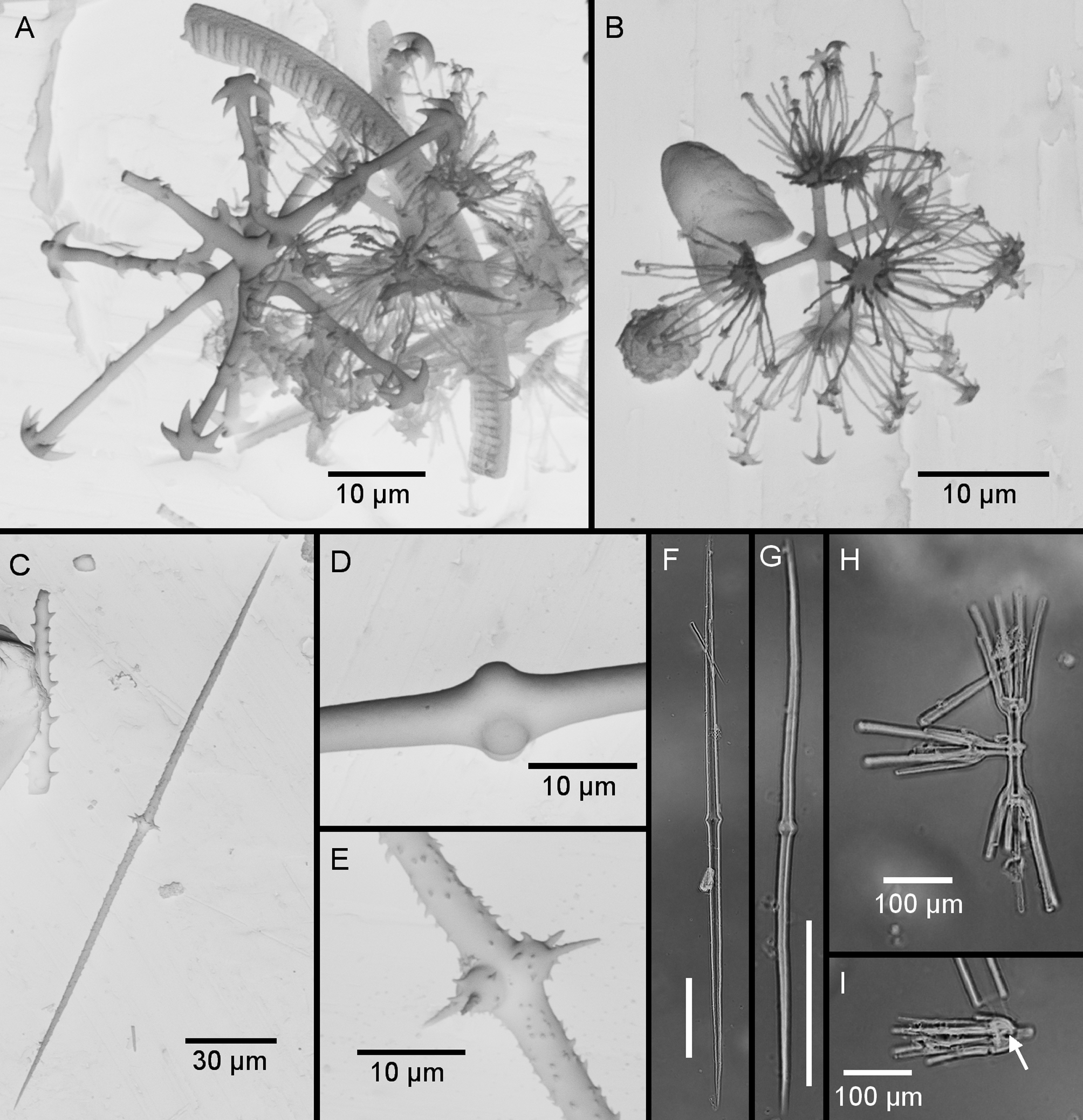
TABLE 3. Spicule sizes of Rossella fibulata Schulze & Kirkpatrick, 1910. Values in μm are given as follows: minimum - mean - maximum (number of spicules measured). For comparison, values from Schulze & Kirkpatrick (1910 b) and Barthel & Tendal (1994) are given.
| parameter | SMF 11906 | SMF 11732 | Schulze & Kirkpatrick Barthel & Tendal (1910b) (1994) |
|---|---|---|---|
| rough Pentactin | |||
| tangential ray (L) proximal ray (L) rough Hexactin (D) | 70–104.3–150 (30) 80–87.5–100 (6) 105–172.1–330 (33) | 80–112.5–180 (30) 70 (1) 150–195.2–250 (33) | 146 115 |
| Stauractin ray (L) Oxyhexactin (D) | 90–134.2–240 (30) | 90–144–220 (35) | 100–120 |
| Oxyhexaster (D) | 75–100.3–120 (30) | 70–93.5–125 (33) | 80 |
| Heterodiactin (L) Discohexactin (D) Microdiscohexaster (D) | 220–649.3–1150 (30) 25–33.2–42.5 (31) | 150–517.1–960 (34) 25–31–37.5 (30) | 160–600 <=600 40 |
| Mesodiscohexaster (D) Calycocome | 52.5 (1) | 42.5 (1) | |
| (D) | 164 164–350 | ||
| complete ray (L) primary ray (L) middle piece (L) | 180 (1) 15 (1) 22.5 (1) | 16.5 16 10 10–25 | |
| secondary ray (L) number of sec. rays | 142.5 (1) 4 (1) | 4–7 | |
| Curvature | S: 1 |
No known copyright restrictions apply. See Agosti, D., Egloff, W., 2009. Taxonomic information exchange and copyright: the Plazi approach. BMC Research Notes 2009, 2:53 for further explanation.