
Scottia birigida, Smith, Robin J., Matzke-Karasz, Renate, Kamiya, Takahiro & Ikeda, Yoshinori, 2002
|
publication ID |
https://doi.org/ 10.5281/zenodo.156019 |
|
DOI |
https://doi.org/10.5281/zenodo.6277509 |
|
persistent identifier |
https://treatment.plazi.org/id/633787C8-FFD6-FFC1-FE80-F4E37ADFF8BC |
|
treatment provided by |
Plazi |
|
scientific name |
Scottia birigida |
| status |
sp. nov. |
Scottia birigida View in CoL sp. nov.
Type locality: The area directly adjacent to an artificial pond, Shinkai Pond, with a dam at one end within the grounds of Kanazawa University, Kakuma, Kanazawa, Ishikawa Prefecture, Japan (N36° 32’ 31.6” / E136° 42’ 12.4” (Tokyo map datum), map: NJ 53 12 5.9, Kanazawa, 1: 50 000). The water level falls a couple of meters during the spring when the water is used to flood rice fields. The small area immediately adjacent to the pond is flat, often water logged and covered with many fallen leaves during the autumn and winter. A small stream from a spring higher up the slope flows across the flat ground and feeds the pond. The surrounding slopes are thickly wooded with deciduous trees and small bamboo, which provide ample shade. The temperature of the area ranges from a high of 30 35 °C at the height of summer to 5 0 °C during the winter and the area is often covered with snow. Specimens of this species can be collected on the surface of wet fallen leaves and from the soil surface throughout the year.
Type material: All material was collected by R.J.S. and T.K. from the type locality.
Holotype: a male, with soft parts dissected in glycerine and sealed in a glass slide, and valves stored in a cavity slide. (Number: UMUT RA 28198).
Allotype: a female, with soft parts dissected in glycerine and sealed in a glass slide, and valves stored in a micropalaeontological slide. (Number: UMUT RA 28201).
Paratypes: three females, with soft parts dissected in glycerine and sealed in a glass slide, and valves stored in a cavity slide. (Numbers: UMUT RA 28202 28204).
Two males, with soft parts dissected in glycerine and sealed in a glass slide, and valves stored in a cavity slide. (Numbers: UMUT RA 28199 28200)
Two whole carapaces of undetermined sex. (Numbers: UMUT RA 28207 28208)
Two freeze dried specimens. (Numbers: UMUT RA 28205 28206)
Location of material: All material is deposited in the University Museum, University of Tokyo, 731 Hongo, Bunkyoku, Tokyo 1130033, Japan.
Derivation of name: This species is characterised by its two rigid and erect lobes on the hemipenis, the medial and dorsal lobes of the lateral shield, hence birigida from: rigidus (rigid) and bis (twofold).
Diagnosis: Carapace less than 680 µm in length and with highly arched dorsal margin. Dorsal view, left valve with small lobelike extension overlapping right valve towards anterior margin. Male GM claw of antenna with large denticles along one edge and distally sinuously bent. Mandibular branchial plate with 4 long and 1 short setae. Male 5th limb prehensile palps lowly curved. Setae of endopodite of female 5th limbs of different length, 2 longer setae with long setules distally. Zenker organs with 14 17 whorls of spines. Hemipenes with erect projection on dorsal lobe of lateral shield. Internally with large beaklike expansion with parallel sides above bursa copulatrix.
Description of male
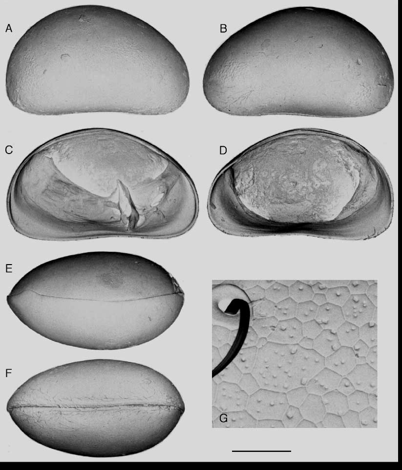
Carapace ( Figs 1 View FIGURE 1 , 2 View FIGURE 2 and 3 View FIGURE 3 ) colour variable, ranging from pale yellow to olive green or brown, but typically grading to grey towards dorsal margin. Lateral view, posterior more rounded than anterior, ventral margin slightly concave, and dorsal margin highly arched. Maximum height situated posterior of middle. Dorsal view, carapace ovoid, with anterior side slightly less rounded than posterior, left valve overlapping right valve. Small lobelike extension of left valve overlaps right valve towards anterior margin. Weakly developed lists (or selvages?) along ventral on both valves. Surface of valves smooth. Under high SEM magnification many small polygons, most of which pitted, covering surface. Hinge adont.
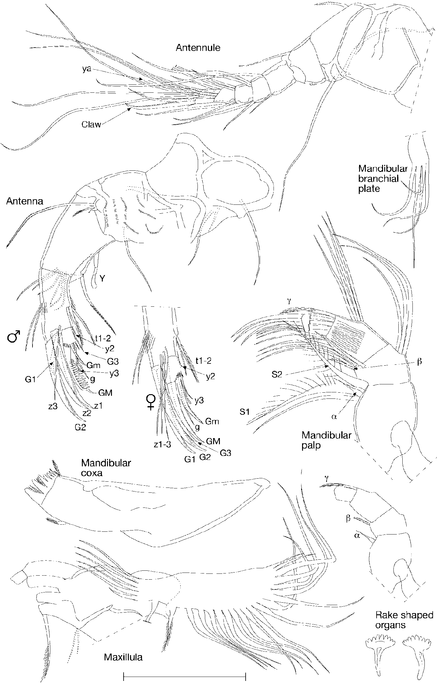
Antennule ( Fig. 4 View FIGURE 4 ) consisting of 7 podomeres. 1st podomere with 1 short dorsal seta and 2 long ventral setae. 2nd podomere with small Rome organ and 1 dorsoapical seta. 3rd podomere with 1 long dorsoapical seta and 1 short ventroapical seta. 4th and 5th podomeres both with 2 dorsoapical setae and 2 short ventroapical setae each. 6th podomere with 3 long and 2 short apical setae. Terminal podomere terminating with 2 long setae, 1 aesthetasc (ya) and 1 robust claw.
Base of antenna ( Fig. 4 View FIGURE 4 ) broad and well developed. Y aesthetasc large and with bulbous end. 6 natatory setae reduced, only reaching approximately 3/4 along length of 3rd podomere. 3rd podomere supports 1 small G1 claw, 3 larger claws, G2, z1 and z2, and 1 seta z3. z2 claw with well developed, rounded denticles towards distal end. Final podomere supports 1 tiny Gm claw, g seta, y3 aesthetasc and GM claw. GM claw set with series of large rounded denticles along one edge and distally sinuously bent.
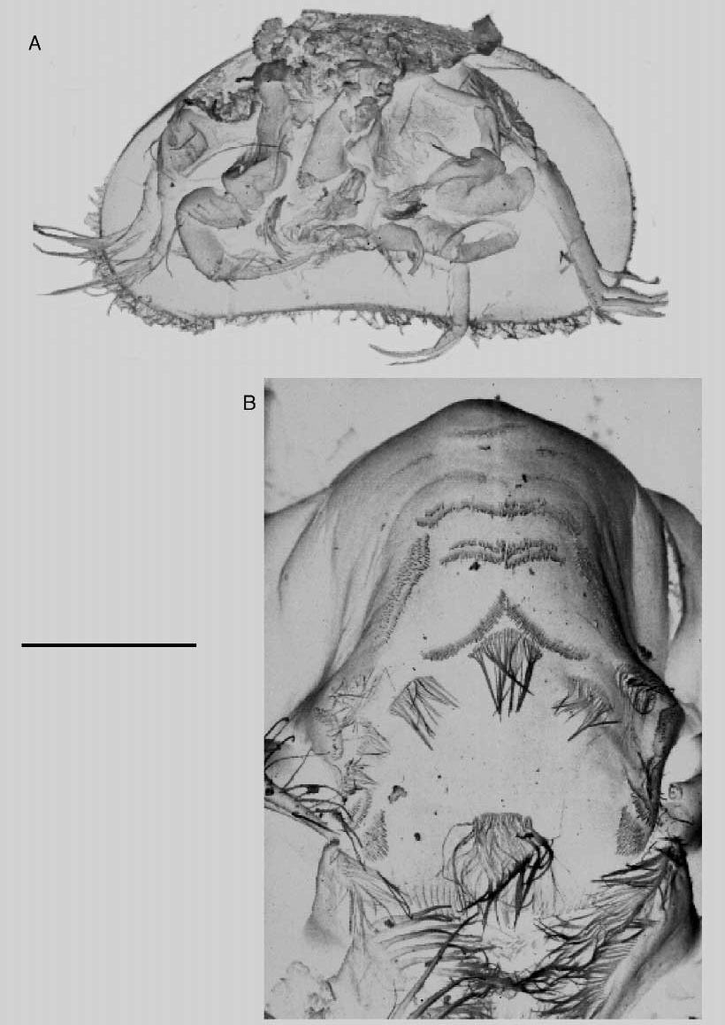
Mandibular coxa ( Fig. 4 View FIGURE 4 ) with 7 teeth and palp consisting of 4 podomeres. s1 and s2 setae with long, robust, evenly spaced setules. Alpha seta short and thin, with inflated base. Beta seta short and thin and accompanied by 4 long setae. Gamma seta thin and curved, and only slightly thicker than accompanying 3 setae. Palp terminates with 3 claws and 3 setae. Branchial plate with elongate base and with 4 long and 1 shorter setae.
Upper lip broad and without ridges ( Fig. 2 View FIGURE 2 ), sparsely covered with patches of pseudochaetae and microspines. Two tiny pores towards posterior edge. Central process small, lateral striations well developed. Hypostome densely covered with long pseudochaetae.
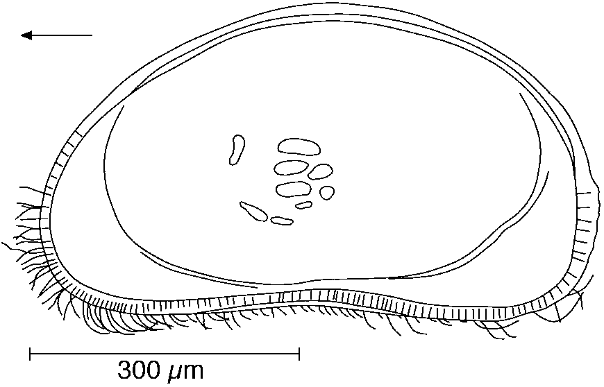
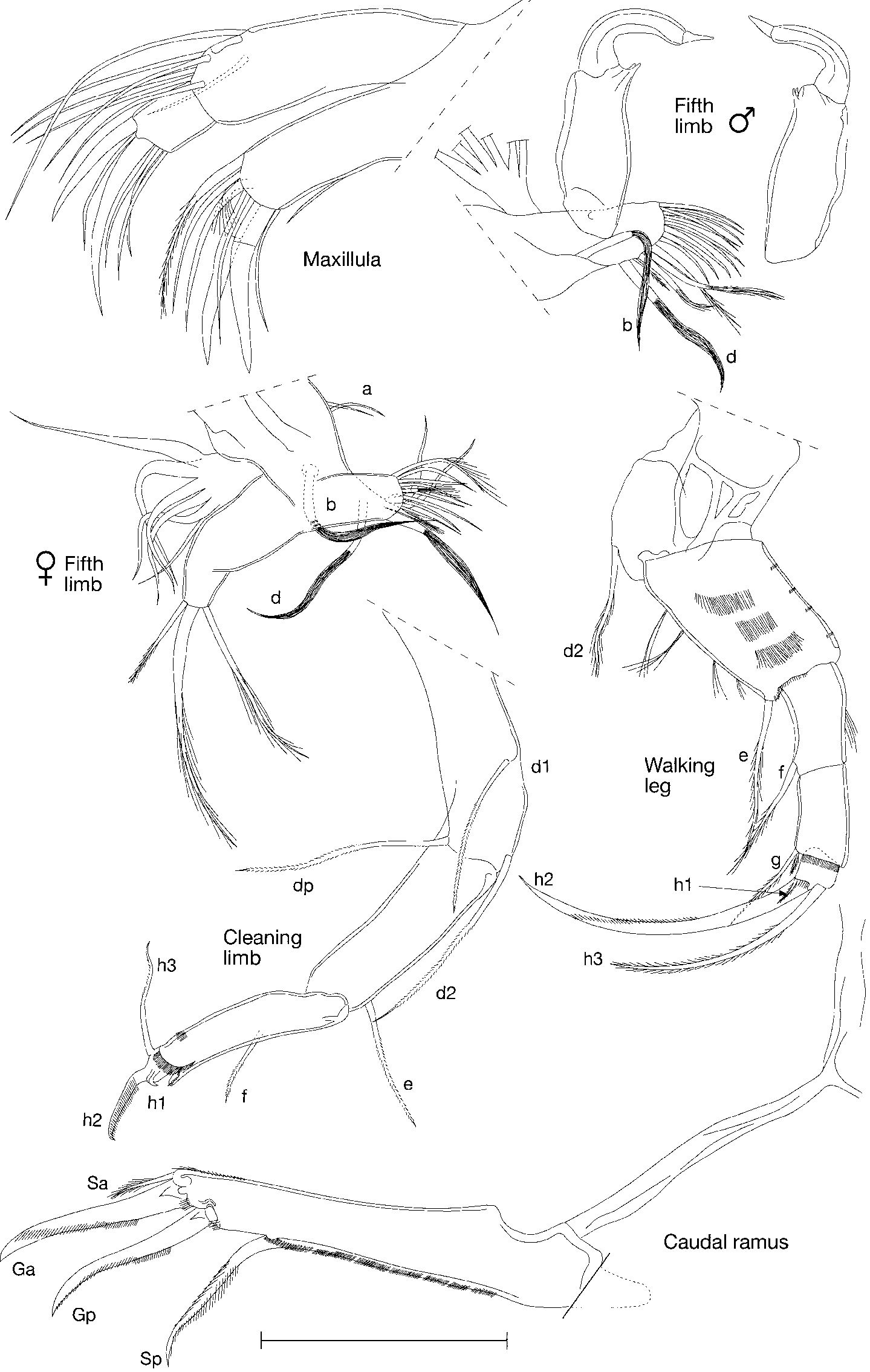
Maxillula ( Fig. 4 View FIGURE 4 ) consists of segmented palp and 3 well developed endites and branchial plate. 1st podomere of palp with 5 apical setae and 2 subapical setae. 2nd podomere with 2 stout claws and 4 setae. 3rd endite with 2 smooth and curved Zahnborsten accompanied by 5 long setae and 1 short, bent seta. 1st and 2nd endites terminate with numerous setae. 1 long, stout seta protrudes from inner edge of 1st endite. Branchial plate consists of 6 reflexed setae and 20 setae along ventral and posterior edges, 2 dorsal most setae reduced.
5th limb ( Fig. 5 View FIGURE 5 ) characterised by 2 setae (b, d) covered with long setules producing 'paintbrush' morphology, located on subapical position and inner apical corner of endite. Additionally, endite with 1 small, 1 medium length and 2 longer setulous setae and group of 10 medium length setae on apical edge. Endopodite elongate with 2 small sensory organs closely situated together in inner apical corner and with 1 large grasping hook. Hooks lowly arched and terminate with large, elongate coneshaped sensory organ. Hooks approximately symmetrical. Branchial plate with 6 setae.
Walking leg ( Fig. 5 View FIGURE 5 ) robust with 6 podomeres. Kneesegment podomere short and rounded and with well developed d2 seta. 2nd podomere elongate with long setules along ventral edge and long e seta. 3rd and 4th podomeres approximately same length and terminate with setae f and g respectively. Additionally, seta g accompanied by 1 tiny seta. Small, quadrate final podomere supports 1 tiny h1 seta, 1 well developed curved claw (h2) and 1 stout, long, curved seta (h3) approximately 75% of length of h2 claw.
Cleaning limb ( Fig. 5 View FIGURE 5 ) robust with long setae d1, d2 and dp on 1st podomere. 2nd podomere supports e seta. 3rd podomere with f seta approximately in mid length position. 4th podomere relatively elongate and supports h3 seta and 1 well developed and broad h2 seta with well developed denticles.
Caudal ramus ( Fig. 5 View FIGURE 5 ) well developed and robust. Claws Ga and Gp approximately same length, although Ga broader. Both claws gently curved. Sa seta short, less than half length of Ga. Seta Sp long, broad and stout. Attachment long and broad, and bifurcates distally, with small ventral branch and much longer and broader dorsal branch.
Zenker organs ( Fig. 6 View FIGURE 6 ) with 14 17 internal whorls of spines.
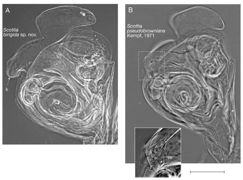
Hemipene ( Fig. 6 View FIGURE 6 ) lateral shield welldeveloped; medial lobe of lateral shield lowly arched and dorsal lobe with well developed projection pointing proximally (k). Ventral lobe of medial shield inflated and medial lobe of ventral shield elongate and sinuously curved. Some specimens with prelabyrinthal inner spermiduct 'S' shaped and lying over coiled postlabyrinthal inner spermiduct, but in other specimens (e.g. holotype) 'S' shaped curve absent. Labyrinth small and connected to coiled postlabyrinthal inner spermiduct by broad duct. Coils of postlabyrinthal inner spermiduct situated below level of dorsal lobe. Inner edge of bursa copulatrix thickened. Beaklike projection with subparallel sides, just above bursa copulatrix.
Description of female
Carapace, antennula, mandible, maxillula, walking leg, cleaning limb and caudal ramus of female similar to those of male.
3rd and 4th podomeres of antenna are sexually dimorphic ( Fig. 4 View FIGURE 4 ). 3rd podomere claws G1 3 gently curved with G3 slightly shorter than other two. Setae z1 3 reach approximately 3/4 of length of G1 claw. Gm claw of 4th podomere approximately 3/4 length of accompanying GM claw. Seta g and aesthetasc y3 similar to those of male.
h1 and h2 setae of 5th limb endopodite both covered distally with long setules, with h1 slightly shorter than h2 ( Fig. 5 View FIGURE 5 ). h3 seta short and with short setules distally.
Measurements:
UMUT RA 28198 male (holotype) L: 622 µm, H: 367 µm
UMUT RA 28199 male (paratype) L: 633 µm, H: 352 µm
UMUT RA 28200 male (paratype) L: 644 µm, H: 363 µm
UMUT RA 28201 female (allotype) L: 652 µm, H: 356 µm
UMUT RA 28202 female (paratype) L: 659 µm, H: 393 µm
UMUT RA 28203 female (paratype) L: 663 µm, H: 381 µm
UMUT RA 28204 female (paratype) L: 630 µm, H: 363 µm
Differential diagnosis: Scottia birigida sp. nov., S. pseudobrowniana and S. audax all have similar carapace shape in lateral view, but differ from each other in size. S. audax is the largest with a length of approx. 1140 µm, S. pseudobrowniana is an intermediate size typically 750 830 µm in length (although Danielopol and Vespremeanu (1964), reported S. pseudobrowniana from Romania with an average length of 650 µm, and US specimens have been reported with a length of 700 µm, ( Cole 1966; Külköylüoglu and Vinyard 2000), see below) and S. birigida is the smallest with a length of 622 663 µm. The carapace of S. audax is densely covered with setae, whereas dense setae are mostly confined to the posterior and anterior regions of the ventral margin in the other two species.
The appendages of S. birigida are generally more similar to those of S. pseudobrowniana than to S. audax , but there are many significant differences: In Scottia birigida the terminal claw of the antennule is distally curved, in contrast to the straight claw of S.
pseudobrowniana . The male GM claw of the antennae of S. birigida is distally sinuously bent, whereas the male GM claw of S. pseudobrowniana is without such a pronounced bend. S. birigida has 5 setae on the mandibular branchial plate whereas S. pseudobrowniana has 7 setae. The arrangement of the setae on the endite of the 5th limbs are different between the two species. The h3 seta of the cleaning limb is shorter than the penultimate podomere in S. birigida (80% or less of length), whereas it is as long or longer than this podomere in S. pseudobrowniana . The Zenker organs of S. birigida have only 1417 whorls of spines, compared with the 1921 whorls of spines of S. pseudobrowniana . The hemipenes are of a different shape in the two species, with S. birigida having a proximally pointing projection on the dorsal lobe of the lateral shield (labelled k in Figs 6 View FIGURE 6 & 7 View FIGURE 7 ), whereas this projection in S. pseudobrowniana is folded under the hemipenis. Additionally, the ventral part of the medial shield is more inflated in S. birigida than in S. pseudobrowniana (labelled m in Figs 6 View FIGURE 6 & 7 View FIGURE 7 ). Internally, the bursa copulatrix is more kinked on the inner edge of S. pseudobrowniana than in Scottia birigida . The beaklike structure above the bursa copulatrix has more parallel sides in Scottia birigida , with this structure being thicker at the base and thinner on the outer margin in S. pseudobrowniana ( Figs 6 View FIGURE 6 & 7 View FIGURE 7 ).
Ecology and life history: The main habitat of Scottia birigida is an area approximately 30 cm wide each side of a small, shallow channel (20 cm wide, 1 cm deep). The area is always wet with a film of water 2 to 3 mm deep. Scottia birigida is found in this area all through the year, both from the soil surface and from fallen leaves. In summer Scottia birigida is not found outside this zone, but in autumn, winter and spring the surrounding area is covered with fallen leaves and this creates a much wider suitable habitat for this species; consequently it can be found over a much larger area than in the summer.
The main breeding season is in the early summer (May to July) and it produces one generation a year (Appendix 1). A8 instars generally appear in May and the number of juveniles increases through June so that the maximum number of juveniles is reached in July (2360 juveniles and 32 adults in an area of 450 cm 2). However, small numbers of early juveniles (A7 onwards) have also been recovered during January. Most juveniles have been recovered from the surface of the soil under the wet leaves within 30 cms of the channel, with lesser numbers of juveniles from the leaves. Further than 30 cms from the channel the majority of individuals are adults, with generally higher numbers living on the surface of the soil rather than the overlying leaves.
Discussion
S. birigida is the first species of Scottia and the subfamily Scottiinae to be reported from Japan and North East Asia.
S. birigida is only the third living species of Scottia to be described, and most closely resembles the western European populations of S. pseudobrowniana (e.g. MatzkeKarasz, 1995). Some descriptions of S. pseudobrowniana from other areas differ from those of western European S. pseudobrowniana . For example, Bronstein's (1947) reports of S. pseudobrowniana (as S. browniana ) from Russia, lack the dorsal lobe on the left valve, have one thin palp on the male fifth limbs and have longer claws on the female antenna, features which, if confirmed, would perhaps set this population apart from S. pseudobrowniana . Külköylüoglu and Vinyard (2000) reported S. pseudobrowniana from Nevada, USA. However, their drawings only have six podomeres in the antennule (in contrast to seven in European S. pseudobrowniana and other American specimens), much shorter and less numerous setae on the antennule, very short natatory setae on the antenna and a finger like projection on the hemipenes.
Such variation in some features previously reported within S. pseudobrowniana could be due to errors in drawings. If such differences are not errors, however, then there is greater variation within reported S. pseudobrowniana than the differences between European S. pseudobrowniana and Scottia birigida . Scottia birigida , however, is clearly a separate species as shown by the unique features of the hemipenes, the Zenker organs and other features revealed by detailed micromorphological investigation. This may indicate that there are more species of Scottia yet to be described and we recommend detailed investigation of material from Russia and the USA to determine if these records are from different species.

| UMUT |
University Museum, University of Tokyo |
No known copyright restrictions apply. See Agosti, D., Egloff, W., 2009. Taxonomic information exchange and copyright: the Plazi approach. BMC Research Notes 2009, 2:53 for further explanation.