
Siconema diducuncinum, Luc, Pham Van, 2012
|
publication ID |
https://doi.org/ 10.5281/zenodo.212402 |
|
DOI |
https://doi.org/10.5281/zenodo.5671809 |
|
persistent identifier |
https://treatment.plazi.org/id/3D0087C1-FFC7-B611-9DD6-FC71C2447458 |
|
treatment provided by |
Plazi |
|
scientific name |
Siconema diducuncinum |
| status |
sp. nov. |
Siconema diducuncinum sp. n.
( Fig. 3 View FIGURE 3 & 4 View FIGURE 4 )
Measurements: Table 2 View TABLE 2
Adult: Body moderately slim, expanding at level of caudal organs and tapering to both ends, covered by folded cuticular membrane apparent in anterior region. Cuticle thin, smooth. Lateral fields ca. 23–27 µm wide at mid-body, displaced dorsally in posterior region; smooth, margins marked by 2 rows of ladder-like ‘stitches’ of modified cuticle 3–4 µm wide. Head inclined dorsally bearing pair of dorsally directed head hooks. Hook base thick; embedded in head tissue; divided into left and right parts. Hook blades widely spaced, proximally curved but distally directed almost parallel to hook base; thin, conical, with rounded tips. Mouth confined in heavily cuticularised tube ca. 3–5 µm high and 2–3 µm in diam. situated between hooks. Amphids situated closely to hook base; pocket-like with small pouches and half-moon-like apertures surrounded by thickened rim ca. 1 µm thick. Pharynx reaching bottom of mouth tube; comprising long, curved, uniformly broad, very finely muscled corpus and twice shorter and wider glandular, pear-shaped, displaced dorsally bulb. Isthmus not pronounced. Thick nerve ring crossing corpus anterior to bulb. Excretory pore situated on eminence opposite anterior part of bulb. Excretory duct heavily cuticularised, ca. 70 µm long and 2 µm wide. Excretory gland extending from proximity of excretory pore at least to mid-body. Excretory channels weakly cuticularised, ca. 5 µm wide. Intestine transparent. Cardia small. Caudal organs paired, causing swelling of middle part of tail; longitudinally elliptical, flat or hollow-like, symmetrically disposed. Each organ externally surrounded by thin rim. Posterior to caudal organs, tail narrowing to conical tip.
Female: Total hook height 17.3 ± 2.3 (16–20) µm. Blades 12.3 ± 1.5 (11–14) µm long. Stomatal tube 3.5 ± 0.7 (3–4) µm high and 3 ± 1.4 (2–3) µm wide. Amphids ca. 5– 8 x 2–4 µm in size. Anus and rectum not discernible. Prodelphic, monodelphic. Ovary tip located near tail proximity. Ovary rounding caudal organs on dorsal side and making several twists between caudal organs and vulva, then running anteriad and turning back in ca. 100 µm posterior to pharynx. Spermatheca 173.3 ± 45.1 (130–220) µm long and 20 ± 2 (38–42) µm wide, not offset but morphologically distinct from oviduct. Oviduct long, thick-walled; uterus thin-walled. Small appendage ca. 25–30 µm long (nonfunctioning post-uterine sac?) present posterior to vulva. Vagina ca. 27 µm long, straight or slightly inclined posteriad. Vulva lips very slightly inflated. Eggs prolate-oval, with two slightly protruding knobs ca. 7 µm in diam. Eggshells 1.7 ± 0.6 (1–2) µm wide, covered in small dots more densely distributed on poles. Caudal organs elliptical, 108.3 ± 7.6 (100–115) µm long and 51.7 ± 10.4 (40–60) µm wide.
Male: About twice shorter than females. Anterior end similar in structure to female. Stomatal tube 5 ± 1.4 (4–6) µm high and 3.5 ± 0.7 (3–4) µm wide. Total hook height 16.5 ± 1.7 (15–19) µm. Blades 13.3 ± 0.3 (13–14) µm long. Intestine transparent, thin. Testis thin, reflexing in 528 ± 38 (478–564) µm from anterior, flexure 176 ± 22 (143–190) µm long. Spermatocytes in 2, then 3 rows, spherical. Spermatids ca. 6– 8 x 7–10 µm in size. Immature sperm spherical; ca. 5 µm in diam. Vas deferens set off by constriction. Cloaca encircled by thickened copulatory disc ca. 30 µm in diameter. Tail elongated and slightly swollen anterior to its middle because of caudal organs. These are longitudinally oval to circular spots lacked cuticle with a centrally positioned pore not opened on surface. One pair of short ventral papillae on level of anterior third of caudal organ. Portion of tail posterior to caudal organs long, narrowly conical.
Female Male Type-material. Holotype female (on the slide accession No. 1127) and paratype male and female in copula (on the slide accession No. 1128) deposited in the Museum of the Helminthological Collections of the Centre of Parasitology at the Severtsov Institute of Ecology and Evolution, Moscow. Three paratype males and 3 paratype females, each on a separate slide, are in the senior author’s collection.
Type-host and locality. Amynthas tuberculatus collected in Pu Mat Nature Reserve, prov. Nghe An, Vietnam, November 2008, by S. Spiridonov.
Etymology. The species name is derived from Latin words ‘ diduco’ —spread apart and ‘ uncinus’ —small hook and reflects the particular appearance of cephalic hooks.
Diagnosis and relationships. Siconema diducuncinum sp. n. is characterised by a stoma confined in the tube protruding between long, thin cephalic hooks with widely distributed blades, caudal organs situated at a long distance from the tail extremity, eggs shaped similarly to a double-poled elongated lemon and a copulatory disc in males.
By having similar body size and shape, S. diducuncinum sp. n. resembles S. sinense Timm, 1966 , S. ovicoronatum Timm, 1966 , S. bahli Spiridonov & Danilova, 1986 , and S. neozelandicum Yeates & Spiridonov, 1996 .
It is most similar to S. sinense due to its similar body and cephalic hooks size, the caudal organs and vulva position and long, similarly shaped cephalic hooks. It differs from S. sinense by having a rounded vs elongate pharyngeal bulb, thinner (1 vs 5 µm) and smoother eggshells with more pronounced polar caps, a twisted vs straight ovary and a copulatory disc in males. The original description of S. sinense had presented no indication to whether cephalic hooks were diverged or closely placed but from an illustration it can be assumed that the latter is more probable.
From S. ovicoronatum the present species can be distinguished by a twisted vs straight ovary, smaller (av. 57.7 x 26.3 µm vs av. 70 x 40 µm) eggs with differentiated poles vs not differentiated and much thinner eggshells (ca. 1.7 vs 6–7 µm thick) with different pattern of ornamentation (thinly mamillate vs mamillate, marked with thin irregular filamentous rods and the shorter, tapering tail spike (av. 215.7 µm vs av. 320 µm long).
By similar body size and conical tails S. diducuncinum sp. n. shares similarities with S. bahli but differs by having longer, diverging hook blades with a thinner base, more posteriorly placed caudal organs and the different ornamentation of eggshells (punctated with two polar caps vs papillate with a single polar cap).
By having an elongated tail spike and bristling hook blades, S. diducuncinum sp. n. shares similarities with S. neozelandicum . However, in the latter the base of hooks is solid vs consisting from two not joined parts and mouth is not confined into a projecting tube but reduced; eggshells tuberculate vs punctate and without prominent polar differentiation vs with polar differentiation. Males of S. diducuncinum sp. n. can be distinguished from S.
neozelandicum by the presence vs absence of the copulatory disk and the absence vs presence of an aperture in the caudal organ.
Remarks. Males of Siconema are characterised as being of smaller size than females (from only slightly smaller to nearly twice as that) and different from female shape of caudal region and caudal organs while retaining the same structure of anterior end. Owing to the fact that males were described for not all members of the genus, we present a key to Siconema based of female characters only. Main diagnostic characters of Siconema females include the size and shape of head hooks, the size, shape and ornamentation of eggshells, the shape of a posterior body portion determined by the presence of caudal organs, the location and appearance of caudal organs and the arrangement of the ovary. Such important features as head hooks were less used in the key because many of original descriptions illustrate head hook structure in lateral position only. The natural dorsal inclination of a nematode’s anterior end and dorsally directed head hooks rarely permit mounting nematodes in a way showing en face or dorsal view of hooks unless provided by occasional contortion of body.
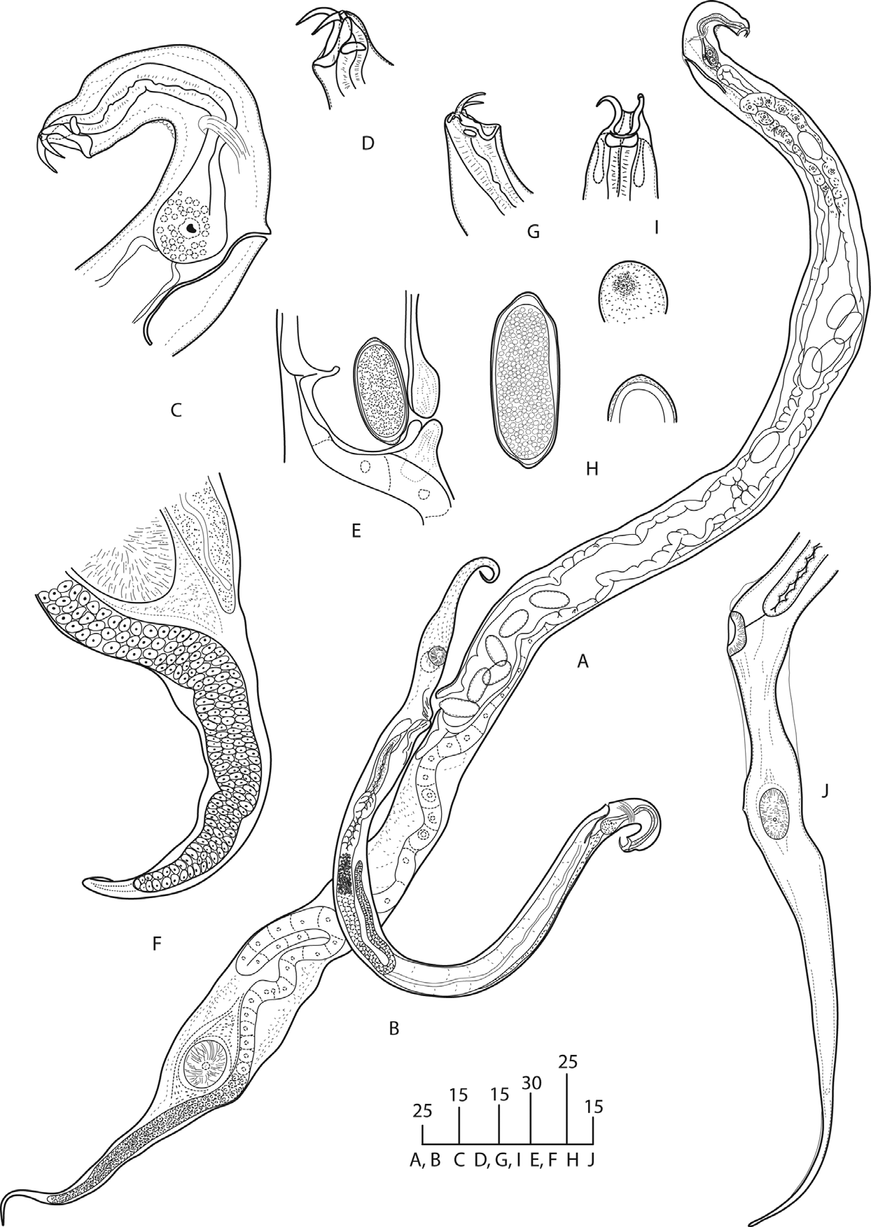
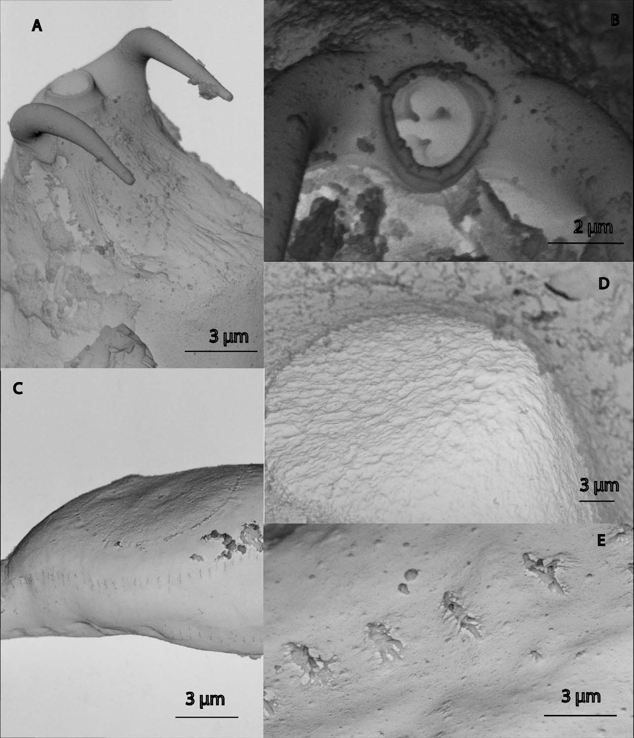
TABLE 2. Morphometrics of Siconema diducuncinum sp. n. Measurements are ranges in µm.
| Holotype | Paratypes | Paratype |
|---|---|---|
| n | 3 | 4 |
| L 2080 | 2246±169 | 1228±162 |
| a 13.9 | 24.2±15 | 24.3±3.3 |
| b 13.9 | 4.4±1 | 8.4±0.7 |
| c | 4.5±0.3 | |
| V 57.7 | 60.9±1.3 | |
| Mid-body diam. 150 | 94±12 | 47±2 |
| Length of pharynx 150 | 150±6 | 150±9 |
| Distance from head to excretory pore 113 | 124±10 | 131±5 |
| Distance from head to nerve ring 90 | 103±5 | 88±5 |
| Length of tail | 273±53 | |
| Egg length 58 | 58±3 | |
| Egg diam. 25 | 26±1 |
No known copyright restrictions apply. See Agosti, D., Egloff, W., 2009. Taxonomic information exchange and copyright: the Plazi approach. BMC Research Notes 2009, 2:53 for further explanation.