
Simulium (Nevermannia) subratai, Takaoka, Hiroyuki, Thapa, Sachin & Henry, Willie, 2011
|
publication ID |
https://doi.org/ 10.5281/zenodo.207675 |
|
DOI |
https://doi.org/10.5281/zenodo.6192690 |
|
persistent identifier |
https://treatment.plazi.org/id/03F487D6-FFB2-FFDD-FF07-BF1BFE24FBF3 |
|
treatment provided by |
Plazi |
|
scientific name |
Simulium (Nevermannia) subratai |
| status |
sp. nov. |
Simulium (Nevermannia) subratai View in CoL sp. nov.
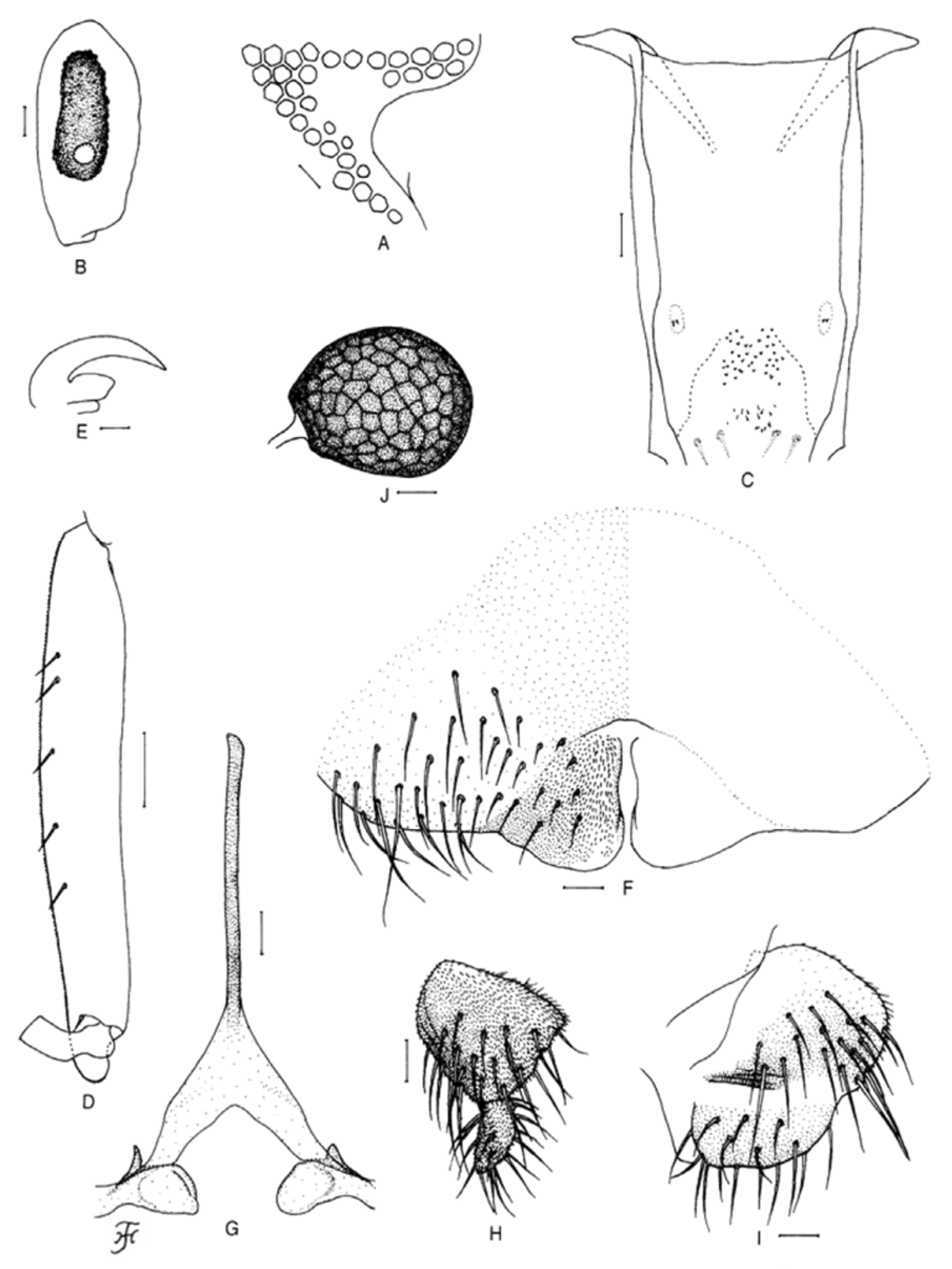
Description. Female (Photo. 1). Body length 2.4 mm. Head. Slightly narrower than thorax. Frons dark brown, densely covered with whitish-yellow recumbent hairs interspersed with several dark brown longer and stouter hairs along each lateral margin. Frontal ratio 1.81:1.00:2.57. Frons-head ratio 1.00:4.96. Fronto-ocular area ( Fig. 1 View FIGURE 1 A) well developed, directed laterally and slightly upward, and with rounded apex. Clypeus dark brown, densely covered with whitish-yellow recumbent hairs (except portion near upper margin bare) intermixed with several dark longer and stouter hairs on each side. Labrum 1.11 times as long as clypeus. Antenna composed of scape, pedicel and 9 flagellomeres, dark brown except scape, pedicel and basal 1/3 of 1st flagellomere yellow. Maxillary palp consisting of 5 segments, proportional lengths of 3rd, 4th, and 5th segments 1.00:0.83:1.41; 3rd segment ( Fig. 1 View FIGURE 1 B) much enlarged; sensory vesicle ( Fig. 1 View FIGURE 1 B) elongate, 0.59 times as long as 3rd segment, with medium-sized opening. Maxillary lacinia with 9 inner and 12 or 13 outer teeth. Mandible with 20 inner teeth (of which few basal teeth are very tiny) and lacking outer teeth. Cibarium ( Fig. 1 View FIGURE 1 C) with 43 minute conical processes with pointed apices as well as several minute spinous processes near lower margin. Thorax. Scutum yellow, densely covered with whitish-yellow recumbent hairs intermixed with several dark brown upright hairs on prescutellar area. Scutellum yellow, densely covered with whitish-yellow hairs interspersed with dark brown hairs. Postnotum yellow with somewhat darkened area and bare. Pleural membrane yellow and bare. Katepisternum longer than deep, yellow and bare. Legs. Foreleg: coxa and trochanter yellow; femur yellow with apical cap dark brown; tibia dark brown except median large portion of outer surface light brown; tarsus dark brown; basitarsus slender, slightly dilated, 7.33 times as long as its greatest width. Midleg: coxa yellow except posterolateral surface medium to dark brown; trochanter whitish-yellow; femur whitish-yellow with apical cap medium brown; tibia light to dark brown except medial large portion on anterior and lateral surface light grayish; tarsus dark brown. Hind leg: coxa and trochanter yellow; femur whitish-yellow with apical cap medium brown; tibia light to dark brown except medial large portion on anterior and lateral surfaces light grayish; basitarsus light grayish except base medium brown; rest of tarsus dark brown except basal 1/2 of 2nd tarsomere light grayish; basitarsus ( Fig. 1 View FIGURE 1 D) nearly parallel-sided from base to middle, then slightly narrowed toward apex 5.74 times as long as its greatest width, and 0.83 and 0.68 times as wide as hind tibia and femur, respectively; calcipala ( Fig. 1 View FIGURE 1 D) well developed, slightly shorter than its basal width, and 0.49 times as wide as greatest width of basitarsus; pedisulcus ( Fig. 1 View FIGURE 1 D) well developed. Claw ( Fig. 1 View FIGURE 1 E) with large basal tooth 0.46 times as long as claw. Wing. Length 2.4 mm. Costa with 2 parallel rows of dark brown spinules as well as dark brown hairs. Subcosta with dark brown hairs except near apex bare. Basal portion of radius fully haired; R1 with dark brown spinules and hairs; R2 with dark brown hairs. Hair tuft on stem vein dark brown. Basal medial cell absent. Abdomen. Basal scale whitish-yellow, with fringe of whitish-yellow long hairs intermixed with 4 dark brown hairs. Segments 2 and 3 whitish-yellow, moderately covered with whitish-yellow short hairs interspersed with dark brown hairs dorsally, segments 4–9 whitish-yellow to light brown, widely mottled on dorsolateral and/or lateral surfaces with reddish-brown pigment markedly on segments 5 and 6, moderately on segment 4 and weakly on segment 7; segments 4–9 moderately covered with dark brown hairs; tergites 6–8 appearing very faintly shiny when illuminated at certain angle of light; ventral surface of segment 7 with large sternal plate medially. Genitalia. Sternite 8 ( Fig. 1 View FIGURE 1 F) wide, bare medially but furnished with 29 short to long hairs on each side. Ovipositor valves ( Fig. 1 View FIGURE 1 F) triangular (though posteromedial corner rounded), thin, membranous except inner margin narrowly sclerotized, densely covered with microsetae interspersed with 6 or 7 short to medium-long hairs; inner margins sinuous, narrowly separated from each other. Genital fork ( Fig. 1 View FIGURE 1 G) of inverted Y-form, with well sclerotized stem and relatively wide arms; each arm with lateral plate bearing round lobe-like projection directed medioposteriorly and short narrow stout projection directed anterodorsally. Paraproct in ventral view ( Fig. 1 View FIGURE 1 H) subquadrate, slightly longer than its greatest width; anteromedial surface nearly transparent, with 5 or 6 sensilla; paraproct in lateral view ( Fig. 1 View FIGURE 1 I) somewhat protruded ventrally beyond ventral margin of cercus, and with 19–21 medium to long hairs on ventral and lateral surfaces. Cercus in lateral view ( Fig. 1 View FIGURE 1 I) rounded posteriorly, short, 0.38 times as long as basal width. Spermatheca ( Fig. 1 View FIGURE 1 J) nearly ovoidal, 1.23 times as long as its greatest width, strongly sclerotized except small area around juncture with duct and duct itself unsclerotized, with distinct reticulate surface pattern and without internal setae; main spermathecal duct narrow, somewhat narrower than both accessory ducts.
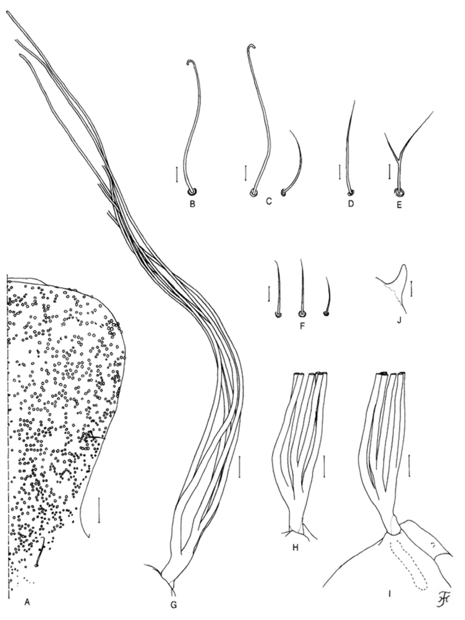
Pupa. Body length 3.0 mm. Head. Integument yellow to light brown, moderately covered with round tubercles of various sizes (0.003–0.008 mm in diameter) on frons ( Fig. 2 View FIGURE 2 A) and sparsely covered with similar tubercles of various sizes on lateral surface of face; antennal sheath bare; frons with 2 short simple trichomes ( Fig. 2 View FIGURE 2 A) on each side; face with 1 medium-long simple trichome ( Fig. 2 View FIGURE 2 A) on each side. Thorax. Integument yellow to light brown, moderately covered with round tubercles of different sizes (0.003–0.006 mm in diameter) on dorsal and lateral surfaces of anterior 1/2 and sparsely covered with smaller ones on dorsal surface of posterior 1/2; thorax on each side with 3 long simple trichomes with coiled apices mediodorsally ( Fig. 2 View FIGURE 2 B), 2 simple trichomes (1 long with coiled apex, 1 medium-long with uncoiled apex) anterolaterally ( Fig. 2 View FIGURE 2 C), 1 medium-long trichome with uncoiled apex (simple on right side—Fig. 2D, bifid on left side—Fig. 2E) mediolaterally and 3 simple trichomes with uncoiled apices (2 medium-long, 1 short) ventrolaterally ( Fig. 2 View FIGURE 2 F). Gill ( Fig. 2 View FIGURE 2 G–I) with 6 long thread-like slender filaments, arranged in 3 pairs (1 inner dorsal, 1 outer dorsal and 1 outer ventral); 2 outer pairs sharing very short stalk; all pairs short-stalked, stalks of outer pairs subequal in length to each other and longer than that of inner dorsal pair; common basal stalk short; all filaments grayish-brown, tapered toward tip, extending close together anteriorly, subequal in length (3.2–3.4 mm) and thickness to one another (though filaments of inner dorsal pair very slightly thicker than others when compared basally); cuticular surface with distinct annular ridges and furrows, and moderately covered with minute tubercles of different sizes (larger ones on ridges and smaller ones on interridges). Abdomen. Dorsally, segments 1–4 weakly sclerotized and light brown; segment 1 moderately covered with minute tubercles, with 1 medium-long slender simple seta on each side; segment 2 moderately covered with minute tubercles and with 1 medium-long slender simple seta and 5 very short spinous setae on each side; segments 3–8 without minute tubercles; segments 3 and 4 each with 4 hooks and 1 very short spinous seta on each side; segment 5 bare; segments 6–8 each with spine-combs directed backward in transverse row and comb-like groups of minute spines on each side; segment 9 yellow, with pair of distinct conical terminal hooks ( Fig. 2 View FIGURE 2 J) as well as comb-like groups of minute spines. Ventrally, segments 3–8 with comb-like groups of minute spines; segment 4 with 1 simple hooklet and few short simple slender setae on each side; segment 5 with pair of bifid hooks submedially and few very short slender setae on each side; segments 6 and 7 each with pair of bifid inner and simple outer hooks and few slender very short setae on each side. Cocoon. Simple, wall-pocket shaped, compactly woven without open spaces in web, with anterior margin thickly woven, without anterodorsal projection or bulge, and widely extending ventrolaterally; individual threads invisible; 4.0 mm long by 2.5 mm wide.
PHOTO 1. The whole body of an adult female (lateral view) of Simulium (Nevermannia) subratai sp. nov. Preserved in 80% ethanol after reared from a pupa, and photographed by a digital imaging system of Olympus DP25. Body length 2.4 mm.
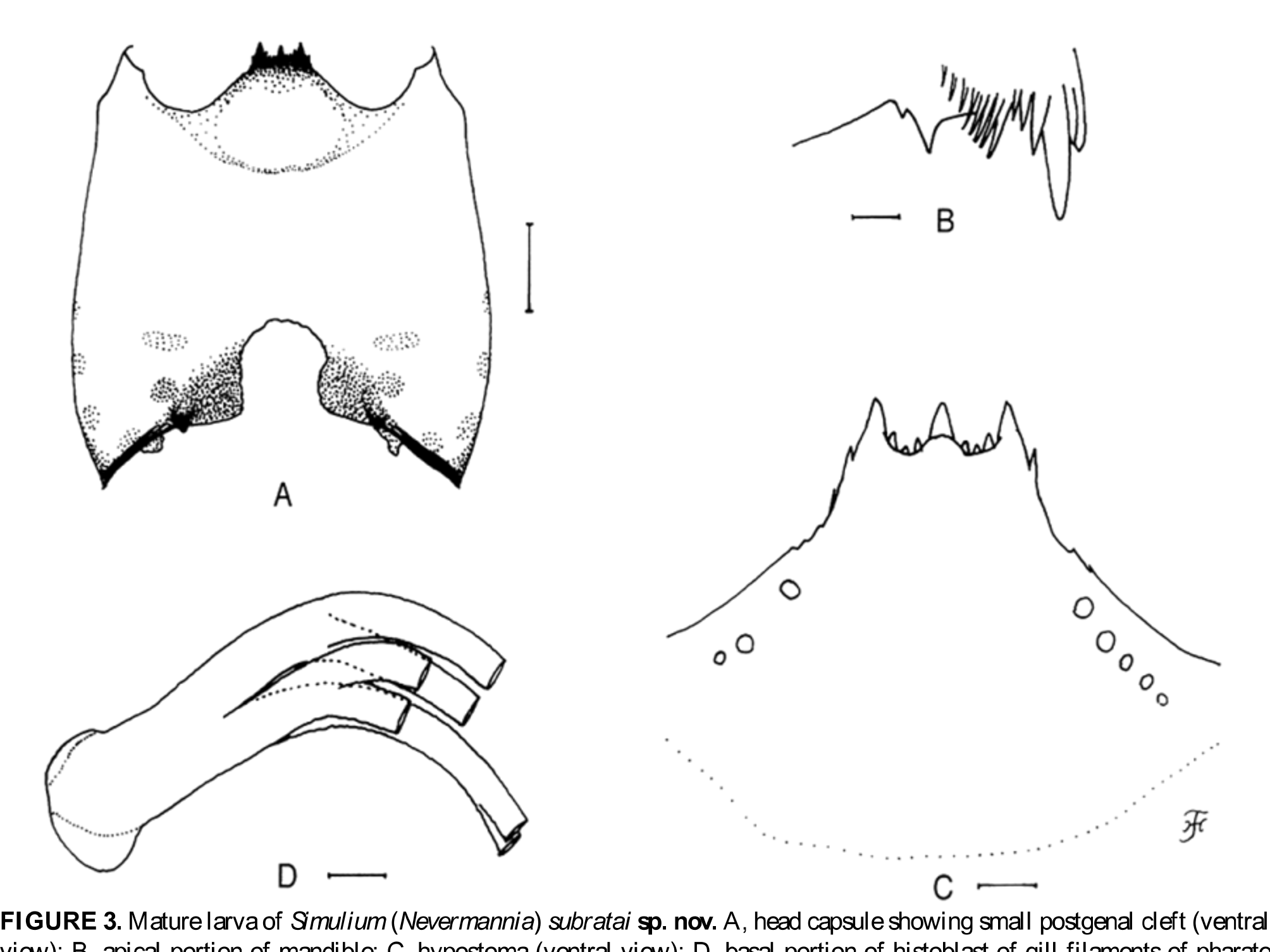
Mature larva. Body length 5.1 mm. Body creamy, with grayish band or area on thoracic segments 1 and 3 and abdominal segments 1–4. Cephalic apotome whitish-yellow to yellow; head spots very faintly positive. Lateral surface of head capsule yellow except eye-spot region white; eyebrow indistinct though 1 dark spot present medially; 2 large spots and 1 small spot near posterior margin and 2 small spots below eye-spot region faintly to moderately positive. Ventral surface of head capsule ( Fig. 3 View FIGURE 3 A) yellow except basal area on each side of postgenal cleft dark brown; 1 elongate and 1 round spot on each side of postgenal cleft markedly positive. Cervical sclerite composed of 2 faint small elliptical pieces, not fused to occiput, widely separated medially from each other. Antenna consisting of 3 segments and apical sensillum, much longer than stem of labral fan; proportional lengths of 1st, 2nd, and 3rd segments 1.00:0.76:0.92. Labral fan with 19 main rays. Mandible ( Fig. 3 View FIGURE 3 B) with mandibular serrations consisting of 2 teeth (1 large and 1 small); large tooth at obtuse angle with mandible on apical side; comb-teeth composed of 3 teeth shortened from 1st to 3rd; supernumerary serrations absent. Hypostoma ( Fig. 3 View FIGURE 3 C) with 9 apical teeth in row; median and corner teeth well developed; lateral margin nearly smooth except apical portion with 2 or 3 weakly developed teeth; 3 or 5 hypostomal bristles lying slightly divergent posteriorly from lateral margin on each side. Postgenal cleft ( Fig. 3 View FIGURE 3 A) small, 0.73 times as long as postgenal bridge. Thoracic cuticle bare. Histoblast of pharate pupal gill ( Fig. 3 View FIGURE 3 D) with 6 slender thread-like filaments arranged in 3 pairs as in pupal gill filaments except relative length of stalks of 3 pairs: i.e., stalk of outer ventral pair is much longer than that of outer dorsal pair, which is slightly shorter than that of inner dorsal pair. Abdominal cuticle bare except both sides of anal sclerite moderately covered with simple colorless setae and each lateral surface just above ventral papilla also sparsely covered with simple colorless setae. Rectal scales minute and colorless. Rectal organ compound, with 8–10 fingerlike secondary lobules per lobe. Anal sclerite of usual X-form, with anterior arms nearly as long as posterior ones, broadly sclerotized at basal juncture; sensilla absent on and just posterior to basal juncture area; accessory sclerite absent. Last abdominal segment much expanded ventrolaterally forming large ventral papilla on each side. Posterior circlet with 82 rows of up to 14 hooklets per row.
Male. Unknown.
Type specimens. Holotype female (associated with pupal exuviae and cocoon) (in 80% ethanol), collected from a moderately-running trickle (width 12–13 cm, depth 2–3 cm, water temperature 15–16˚C, completely shaded, altitude 2,354 m, 27˚23’25.3” N, 88˚25’68.5” E), Dali, Darjeeling, India, 18.VII.2010, by S. Thapa. Paratype: 1 mature larva, same data as those of holotype except date, 28.VII.2010.
Biological notes. The pupa and larva of this new species were taken from small pebbles in the water. Associated species were S. (G.) sachini Takaoka & Henry , S. (G.) williei Takaoka & Thapa, S. (Montisimulium) sp. and S. (N.) sp. ( feuerborni species-group).
Etymology. The species name subratai is in honor of Prof. Subrata Dey, who was a Ph.D. supervisor for W.H.
Remarks. Simulium (N.) subratai sp. nov. is readily assigned to the feuerborni species-group, redefined by Takaoka (2003), by the combination of the following characteristics: both pleural membrane and katepisternum bare, the basal portion of the radial vein haired and claw with a large basal tooth in the female, the pupal gill with six long thread-like filaments per side ( Fig. 2 View FIGURE 2 G); and the larval head capsule with a small short postgenal cleft ( Fig. 3 View FIGURE 3 A).
This new species is distinctive in having the female thorax entirely yellow (Photo. 1), a characteristics infrequently occurring in the 2,101 described species of the family Simuliidae , such as five species of the genus Prosimulium Roubaud from North America ( Adler et al. 2004).
Simulium (N.) subratai View in CoL sp. nov. is also exceptional within the feuerborni View in CoL species-group in lacking any colored markings on the larval abdomen. Among 18 described species of this species-group, of which the larval stage is known, only S. (N.) saitoi Takaoka described from Japan has no colored markings on the larval abdomen ( Takaoka & Saito 2000). However, S. (N.) saitoi differs from the present new species by the following characteristics: the apex of the female mandible serrated on both sides, both female postnotum and katepisternum light brown (though the scutum is yellow), the common basal stalk of the pupal gill medium-long and the larval postgenal cleft much shorter, 0.36 times as long as the postgenal bridge ( Takaoka & Saito 2000).
The gill filaments of the pupa of this new species are arranged in three pairs with short stalks and extend forward in the form of a complete bundle, resembling those of the pupal exuviae collected in June from Dyushambinka River, Tadzhikistan, which was thought to be a different form of S. (N.) lepnevae Rubtsov View in CoL , described on the basis of male, pupal and larval specimens collected in October from the same river ( Rubtsov 1959 –1964). However, tubercles on the annular ridges of the pupal gill filaments are apparently not enlarged in the different form of S. (N.) lepnevae View in CoL according to the illustration ( Rubtsov 1959 –1964). The typical form of the pupa of S. (N.) lepnevae View in CoL differs from the pupa of this new species by the gill filaments arranged in three pairs with no or very short stalks, and the cocoon with a short anterodorsal bulge ( Rubtsov 1959 –1964). The mature larva of S. (N.) lepnevae View in CoL has a chocholate-brown body, differing from that of S. (N.) subratai View in CoL sp. nov.
The pupa of this new species is very similar to that of S. (N.) wichaii Takaoka View in CoL , which was described from male, pupal and larval specimens collected from Thailand ( Takaoka & Srisuka 2010) in having the similar arrangement of the pupal gill filaments. However, S. (N.) wichaii View in CoL differs by having the pupal frons and thorax covered with dark, relatively enlarged tubercles (0.008–0.011 mm in diameter) and the presence of reddish-brown markings on the larval abdomen.
The five species of the feuerborni View in CoL species-group recorded from India are readily distinguished from the present new species by the following characteristics: S. (N.) rufithorax View in CoL , described from both female and male adult specimens, differs by having the reddish-brown scutum in both sexes ( Brunetti 1911); S. (N.) senile View in CoL , described from a single male specimen, has the black scutum in the male ( Brunetti 1911) and probably the same colored scutum in the female; S. (N.) praelargum View in CoL , which was described from female, male, pupal and larval specimens, differs in the female by the orange-red scutum, in the pupa by the cocoon with a long anterodorsal projection as well the gill filaments arranged as 2+1+1+2 filaments from dorsal to ventral, and in the larva by possessing reddish-brown markings on the abdomen and the small postgenal cleft which is 0.32 times as long as the postgenal bridge ( Datta 1973); S. (N.) sp. A, which was described from a single female, has an orange-red scutum ( Datta 1973); S. (N.) sp. D, reported from pupal specimens, has the arrangement of the pupal gill filaments similar to that of S. (N.) praelargum View in CoL according to Datta et al. (1975).
No known copyright restrictions apply. See Agosti, D., Egloff, W., 2009. Taxonomic information exchange and copyright: the Plazi approach. BMC Research Notes 2009, 2:53 for further explanation.
|
Kingdom |
|
|
Phylum |
|
|
Class |
|
|
Order |
|
|
Family |
|
|
Genus |