
Solenozetes makokouensis, Fernandez & Theron & Rollard, 2013
|
publication ID |
https://doi.org/ 10.5252/z2013n2a1 |
|
persistent identifier |
https://treatment.plazi.org/id/0F409E28-5810-1B73-FCAA-FD564CF80378 |
|
treatment provided by |
Felipe |
|
scientific name |
Solenozetes makokouensis |
| status |
sp. nov. |
Solenozetes makokouensis View in CoL n. sp.
HOLOTYPE. — Makokou, northeastern province of Ogoové-Ivindo, 500 m. alt., dense evergreen humid forest, I.1974, Y. Coineau, 1 ♀ (MNHN-Ac1165).
PARATYPES. — Same data as holotype, 9 ♀♀ (3 in MNHN, MNHN-Ac1166; 3 in MNHG; and 3 in NMP). All specimens are preserved in 70% ethanol .
TYPE LOCALITY. — Makokou , province of Ogoové-Ivindo, northeastern Gabon ; situated at 0º34’0”N, 12º52’0”E.
ETYMOLOGY. — Named after type locality.
DIAGNOSIS (ADULT FEMALE). — Characterised by the following combination of character states: cerotegument, amorphous layer: prodorsum, genital plate, surrounding anal opening; slightly tuberculate layer:internal bothridia; mixed layer: infracapitulum, epimeres, lateral body; ro seta simple, inflated in middle zone, inserted on protuberance with forward extending basal visor; in seta inserted on large protuberance; only apical part of seta visible; in seta wrapped in cerotegumental granulate layer; prodorsal anterior zone covered with complex irregular protuberances; incision narrow, rounded posteriorly; bothridium horseshoe-shaped; internal bothridial rings gear-like; sensillus filiform; semicircular anterior notogastral furrow and depressed zone; macropores opening on foveae. Microsculpture behind depressed zone irregularly foveate; opisthosomal gland apophysis conical, rounded tip; sejugal apodeme present; sejugal furrow deep; aggenital setae absent; simple pore opening podocephalic canal; notogastral setae five pairs; genital setae six pairs; setae d on tibia I small, associated with φ 1; larval scalp with three gibbose areas separated by transverse furrows; two anterior, rounded; posterior, large extended irregular multi-gibbous dorsal surface, tritonymphal scalps not incised.
REMARK
In several of our samples we found males of very different size; at this moment we work with new samples, to understand the situation.
ADULT DESCRIPTION
Measurements
Total length 250 Μm (232-279 Μm); notogastral width 133 Μm (111-157 Μm) (light microscopy measurements).Total length 233 Μm (221-247 Μm); notogastral width 152 Μm (150-183 Μm) (SEM measurements).
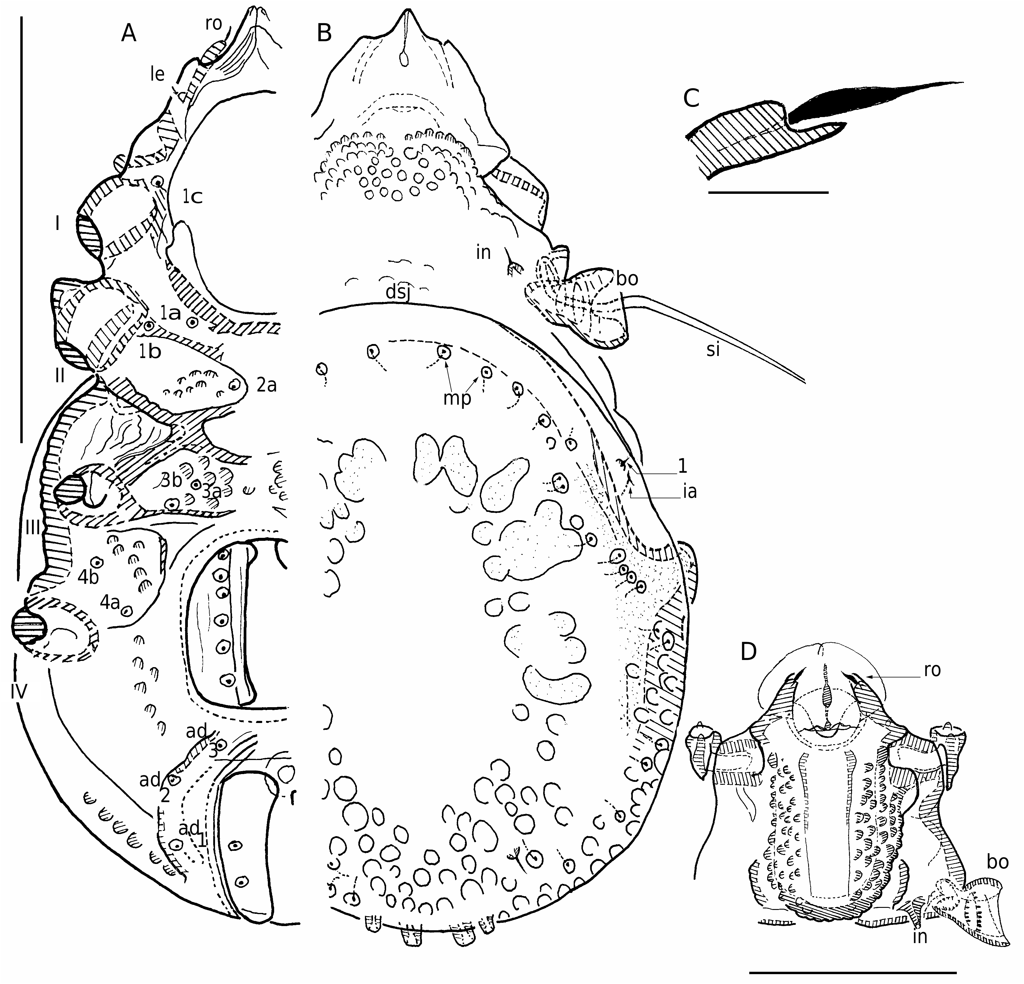
Shape: specimens with scalps ovoid in dorsal view ( Fig. 1A View FIG ), medial superior zone irregularly gibbous (larval scalps); in lateral view pyramidal ( Fig. 6A View FIG ). Specimens without scalps ( Figs 3B View FIG ; 5A, B, C, E View FIG ): prodorsum flat to slightly globose; posterior zone of notogaster globular; anterior zone with well defined furrow.
Colour: specimens without cerotegument and scalps, dark yellow to medium brown.
Cerotegument (scalps not considered)
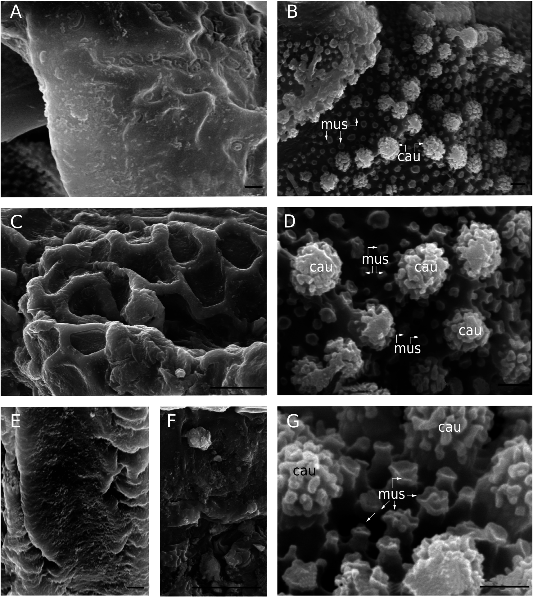
Thick complex layer, composed of wax layer and amorphous cement layer ( Norton et al. 1997); present over entire body and legs, with elaborate patterns and structures in four different layer types: amorphous; small tuberculate, mixed and granulate (see below).
Amorphous layer: prodorsum, bothridial external zone, seta in on tubercle ( Figs 2A View FIG ; 4B, C View FIG ), genital plate and surrounding zone, anal plate and surrounding zone.
Small tuberculate layer: internal bothridial zone ( Fig. 4C, E View FIG ).
Mixed layer: constituted by mushroom-like microtubercles (mus) associated with cauliflower-like microtubercles (cau) ( Fig. 2B, D, G View FIG ); mus diameter 0.1-0.9 Μm, height 0.5-2.0 Μm; cau diameter 0.9- 3.1 Μm, height 1.5-2.9 Μm. Distribution: infracapitulum, epimeric zone, lateral body zone.
Granulate layer: wrapping interlamellar setae, only small sections of setae visible ( Fig. 4B, D View FIG ).
Legs covered by amorphous layer, with prominent folds.
Integument
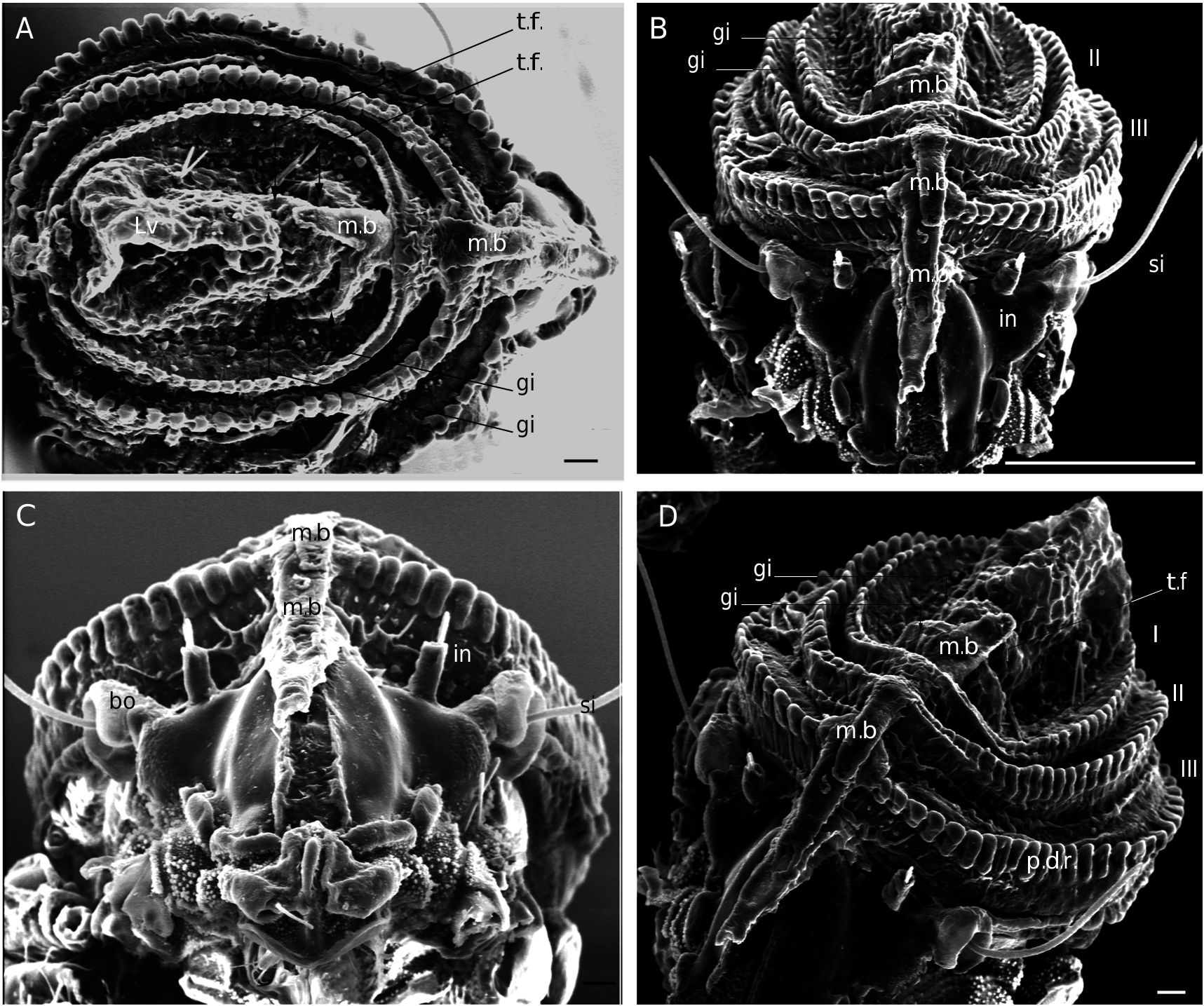
Prodorsum, tuberculate microsculpture ( Fig. 3B View FIG ): particularly in medial zone; the attachment in zone of the exuvial medial band extension (m.b) is attached, tubercles small and only discernible in lateral inclined view, in dorsal view zone smooth ( Fig. 3D View FIG ); anterior zone, tubercles small.
Notogaster, dorsal posterior zone tuberculate ( Figs 3B View FIG ; 5A, C View FIG ); smooth towards bng ( Fig. 5E View FIG ); laterally with ridges separated by furrows ( Fig. 5E View FIG ). Anterior depressed zone situated backwards toward macropores with many parallel ridges separated by furrows ( Fig. 5A, C View FIG ), terminating in front of macropores, clearly visible in inclined lateral view. Zone behind depressed area, irregularly foveate patterns, variable lengths, distributed around central zone ( Fig. 3B View FIG ).
Macropores (mp), varying in size (diameter 0.5- 1.2 Μm), openings situated in small depressed circular zones; circumgastric distribution; anterior zone in single line near dsj; middle posterior zone, numerous macropores irregularly distributed ( Fig. 5A, C, E View FIG ); internal channel clearly visible close to cuticle.
Ventral, tuberculate ( Fig. 3A View FIG ); tubercules of epimeric zone small; posterior zone around genital and anal openings, large tubercles. Genital and anal plates, slightly rugose to smooth.
Setation
Lamellar, notogastral, subcapitular, exostigmatal, epimeric, genital and anal setae spiniform; rostral seta simple, inflated in middle zone ( Fig. 3C View FIG ) (13- 15 Μm); interlamellar setae (10-12 Μm), ( Fig. 4B, D View FIG ) filiform.
Prodorsum
Specimens with scalps ( Figs 1A, B, C, D View FIG ); medial band extension (m.b) on central part up to rostrum. Medial zone around area covered by medial band extension, elevated ( Fig. 1B, C View FIG ), latter area easily discernible ( Fig. 3D View FIG ). Near dsj and medial posterior part, flat to slightly gibbose ( Fig. 5B View FIG ), microtuberculate.
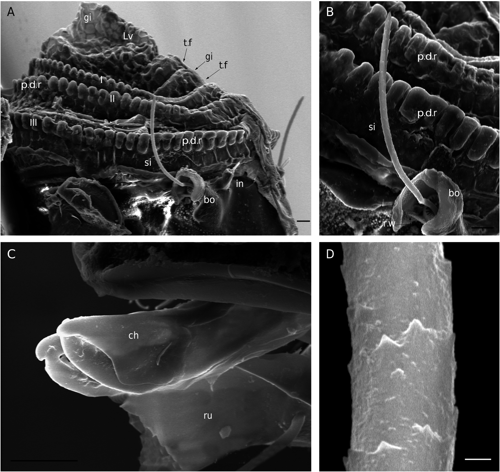
Interlamellar seta (in), inserted near bothridial base on large protuberance, extending upwards ( Figs 1B, C, D View FIG ; 4B, D View FIG ); lamellar seta (le) small, inserted on small protuberance; rostral seta (ro) inserted on basal zone of protuberance with forward-extending visor present ( Fig. 3C View FIG ).
Anterior zone of rostrum complex in structure; zone behind and around insertion of rostral seta with many irregular protuberances ( Fig. 5B View FIG ). Rostrum medially incised ( Fig. 3D View FIG ). Incision narrow, terminating in tooth-like projection; in frontal view the tooth is less pronounced and appears rounded; in dorsal view ( Fig. 3B View FIG ) posterior incision end rounded.
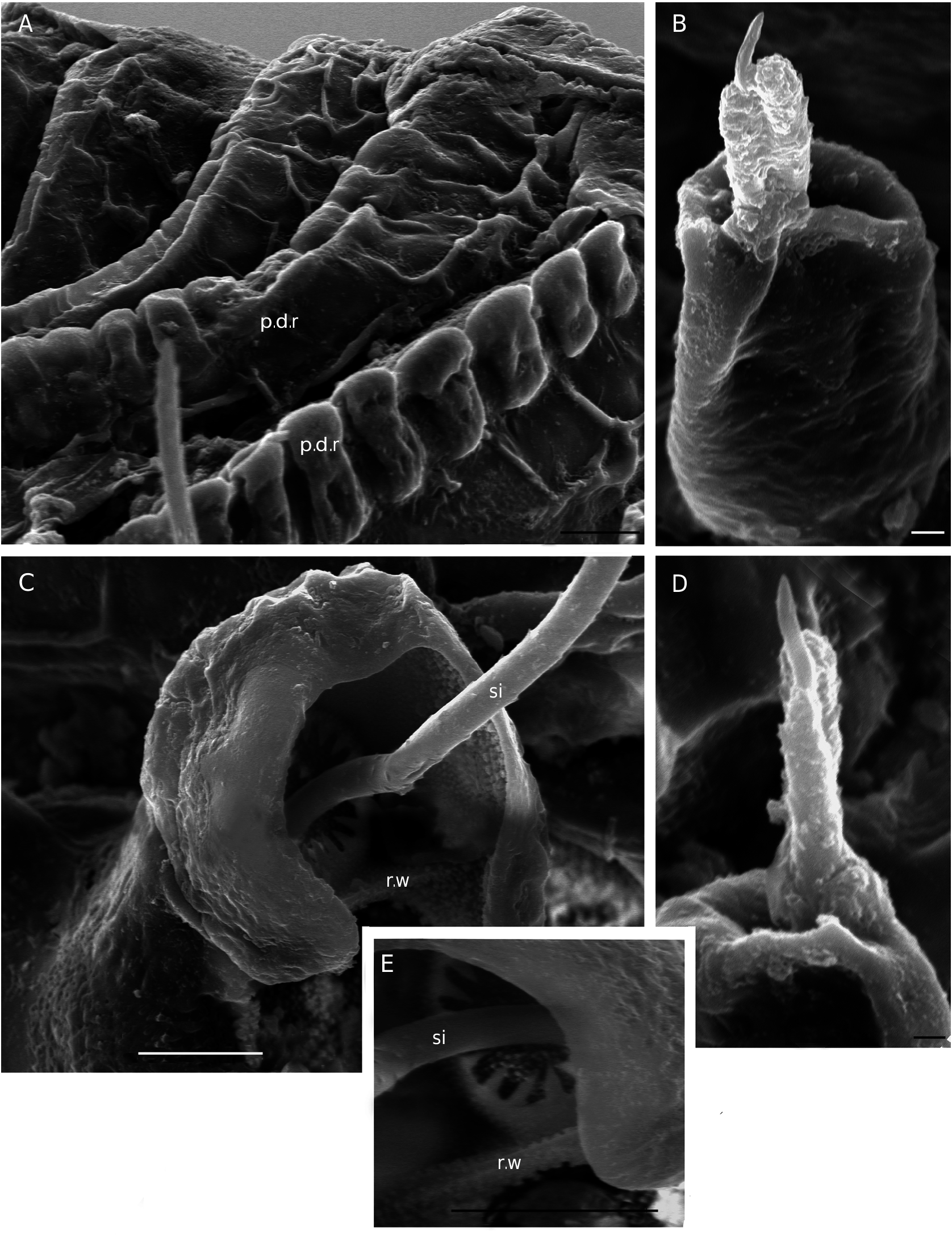
Bothridium ( Fig. 1C View FIG ) prominent, opening directed sideways ( Fig. 4C View FIG ), horseshoe-shaped; bothridial rim dorsally semicircular, basally incised, with thin rectilinear wall running across incision (r.w). Internal bothridial rings gear-like ( Fig. 4C, E View FIG ).
Sensillus filiform (86-112 Μm), minutely barbed ( Figs 5B View FIG ; 6A, B, D View FIG ); diameter of pedicel 0.6-1 Μm; exostigmatal seta (ex) small ( Fig. 5B View FIG ).
Notogaster
Near dsj, aligned macropores (mp), opening inside foveae; mp varying in size (diameter 0.4-0.8); internal channel close to cuticle.Distribution: circumgastric
( Fig. 3B View FIG ); anteriorly in a simple ring; in front of and behind the opisthosomal gland several alignments and numerous pores ( Fig. 5A, C, E View FIG ).
The anterior zone, near dsj, and posterior to macropores a well defined semicircular furrow (s.c.f), clearly visible in inclined lateral view ( Fig. 5C View FIG );
s.c.f extending to a third of anterior notogaster and delimiting a prominent depressed zone crossed by many more or less parallel ridges, separated by furrows ( Fig. 5A, C View FIG ).
Apophysis of opisthosomal gland (apo.gla) (opisthonotal gland sensu Norton and Behan-Pelletier 2009) conical, round tipped, positioned laterally ( Figs 3B View FIG ; 5A, E View FIG ) extending forward.
Five pairs of notogastral setae, one anterior and four posterior; intraspecific setae variability between five to six pairs, last additional pair small, dorsally to seta 5, situated in macropore zone. Five pairs of lyrifissures (ia, im, ih, ips, ip) ( Fig. 5A, E View FIG ).
Ventral region
Specimens with cerotegument:epimeral furrows easily discernible, with mushroom-like microtubercules (mus) and cauliflower-like microtubercules (cau) ( Fig. 2B, D, G View FIG ). Specimens without cerotegument ( Fig. 3A View FIG ): cuticle smooth.Epimeral furrows very flat, only bo.sj. deeper; in medial epimeric zone depression more or less triangular in shape. Apodemes I and II close to each other; sj thin; III small, divided; IV less defined. Epimeral setal formula (3-1-2-2) ( Fig. 3A View FIG ). Genital setation six pairs, in a single longitudinal row; aggenital seta absent; adanal setae three pairs; anal setae two pairs; lyrifissure (ian) not visible.
Lateral region
Exobothridial seta (ex) small but clearly discernible ( Fig. 5B View FIG ); two macropores: one below ex; another opening in front of leg I ( Fig. 5B View FIG , indicated with mp and arrow). Opening of podocephalic canal only discernible as simple pore (p.c) ( Fig. 5B View FIG ). Tubercle of interlamellar seta more or less cylindrical, depressed in apical central zone around setae insertion ( Fig. 4B, D View FIG ).
Seta ro inserted on prominent tubercle ( Fig. 3C View FIG ). Rostral tubercle with forward extending anterior ventral visor ( Fig. 3C View FIG ). Behind tubercle of rostral seta and extending behind seta le a well defined zone of many elevated, more or less aligned cuticular thickenings ( Fig. 5B View FIG ). Rostral margins smooth, cuticle more or less hyaline ( Fig. 3D View FIG ).
Sejugal depression deep, easily discernible ( Fig. 5B View FIG ). Behind acetabulum III and below the level of acetabulum IV, a prominent cuticular thickening. Between anal opening and level of genital opening, many cuticular thickenings equidistantly aligned ( Fig. 5B View FIG ). Lyrifissures ia, im, ih,ip, ips, clearly visible ( Fig. 5A, C View FIG ). Notogastral setae on small protuberances ( Fig. 5A, C View FIG ).
Subcapitulum suctorial with short tube.Subcapitular setae large. Chelicerae without setae ( Fig. 6C View FIG ); Trägårdh’s organ large.
Legs
Setal and solenidial formulae (trochanter to tarsus): I (1-5-5-5-20-1) (1-2-2); II (1-5-4-4-14-1) (1-1-2); III (2-3-2-4-13-1) (1-1-0); IV (1-3-2-4-13-1) (0-1-0).
Scalps
Exuviae of immature stases adhering one on top of the other, creating a multilayered structure; anteriorly each scalp extends in a medial band (m.b) ( Fig. 1A, B, C, D View FIG ). Medial band covering the central zone, firmly adhering on prodorsal surface ( Fig. 1C View FIG ) and extending to the rostrum. Scalps (medial band included) firmly attached. Cerotegumental layer: medial band covered by thick amorphous layer ( Fig. 2E, F View FIG ); rest of scalp covered by layer with network of round to polygonal depressions patterns ( Fig. 2C View FIG ). Shape of larval scalp different from that of other stases, broad and elevated, with three gibbose areas (gi) separated by transverse furrows (t.f) ( Fig. 1A, D View FIG ); the anterior two rounded and more or less similar in shape and length; posterior one large, extended with irregular multi-gibbous dorsal surface ( Fig. 1A, D View FIG ). In lateral view shaped like a chinese hat ( Fig. 6A View FIG ).
Nymphal scalps with dentate peripheral ridge (p.d.r) ( Figs 1 View FIG A-D; 4A; 6A, B). Setae hardly discernible. Scalps simple without anterior tuft of filaments and without anterior indentation.
REMARKS
Legs chaetotaxy, the only difference between S. makokouensis and Malgachebates peyrierasi is seta d associated with solenidion φ 1 on tibia I very small and attached to solenidion.
No known copyright restrictions apply. See Agosti, D., Egloff, W., 2009. Taxonomic information exchange and copyright: the Plazi approach. BMC Research Notes 2009, 2:53 for further explanation.
|
Kingdom |
|
|
Phylum |
|
|
Class |
|
|
Order |
|
|
Family |
|
|
Genus |