
Solenysa retractilis, 2011
|
publication ID |
https://doi.org/ 10.1111/j.1096-3642.2010.00640.x |
|
persistent identifier |
https://treatment.plazi.org/id/038887F0-722F-FFB5-FE86-F8FBFC1DE0ED |
|
treatment provided by |
Valdenar |
|
scientific name |
Solenysa retractilis |
| status |
|
SOLENYSA WULINGENSIS View in CoL GROUP
Composition: Four species, S. geumoensis Seo, 1996 , S. retractilis Tu sp. nov., and S. wulingensis Li & Song, 1992 , and S. tianmushana Tu sp. nov.
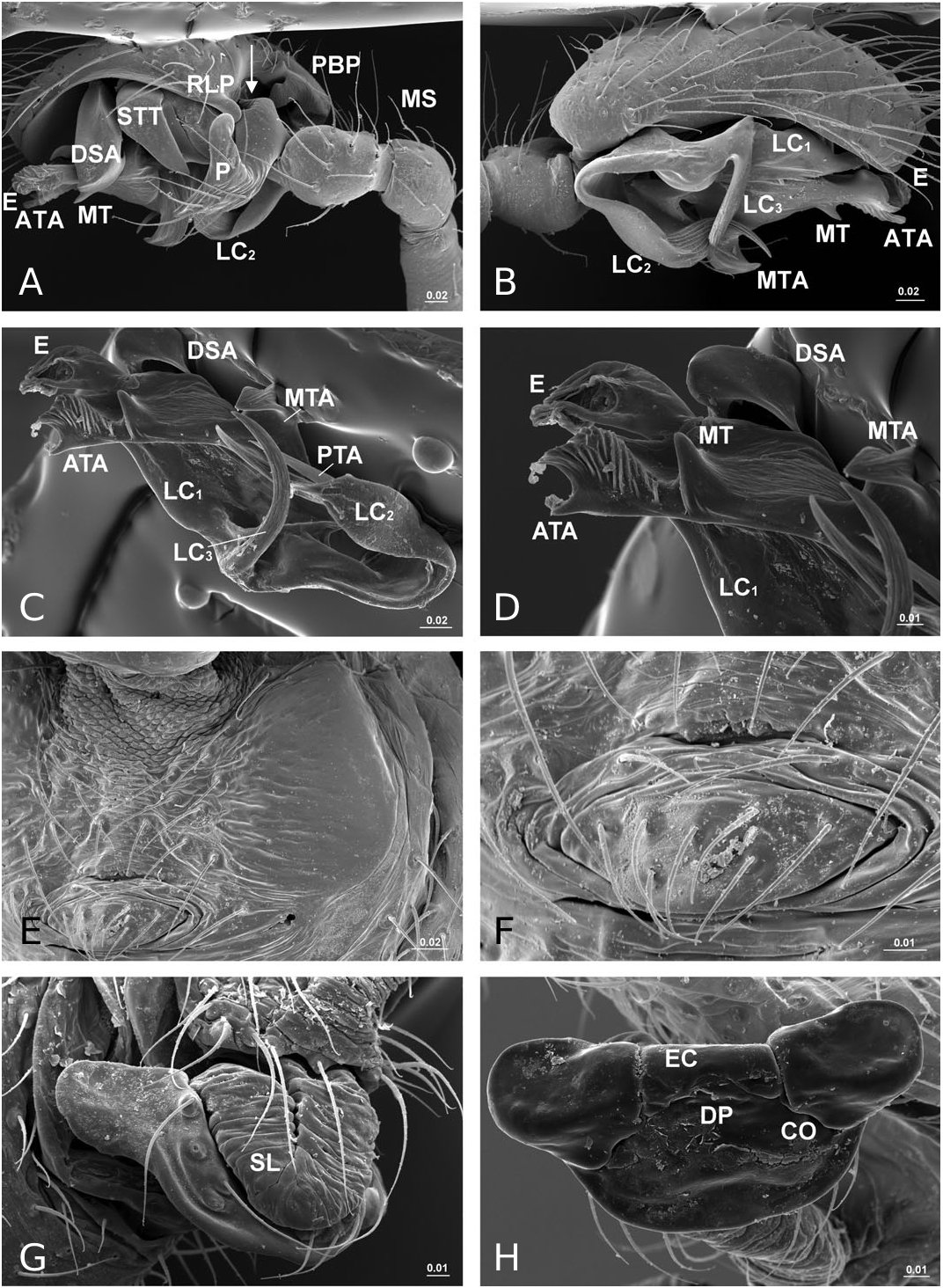
Diagnosis: Males of this group can be distinguished by the hidden proximal cymbial apophysis ( Fig. 21A View Figure 21 ), except for S. tianmushana in which the proximal cymbial apophysis projects outwards ( Tu & Li, 2006b: fig. 4), by the additional paracymbial curve, orientated anteriorly ( Fig. 21A View Figure 21 ) and by the membranous embolus ( Fig. 21D View Figure 21 ). The twisted solenoid forming a loop on the ventral surface of epigynum is diagnostic for this species group ( Fig. 21G View Figure 21 ), except for S. tianmushana , the female of which is unknown.
Description: Somatic characters as in genus description. Male palp ( Fig. 21A–D View Figure 21 ): tibia unmodified. Cymbial probasal process hidden in large cymbial basal excavation. Paracymbium with additional proximal curve anteriorly orientated. Radix small, embedded in a membranous area that connects lamella characteristica and terminal apophysis. Lamella characteristica tripartite, anterior branch handleshaped, well sclerotized, extending forwards; second one largest, ribbon-like, dragging backwards, then turning forwards, tapering off distally; posterior one aciniform, well sclerotized. Terminal apophysis with three free ends: anterior one elongated forwards, companied with embolus, rounded apex with two apical processes; posterior one thin and sharp, translucent; median one slightly rolling distally. Embolus entirely membranous, without chitinized proper.
Epigynum ( Fig. 21G, H View Figure 21 ): rounded triangular to half rounded, chitinized flat box. Anterior part wider and thicker than posterior part, with two ear-like lateral extensions. Seen in dorsal view, anterior margin trisected, the middle part forming epigynal collar ( Figs 10F, 21H View Figure 21 ). Membranous solenoid connects from ventral side of it, forming a loop on ventral surface of epigynum in nonfunctional stage. Spermathecae triangular shaped, located at each side of epigynal collar. Copulatory ducts extremely short, opening on dorsal surface of epigynum. Fertilization ducts mesially directed.
Remarks: The male and female genitalic structures of S. retractilis , S. wulingensis , and S. geumoensis are very similar, differing only in small details. Comparatively, the male palp of S. tianmushana is more different in the proximal cymbial apophysis and in the morphology of terminal apophysis and lamella characteristic. However, the male of S. tianmushana still shares many characters with the other three species, such as the membranous embolus and the embedded radix. The morphology of the anterior terminal apophysis, with a rounded apex and two apical processes, suggests a close relationship between S. tianmushana and the species of the S. wulingensis group. As the female of S. tianmushana is unknown, we have included this latter species in the S. wulingensis group.
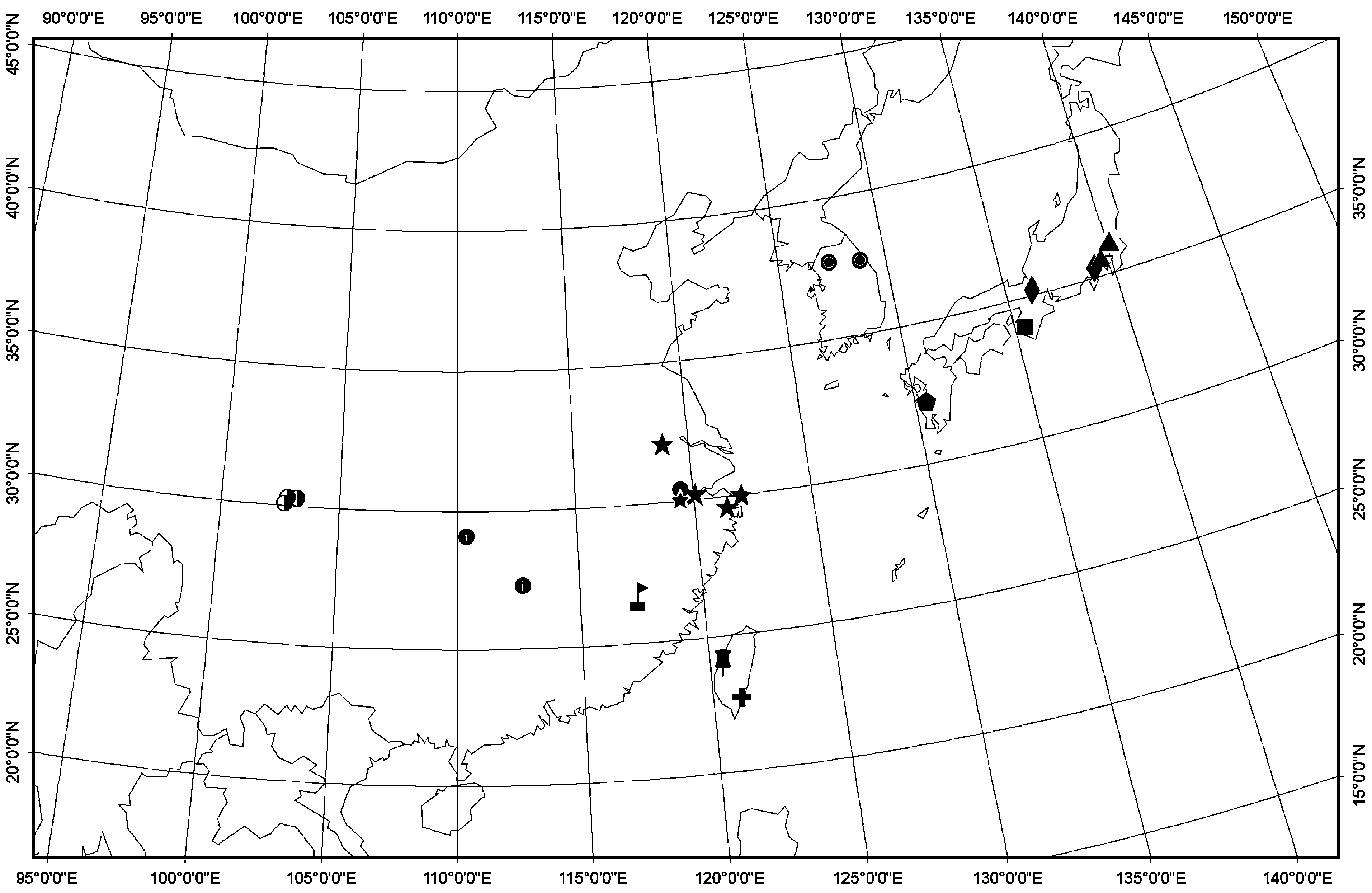
Distribution: China (Sichuan, Hunan, Zhejiang), Korea ( Fig. 22 View Figure 22 ).
No known copyright restrictions apply. See Agosti, D., Egloff, W., 2009. Taxonomic information exchange and copyright: the Plazi approach. BMC Research Notes 2009, 2:53 for further explanation.
|
Kingdom |
|
|
Phylum |
|
|
Class |
|
|
Order |
|
|
Family |
|
|
Genus |