
Sphaerobelum phouloei, Wesener, 2019
|
publication ID |
https://doi.org/ 10.11646/zootaxa.4563.2.1 |
|
publication LSID |
lsid:zoobank.org:pub:CF79B01B-8B5F-4B3A-B642-2CADE4B339AF |
|
DOI |
https://doi.org/10.5281/zenodo.5934524 |
|
persistent identifier |
https://treatment.plazi.org/id/03EB5506-E304-8C3B-A2ED-FDD6FA80FD54 |
|
treatment provided by |
Plazi |
|
scientific name |
Sphaerobelum phouloei |
| status |
sp. nov. |
Sphaerobelum phouloei new species
Figures 6B View FIGURE 6 , 9B View FIGURE 9 , 11 View FIGURE 11 , 12 View FIGURE 12 .
Sphaerobelum sp. II 1 M
Material examined: Type specimen. 1 M holotype (ZMUC00040257) from Laos, Houaphan Province, Phou Loei, way from camp 1 to camp 2, ~ 1200 m, leg. 9.VIII.2008; S. Tarasov.
Diagnosis: S. phouloei n. sp. belongs to a group of Sphaerobelum species in which the mesal margin of the femur is extended in several teeth ( Fig. 11B View FIGURE 11 ). S. phouloei n. sp. shares only with S. denticulatum n. sp. a curved telopoditomere 4 of the posterior telopods which overlaps the immovable finger ( Figs 12E, F View FIGURE 12 ). S. phouloei n. sp. differs in several characters from S. denticulatum n. sp.: male antenna with only 30–35 apical cones (> 50 in S. denticulatum n. sp.), prefemur mesal margin well-rounded (indentated in S. denticulatum n. sp.), tergites covered with short setae (glabrous in S. denticulatum n. sp.), legs orange (brown in S. denticulatum n. sp.).
Description. Measurements: Body length: holotype male: length ca 25.8 mm. Width, of thoracic shield 13.3 mm, of tergite 8 = 14.1 mm (= broadest). Height, of thoracic shield = 8.9 mm, of tergite 8 = 9.3 mm (= highest).
Coloration: in preserved specimens anterior 2/3 of tergites dark olive green, posterior 1/3 black. Head, ventral side, anal shield orange ( Fig. 6B View FIGURE 6 ). Legs and antennae orange.
Head: Eyes with 65–70 ocelli. Aberrant ocellus located inside antennal groove. Antennae short, with rounded joints, extending posteriorly to leg-pair 4. First antennomere with cuticular scales. Lengths of antennomeres: 1=2=3<4<5<<6. All antennomeres densely pubescent, sensilla basiconica surrounding apical disc. Last antennomere thickened, apically widened and well rounded. Apical disc with ca 36/33 apical cones. Organ of Tömösváry located inside antennal groove. Gnathochilarium: structure typical of the order. Palpi sensory cones located in several clusters. Mandibles: not dissected.
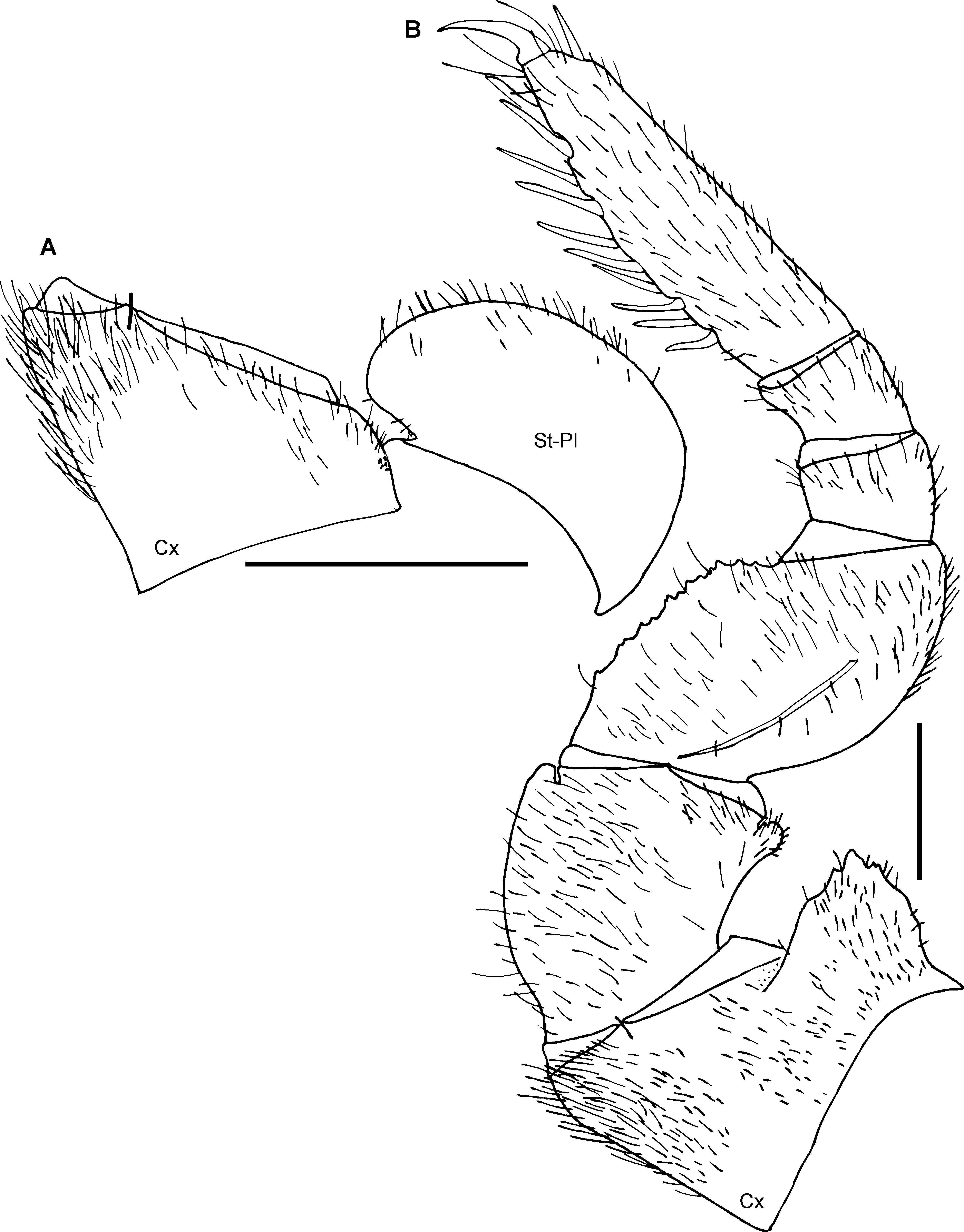
Stigmatic plates: first stigmatic plate widely rounded, apex well-rounded, curved towards coxa 1 ( Fig. 11A View FIGURE 11 ).
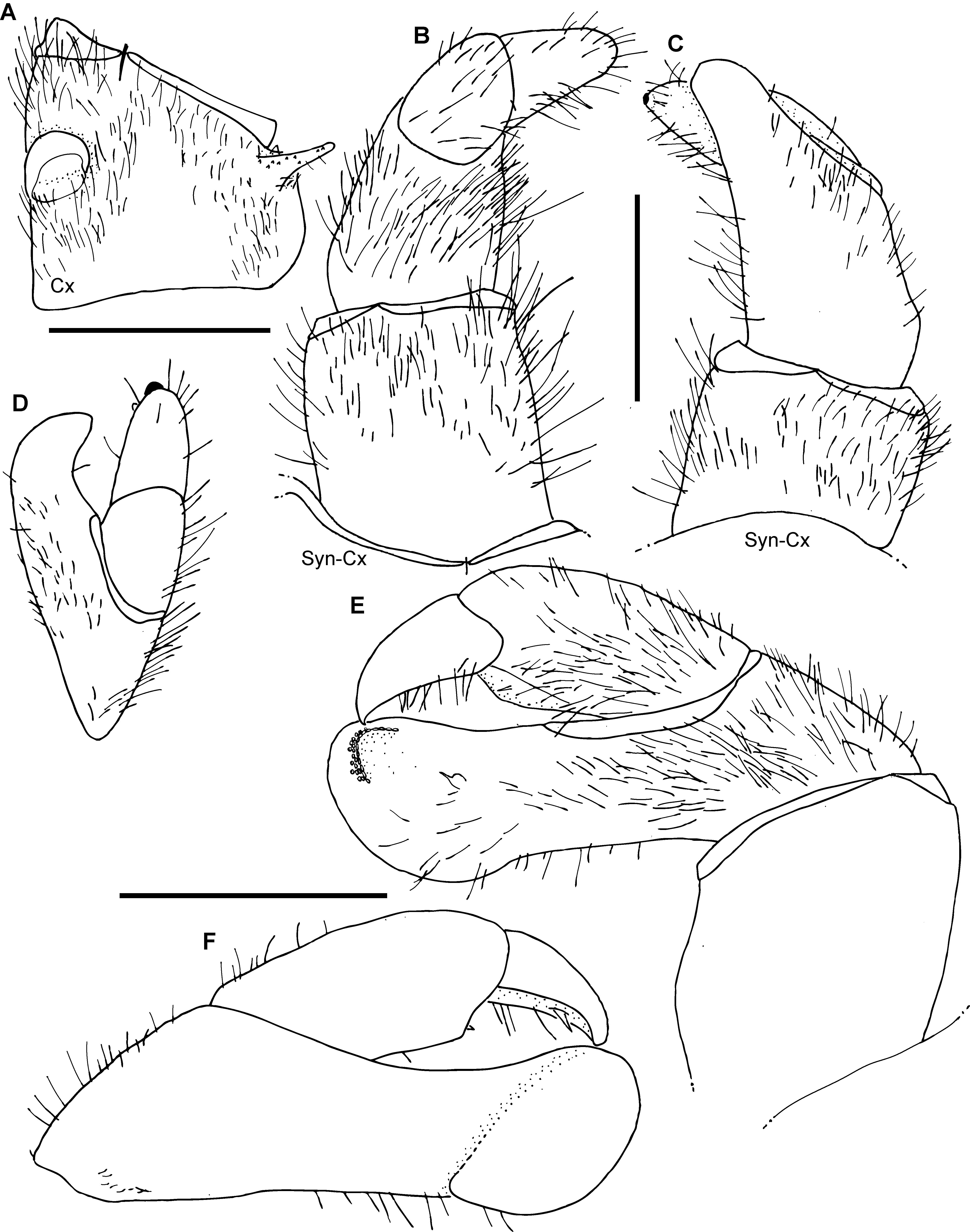
Laterotergites: laterotergite 1 and 2 with a broad, stout projection. Collum: with glabrous surface, margins with few isolated setae. Thoracic shield: surface glabrous unlike tergites, setae only in grooves. Shallow grooves beset with numerous long setae, slope towards groove with 3 weak anterior and 2 or 3 posterior keels. Tergites: anterior 2/3 of surface densely setose with short setae, posterior 1/3 glabrous. Tips of paratergites of midbody tergites projecting posteriorly ( Fig. 6B View FIGURE 6 ). Endotergum ( Fig. 9B View FIGURE 9 ): inner section lacking any spines or setae. Middle area with a single row of large, sparse, elliptical, cuticular impressions. Distance between impressions as wide as their diameter. Apically, 1–2 sparse rows of long marginal bristles, strongly protruding beyond tergal margin. Bristles not smooth, but with numerous small spinicles. Anal shield: large, surface completely covered by tiny setae. Underside with a single, long, black locking carina, located close to last laterotergite. Legs: leg-pair 1 with 2 ventral spines, leg-pair 2 with 5, leg-pair 3 with 6 or 7. First two leg-pairs without an apical spine. Leg pairs 4–21 with 9 or 10 ventral spines and one dorso-apical spine. In leg 9, femur 1.3 times, tarsus 3.7 times longer than wide ( Fig. 11B View FIGURE 11 ). All podomeres densely setose. Coxa with a large and marginally toothed process. Coxa process absent at first leg ( Fig. 11B View FIGURE 11 ) and sharply projecting at second ( Fig. 12A View FIGURE 12 ). Prefemur at apical margin with a projection laterally and mesally. Lateral projection triangular and sharply edged, juxtaposed to coxal process. Femur extended mesally into a dentate margin featuring 12–14 teeth.
Female unknown.
Male sexual characters: gonopore covered with a single, undivided, circular, sclerotized plate ( Fig. 12A View FIGURE 12 ). Anterior telopods ( Figs 12 View FIGURE 12 B–D): consisting of 4 telopoditomeres above syncoxite, but telopoditomere 3 and 4 only divided by thin suture. Telopoditomere 1 rectangular, as long as wide. Telopoditomere 2 large, almost as long as telopoditomere 3 and 4 combined. Process of telopoditomere 2 located posteriorly, not visible in anterior view. Process of telopoditomere 2 wide, projecting to half of telopoditomere 4, apically with a well-rounded tip. Telopoditomere 3 cylindrical, straight, as long as telopoditomere 4. Telopoditomere 4 cylindrical, well-rounded, posterior side with a black sclerotized spot and a small, triangular spine. Telopoditomeres 1–4 in anterior view covered with long setae. In posterior view all telopoditomeres with fewer setae. Posterior telopods ( Figs 12E, F View FIGURE 12 ): telopoditomere 1 elongated, twice as long as wide. Immovable finger (process of telopoditomere 2) shorter than movable finger, consisting of telopoditomeres 3 and 4. Immovable finger with a characteristic, distally swollen apex, well rounded, apex therefore wider than base. Immovable finger in anterior view with a large spine, at tip opposite to tip of telopoditomere 4 with sclerotized spots. Telopoditomere 3 rectangular, towards immoveable finger with a triangular expansion carrying a spine. Telopoditomere 4 reaching 2/3 of length of telopoditomere 3, apically strongly tapering into acute tip, very slightly curved towards immovable finger. Inner margin with single spine towards immovable finger. Telopoditomere 1 at both sides covered by setae. Telopoditomeres 2 and 3 in anterior view covered by long setae, in posterior view mostly glabrous. Telopoditomere 4 only with marginal setae.
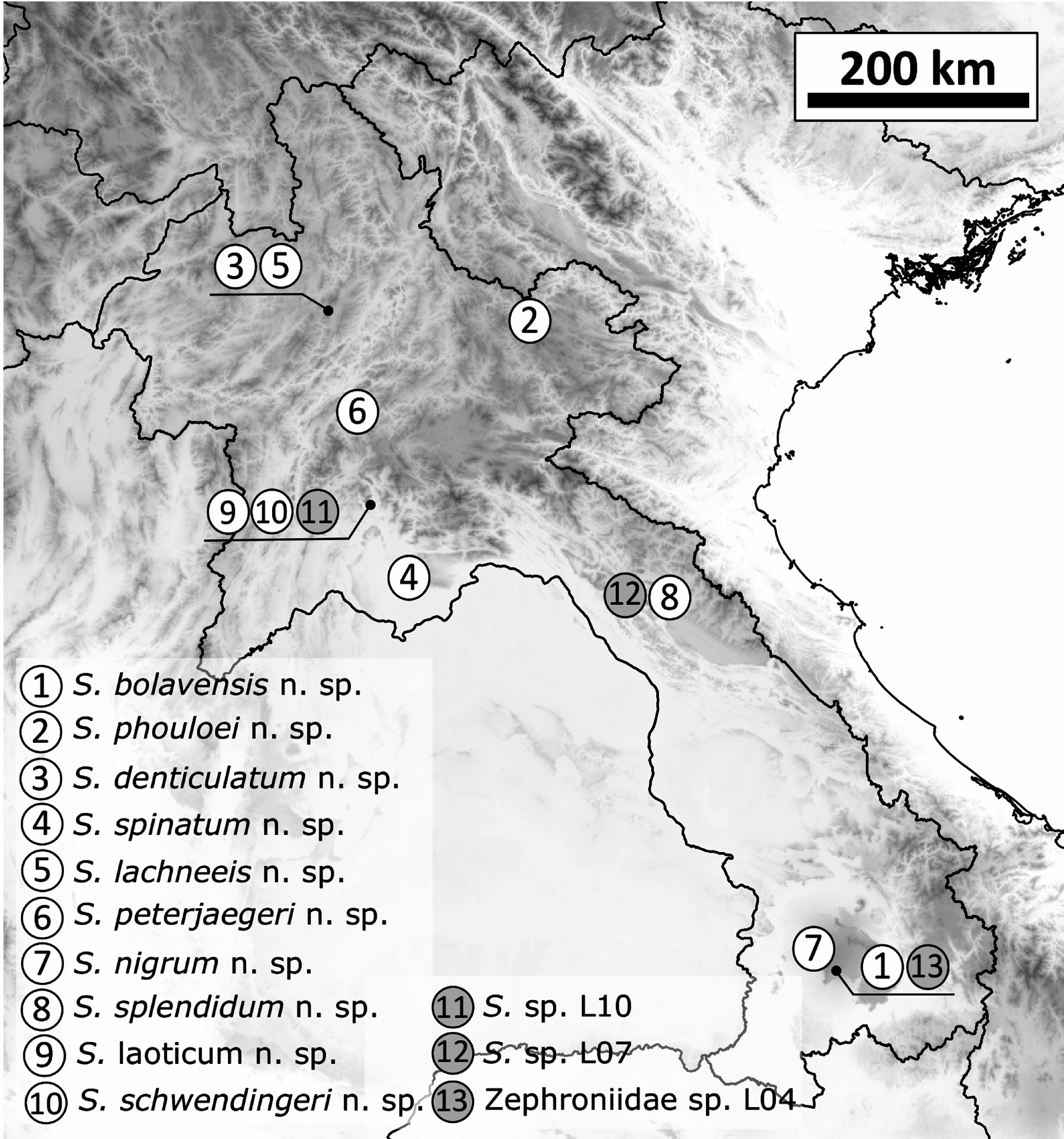
Derivatio nominis: phouloei, noun in apposition, after the Phou Loei Mountain, the type locality ( Fig. 5 View FIGURE 5 ).


No known copyright restrictions apply. See Agosti, D., Egloff, W., 2009. Taxonomic information exchange and copyright: the Plazi approach. BMC Research Notes 2009, 2:53 for further explanation.