
Stylomecynostomum bodegensis, Hooge, Matthew D. & Tyler, Seth, 2003
|
publication ID |
https://doi.org/ 10.5281/zenodo.157080 |
|
DOI |
https://doi.org/10.5281/zenodo.6274967 |
|
persistent identifier |
https://treatment.plazi.org/id/5130080E-FFD2-FF82-FEAE-F4F6B92EFAF3 |
|
treatment provided by |
Plazi |
|
scientific name |
Stylomecynostomum bodegensis |
| status |
sp. nov. |
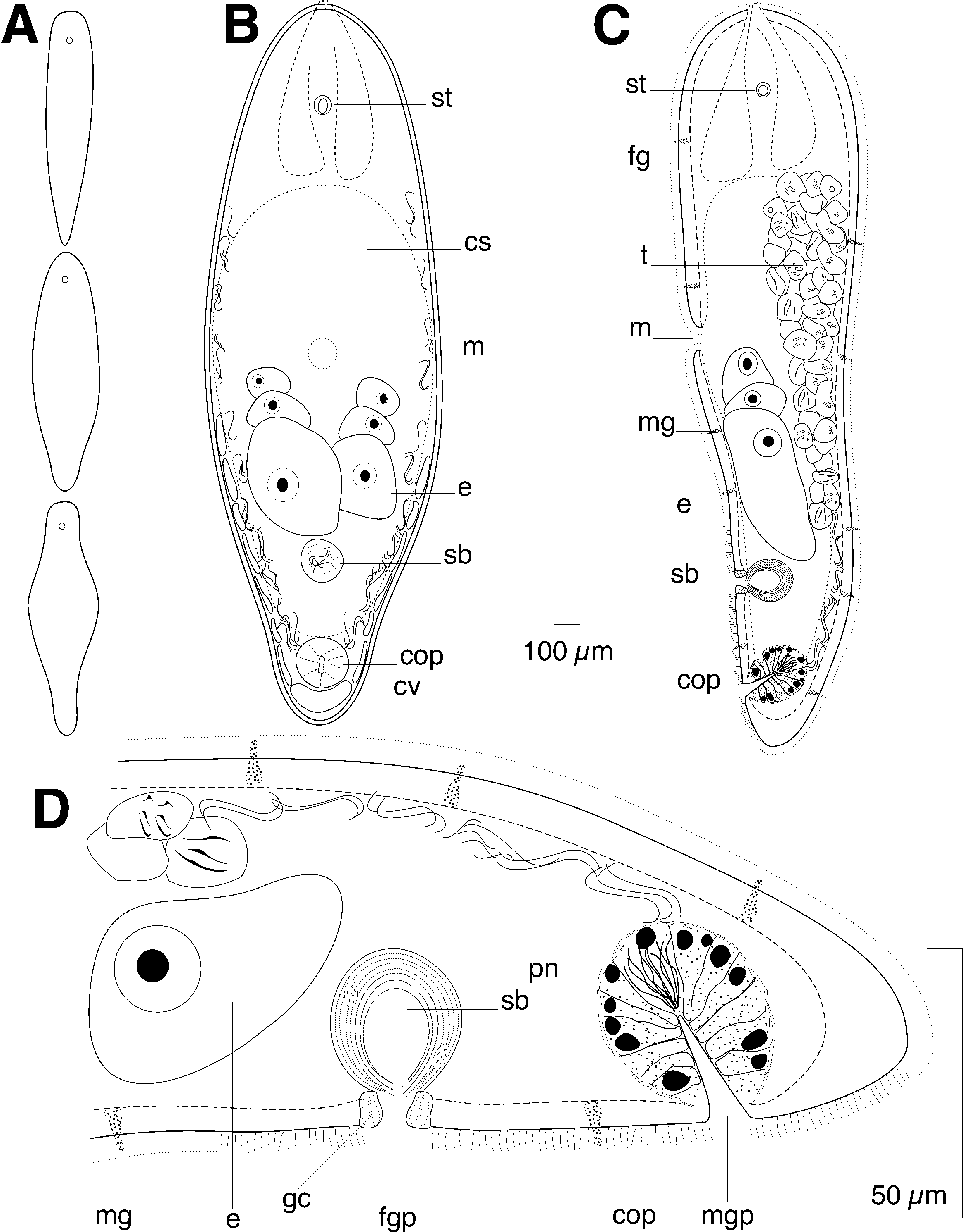
Stylomecynostomum bodegensis View in CoL sp. nov. ( Figs. 56 View FIGURE 5 View FIGURE 6 )
Type Material. Holotype, AMNH PLATY 1639, set of 1.5µmthick serial sagittal sections of epoxyembedded specimen stained with toluidine blue, collected May 2002. Paratypes, AMNH PLATY 1640, and AMNH PLATY 1641 two set of 1.5µmthick serial sections of epoxyembedded specimens stained with toluidine blue.
Living specimens in squeeze preparations; five sets of 1.5µmthick serial sections of epoxyembedded specimens stained with toluidine blue; whole mounts for fluorescence imaging of musculature (3 specimens).
Type Repository. American Museum of Natural History, New York, New York, USA.
Type Locality. Campbell Cove, located at the north side of Bodega Head, Bodega Bay, California, USA (38° 18' 15'' N, 123° 03' 24'' W). Finegrained black sediment at 10 30 cm sediment depth at the low intertidal level.
Etymology. Species name refers the type locality of Bodega Bay, California.
Description. Mature specimens approximately 400 µm long and 130 µm wide ( Figs. 5 View FIGURE 5 B, 5C). Anterior and posterior ends rounded.
Epidermis completely ciliated. Rhabdoids absent. Mucoid glands scattered across dorsal and ventral sides.
Musculature with circular fibers that encircle the body along entire length of animal; straight longitudinal muscles absent between frontal organ and anterior edge of mouth; longitudinal muscles with a longitudinal orientation anteriorly that bend medially to cross diagonally over the body (longitudinalcrossover fibers), present in both dorsal and ventral body wall; anterior end with ventral diagonal muscles positioned between outer circular and inner longitudinal muscles (data not shown).
Frontal organ well developed; cell bodies of frontal glands positioned ~90 µm behind frontal pore ( Fig. 5 View FIGURE 5 C).
Mouth opening on ventral surface, middle of body. Digestive central syncytium extends from frontal glands posteriorly to level of male copulatory apparatus.
Ovaries paired, ventral, extend from level of mouth posteriorly to seminal bursa ( Fig. 5 View FIGURE 5 B, C).
Testes paired, dorsal, separate from eggs. Testes extend anteriorly to frontal glands and posteriorly to level of male copulatory organ ( Fig. 5 View FIGURE 5 C).
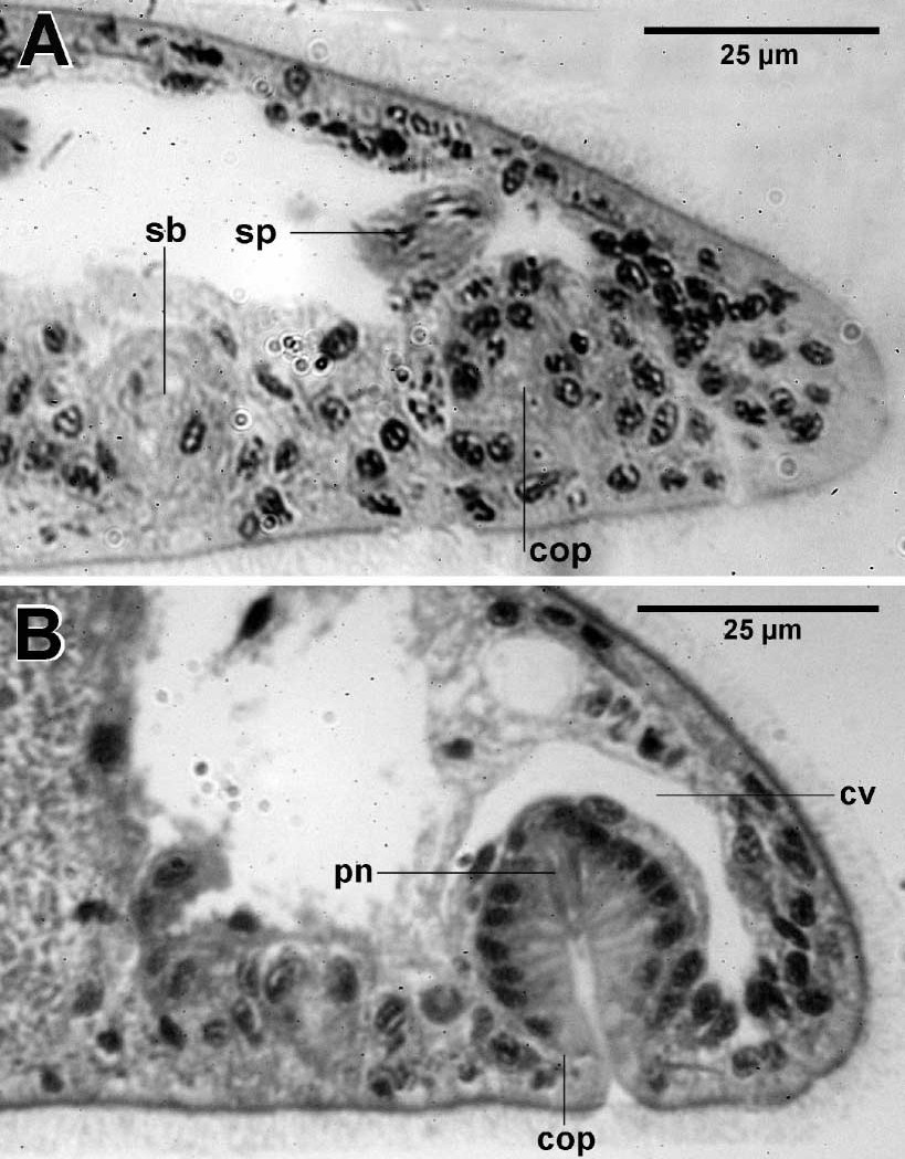
Female gonopore surrounded by large gland cells and opens directly to thickwalled bursa ( Fig. 5 View FIGURE 5 D). Examined specimens did not have sperm in the bursa, and as such, no lumen could be discerned ( Fig. 6 View FIGURE 6 A).
Male gonopore located ventrally at posterior end of body ( Figs. 5 View FIGURE 5 , 6 View FIGURE 6 B). Copulatory organ globular, glandular. Nuclei present along outside edge of copulatory organ. Filamentous penis needles present at proximal end of male duct.
Remarks. The filamentous, brushlike penis needles of Stylomecynostomum bodegensis are distinctly different from the conicallyshaped sclerotized needles found in species of the mecynostomid genera Paedomecynostomum Dörjes, 1968 , and Pseudmecynostomum Dörjes, 1968 . Our species appears to be most similar to Eumecynostomum altitudi Faubel and Regier, 1983 , and E. westbladi (Dörjes, 1968) , both of which are considerably larger than our species (~ 1 mm long), but which have similarly constructed copulatory organs except for their lack of sclerotized penis needles. In line with generic distinctions for mecynostomids, the needlebearing copulatory organ warrants erection of the new genus, Stylomecynostomum . The seminal bursa of this species is similar to the globular bursa of E. altitudi , which also has a bursa wall composed of diffuse tissue. The bursal wall of E. westbladi is composed of gland cells, a condition reminiscent of the prominent gland cells surrounding the female gonopore of our species.
| AMNH |
American Museum of Natural History |
No known copyright restrictions apply. See Agosti, D., Egloff, W., 2009. Taxonomic information exchange and copyright: the Plazi approach. BMC Research Notes 2009, 2:53 for further explanation.