
Sundathelphusa cebu, Husana & Ng, 2019
|
publication ID |
https://doi.org/ 10.11646/zootaxa.4585.2.5 |
|
publication LSID |
lsid:zoobank.org:pub:6E61D5EE-E3AD-44EC-9AB3-C7B3E303BF92 |
|
DOI |
https://doi.org/10.5281/zenodo.5466961 |
|
persistent identifier |
https://treatment.plazi.org/id/FC5AA702-2ED8-4BD4-8133-7549A555889E |
|
taxon LSID |
lsid:zoobank.org:act:FC5AA702-2ED8-4BD4-8133-7549A555889E |
|
treatment provided by |
Plazi |
|
scientific name |
Sundathelphusa cebu |
| status |
sp. nov. |
Sundathelphusa cebu View in CoL sp. nov.
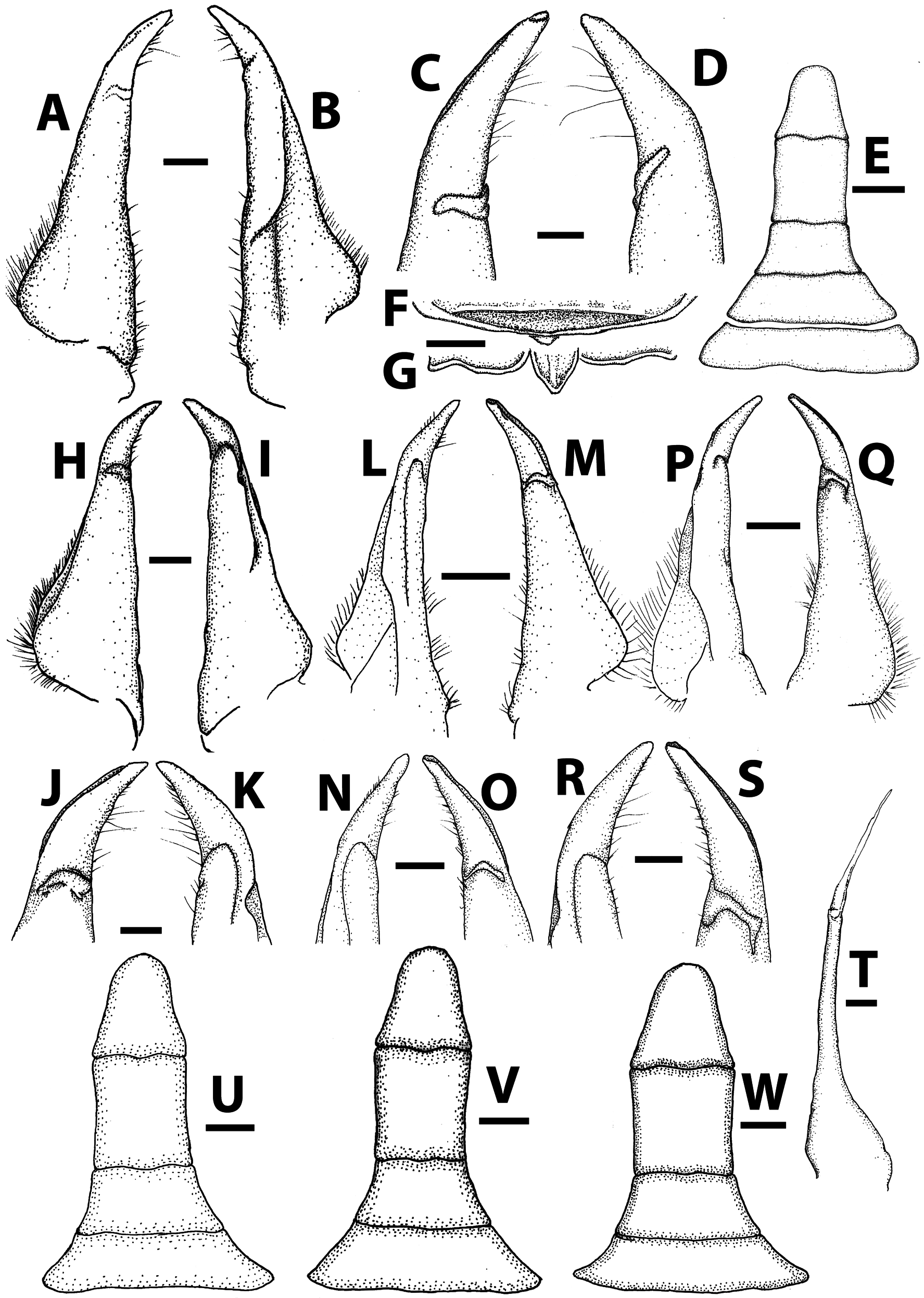
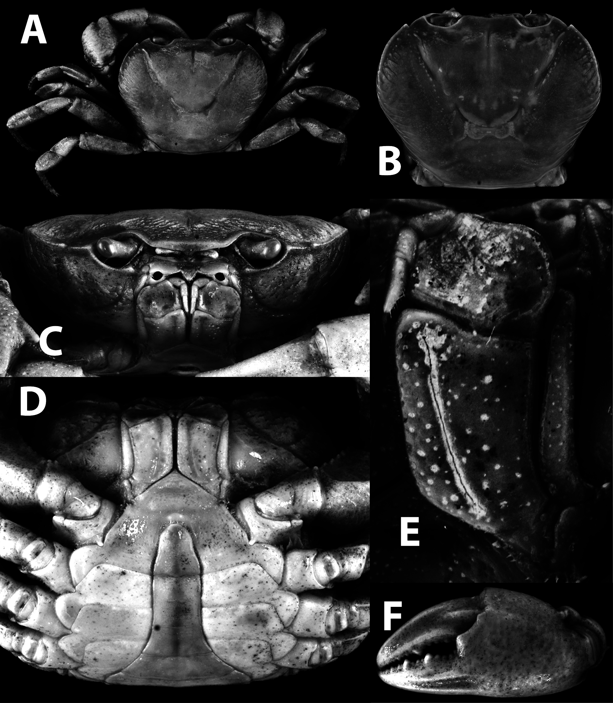
( Figs. 4–5 View FIGURE 4 View FIGURE 5 ; 6B, D, F, H, K, L View FIGURE 6 ; 9C, D View FIGURE 9 )
Sundathelphusa philippina View in CoL — Takeda 1987: 101 (part), 102; Ng & Sket 1996: 697 (discussion) (in part); Mendoza & Naruse 2010: 63 (table 1), 67 (comparative material), (in part). Not S. philippina (von Martens, 1868) View in CoL .
Material examined. Holotype: ♂ (38.1 × 31.2 mm), ZRC 2017.1254 View Materials , Campo Siete (Camp 7, Minglanilla, 450 m asl, Cebu Island , coll. S.I. Ueno, 22 June 1977 . Paratypes: 1 ♂ (32.9 × 26.2 mm), ZRC 2009.0102 View Materials , 4 ♂♂, 5 ♀♀, NSMT-Cr 25887, same data as holotype ; 2 ♂♂ (larger 29.3 × 24.0 mm), 1 ♀ (29.5 × 24.2 mm), ZRC 2017.1061 View Materials , 8 ♂♂, 6 ♀♀, NSMT-Cr 25888, upper stream of Mananga River , Cebu Island, coll. M. Takeda, 29 July 1985 ; 1 ♂ (39.3 × 31.4 mm), 3 young ♂♂, ZRC 2006.0055 View Materials , Cantipla (mislabeled as Caufipla ) Village , near Cebu City, Cebu Island, coll. Y. Cai, 14 December 2000 ; 1 ♂ (37.5 × 30.0 mm), 1 ♀ (52.2 × 40.3 mm), ZRC 2017.1255 View Materials , Malbubog Vilage , near Cebu City, Cebu Island, coll. Y. Cai, 14 December 2000 ; 7 ♂♂ (largest 37.5 × 31.2 mm), 9 ♀♀ (32.6 × 27.5 mm), ZRC 2017.1256 View Materials , Tabunan Village , near Cebu City, Cebu Island, coll. Y. Cai, 14 December 2000 ; 3 ♂♂, 1 ♀, ZRC 2009.0098 View Materials , Kawasan Falls , along Matutinao river , Badian town, Cebu Island, coll. P.K.L. Ng et al., 30 July 2003 ; 1 young ♂, 6 young ♀♀, ZRC 2009.0099 View Materials , Kawasan Falls , Matutinao, Cebu Island, coll. H.- C. Liu, 2 December 2001 ; 2 ♂♂, 5 ♀♀, 3 juveniles, ZRC 2006.0056 View Materials , Kawasan Falls , Matutinao, Cebu Island, coll. Y. Cai, 20 December 2006 ; 2 ♂♂ (36.6 × 30.3 mm, 40.5 × 32.0 mm), ZRC 2011.0002 View Materials , Kawasan Falls , Matutinao, Cebu Island, coll. J.C.Y. Lai, December 2012 ; 1 ♂ (36.5 × 28.9 mm), ZRC 2017.1253 View Materials , 1 ♂, 1 juvenile, NSMT, Kantabako , Cebu Island, coll. M. Takeda et al., 28 July 1985 . All locations in the Philippines .
Description. Carapace trapezoidal, widest at anterior quarter, dorsal surface convex longitudinally, dorsoventrally depressed, regions distinct ( Figs. 4A, B View FIGURE 4 ; 6B, F View FIGURE 6 ). Frontal region sloping anteroventrally; anterolateral regions inflated dorsolaterally, rugose; cervical grooves deep; H-shaped gastric groove deep; epigastric cristae distinct, edges rough, separated by distinct median furrow; postorbital cristae, sharp but low; epigastric and postorbital cristae, confluent; epibranchial teeth and postorbital cristae not confluent, separated by gaps ( Fig. 4A, B View FIGURE 4 ). Frontal margin broadly protruded, two lobes clearly separated with broad median concavity; external orbital tooth produced anteriorly, outer margin longer than inner margin; epibranchial tooth distinct, triangular, well separated from external orbital tooth, margin gently curving dorsally, tapering anteriorly; anterolateral margin convex, crest low but distinct when viewed laterally, not clearly demarcated from posterolateral margin; posterolateral margin gently concave, converging gradually towards posterior margin of carapace ( Figs. 4A, B View FIGURE 4 ; 6B, D, F, H View FIGURE 6 ). Frontal median triangle almost complete; dorsal and lateral margins distinct, smooth; dorsal margin more produced anteriorly than lateral margins ( Fig. 5F View FIGURE 5 ); orbit well demarcated; supraorbital margin smooth; infraorbital margin protruded anteriorly, granulated; outer edge reaching and fused with anterolateral margin; suborbital and subbranchial regions covered with scattered oblique long, short striae; pterygostomial region smooth with oblique ridges on upper outer part ( Figs. 4C View FIGURE 4 ; 6F View FIGURE 6 ). Posterior margin of epistome with three lobes, median lobe large, subtriangular, with notch; lateral lobes wider and protruded ( Figs. 4C View FIGURE 4 ; 5H View FIGURE 5 ; 6F, H View FIGURE 6 ).
Eyes well developed, occupying entire orbit ( Figs. 4A, B, C View FIGURE 4 ; 6B, D, F, H View FIGURE 6 ). Ischium of third maxilliped rectangular, bearing distinct submedian sulcus close to mesial margin; merus quadrate, anteroexternal angle convex, anterior margin slightly concave; tip of exopod reaches midpoint of outer margin of merus, with long flagellum reaching slightly beyond mesial margin ( Fig. 4C, E View FIGURE 4 ).
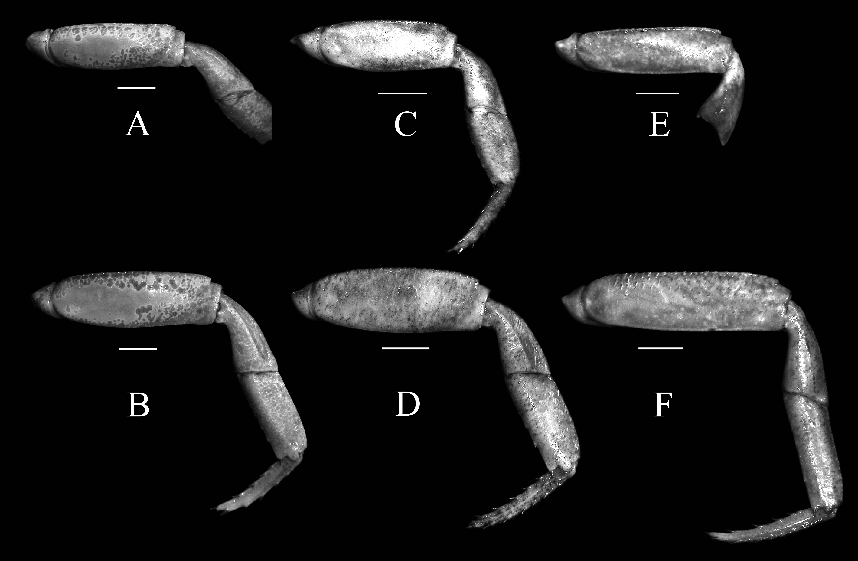
Adult male chelipeds stout, subequal; dorsal margin of merus serrated, dorsal margin with distinct subdistal tooth; carpus with strong distal inner angle, flattened dorsoventrally, laterally fringed with proximal teeth; palm surface smooth, equal in length with finger; ventral margin granulated; fingers robust, cutting edges with teeth of various sizes, largest medially, smaller on distal and proximal parts ( Fig. 4F View FIGURE 4 ).
Ambulatory legs not elongate ( Figs. 4A View FIGURE 4 ; 9C, D View FIGURE 9 ), second leg longest; anterior margin of merus serrated, without subdistal tooth or spine, posterior margins smooth; carpus short, with longitudinal submedian ridge on dorsal and ventral surfaces of all legs except on fourth leg that lacks ventral ridge, dorsal ridge barely visible, widened distally, outer margins indistinctly serrated; propodus with rows of spines on inner and outer margins, shorter on outer margin; dactylus with rows of spines on all margins, spines of both outer and inner margins of dactylus almost equal in length ( Figs. 4A View FIGURE 4 ; 9C, D View FIGURE 9 ).
Male sternopleonal cavity reaching to level of proximal quarter of coxae of chelipeds ( Fig. 4D View FIGURE 4 ). Adult male pleon narrow, T-shaped; somite 1 very short, proximal and distal margins sinuous; somite 2 transversely subrectangular; somites 3–5 narrow gradually; lateral margins of somite 3 convex, lateral margins of somites 4 and 5 slightly concave; somite 6 rectangular, longer than broad, lateral margins slightly concave; telson subtriangular, longer than broad, lateral margin concave medially, rounded distally ( Figs. 4D View FIGURE 4 ; 5F, M View FIGURE 5 ).
G1 relatively slender; subterminal segment, outer margin concave, almost straight, tapering; terminal segment gently bent outwards, outer margin concave, tapering, cylindrical, slightly setose ( Fig. 5 View FIGURE 5 A–D, I–L). G2 subequal in length to G1, distal segment long, about half length of basal segment ( Fig. 5E View FIGURE 5 ).
Etymology. The species is named after the island of Cebu where it is apparently endemic. The name is used as a noun in apposition.
Remarks. Sundathelphusa cebu sp. nov. has long been confused with S. philippina sensu stricto because of their close morphological resemblance (e.g. Takeda 1987; Ng & Sket 1996). An initial unpublished DNA analysis of the large subunit rRNA (16S) of this group of species done by the first author suggests that the species populations from the islands of Samar and Cebu represent distinct genetic groups. The detailed study of the many specimens of both species on hand shows that there is a suite of small but distinct morphological characters that can be used to separate S. philippina sensu stricto and S. cebu sp. nov. ( Fig. 6 View FIGURE 6 A–J).
When viewed laterally, the side of the external orbital tooth gently curves dorsally in S. cebu sp. nov. ( Fig. 6D View FIGURE 6 ) while in S. philippina , the entire tooth and margin is almost flat ( Fig. 6C View FIGURE 6 ). The crest along the anterolateral margin is relatively sharper and more clearly defined in S. cebu sp. nov. ( Figs. 4A, B View FIGURE 4 ; 6B View FIGURE 6 ) than in S. philippina ( Figs. 1A, B View FIGURE 1 ; 6A View FIGURE 6 ). When viewed frontally and laterally, the branchial and gastric regions of the carapace of S. cebu sp. nov. are also proportionately higher than those of S. philippina ( Fig. 6D, F View FIGURE 6 vs. Fig. 6C, E View FIGURE 6 ) of comparable sizes. The oblique striations on the anterolateral regions are generally more pronounced in S. cebu sp. nov. ( Fig. 6B View FIGURE 6 ); in S. philippina , these are relatively lower and the surface appears much smoother ( Fig. 6B View FIGURE 6 ). These characters are consistent across all size-groups and for both sexes examined.
Another useful character that works in most cases is the structure of the median lobe of the posterior margin of the epistome. In S. cebu sp. nov., the lateral margins are prominently cristate and where they meet at the tip, there is almost always a deep notch, giving it a bilobed appearance ( Figs. 4C View FIGURE 4 ; 5G View FIGURE 5 ; 6F, H View FIGURE 6 ). In S. philippina , the lateral margins of the median lobe are not as prominent and where they meet at the tip, they appear to be more confluent, hardly appearing bilobed ( Figs. 1C View FIGURE 1 , 2G View FIGURE 2 , 6G View FIGURE 6 ). The G1s differ slightly in the curvature and shape, with that of S. philippina being usually relatively straighter ( Figs. 2A, B, H, I View FIGURE 2 ; 3F View FIGURE 3 ; 6I, J View FIGURE 6 ) while in S. cebu sp. nov., it is relatively more curved ( Figs. 5A, B, I, J View FIGURE 5 ; 6K, L View FIGURE 6 ). The differences in the G1 structures, however, are not always reliable as in some specimens of S. philippina , the G1 also appears to be relatively more curved ( Fig. 2L, M, P, Q View FIGURE 2 vs. Figs. 5A, C, G, H View FIGURE 5 ; 6J View FIGURE 6 ).
The maximum adult sizes of S. cebu sp. nov. and S. philippina appear to differ. The largest specimen of S. philippina examined is a male 53.4 × 42.5 mm (ZRC 2009.0409) from Binangkaan river in Camarines Sur, Luzon Island and adult females can also exceed 50 mm in carapace width. The largest male specimen of S. cebu sp. nov. measured only 40.5 × 32.0 mm (ZRC 2011.0002), from Kawasan Falls, Matutinao, Cebu Island, but specimens up to 30 mm in carapace width are already mature. The largest female examined is 52.2 × 40.3 mm (ZRC 2017.1255), from Malbubog Vilage, near Cebu City, Cebu Island.
The distributions of the three species treated in this paper generally correspond to biogeographic zones recognized in Ong et al. (2002: 2, fig. 4). Sundathelphusa philippina is known from the islands of Samar, Leyte and southernmost Luzon. Samar and Leyte are part of Greater Mindanao Region, and while southern Luzon belongs to the Greater Luzon Region, its proximity to Samar means that these areas were probably connected during the last ice age 15,000–20,000 years ago. Sundathelphusa cebu sp. nov. occurs in the Greater Negros-Panay Region, while S. quirino sp. nov. is in the Greater Luzon Region. In any case, a number of Sundathelphusa species from Bohol are known to have rapid speciation rates (see Klaus et al. 2013a) and this is likely to be true for species in the other islands.


| NSMT |
National Science Museum (Natural History) |
No known copyright restrictions apply. See Agosti, D., Egloff, W., 2009. Taxonomic information exchange and copyright: the Plazi approach. BMC Research Notes 2009, 2:53 for further explanation.
|
Kingdom |
|
|
Phylum |
|
|
Class |
|
|
Order |
|
|
Family |
|
|
Genus |
Sundathelphusa cebu
| Husana, Daniel Edison M. & Ng, Peter K. L. 2019 |
Sundathelphusa philippina
| Mendoza, J. C. E. & Naruse, T. 2010: 63 |
| Ng, P. K. L. & Sket, B. 1996: 697 |
| Takeda, M. 1987: 101 |