
Symplanella nigricans Gong & Chen, 2020
|
publication ID |
https://doi.org/ 10.11646/zootaxa.4801.2.9 |
|
publication LSID |
lsid:zoobank.org:pub:1C2A615C-26BA-4FC3-977E-4E60FD08D38F |
|
DOI |
https://doi.org/10.5281/zenodo.5920032 |
|
persistent identifier |
https://treatment.plazi.org/id/AE7987AA-9E75-FFFF-D396-F9F9FD88FE03 |
|
treatment provided by |
Plazi |
|
scientific name |
Symplanella nigricans Gong & Chen |
| status |
sp. nov. |
Symplanella nigricans Gong & Chen View in CoL , sp. nov.
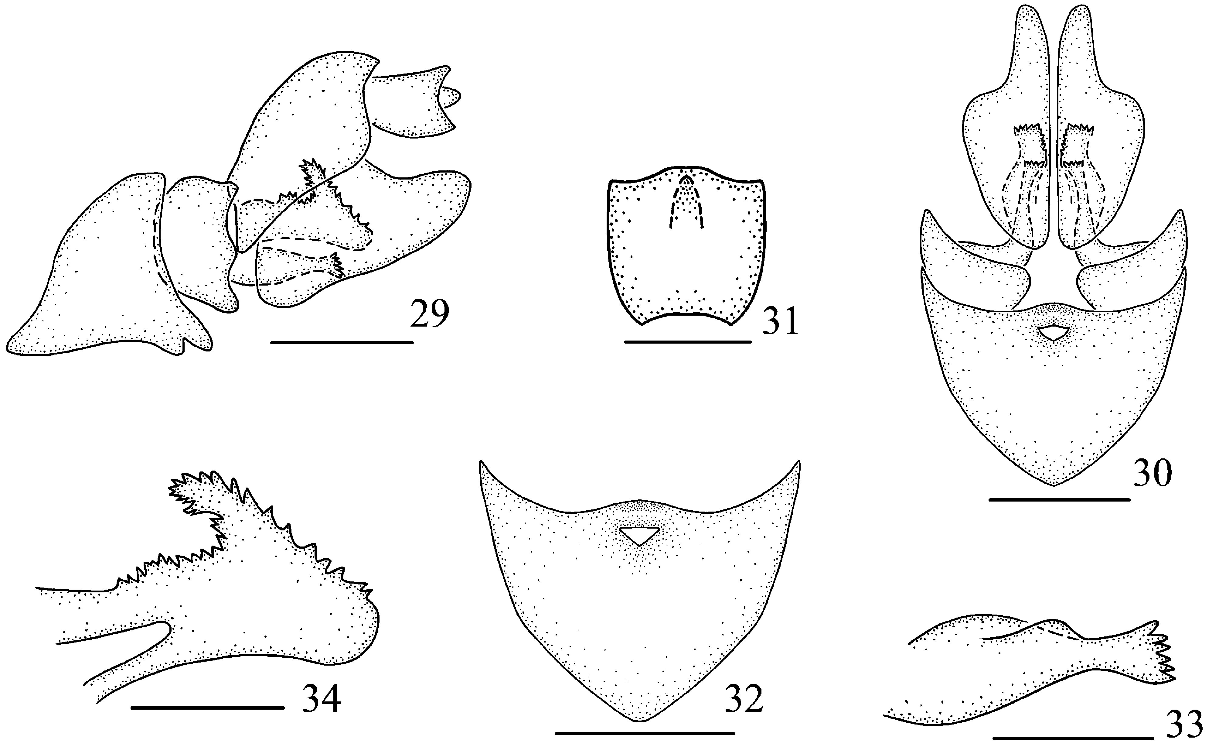
Figs 15–34 View FIGURES 15–28 View FIGURES 29–34
Measurements. Body length including forewing: male 6.1–6.2 mm (N = 3); forewing length: male 5.1–5.2 mm (N = 3).
Diagnosis. Anal segment very short ( Figs 22–23 View FIGURES 15–28 ); posterior margin of pygofer in lateral view broadly concave, one finger-like process at dorsal third, small ( Fig. 23 View FIGURES 15–28 ); genital style with two processes ( Fig. 25 View FIGURES 15–28 ); aedeagus tubular, slender and long, without process, opening dorsally at apex half, apex divided into two lobes ( Figs 27–28 View FIGURES 15–28 ).
Description. Coloration. Body mainly black brown with somewhat yellow ( Figs 15–19 View FIGURES 15–28 ). Ocelli ( Fig. 19 View FIGURES 15–28 ) slightly red, semi-translucent; eyes ( Figs 17–19 View FIGURES 15–28 ) taupe. Antennae ( Figs 18–19 View FIGURES 15–28 ) with one black spot at apex of pedicel. Frons ( Fig. 18 View FIGURES 15–28 ) with areas between sublateral carinae mostly black brown, lateral areas tawny. Clypeus ( Fig. 18 View FIGURES 15–28 ) black. Forewings ( Figs 15–16 View FIGURES 15–28 ) subhyaline, veins brown. Hind wing hyaline, veins slightly brown. Abdominal sternites black brown with somewhat green. Tergites black brown. Spines of legs with black apices.
Head and thorax. Width of vertex including eyes as wide as pronotum. Vertex ( Fig. 17 View FIGURES 15–28 ) as long as wide. Frons ( Fig. 18 View FIGURES 15–28 ) 1.5 times longer in middle line than widest part. Pronotum ( Fig. 17 View FIGURES 15–28 ) shorter in middle line than vertex (1:1.1). Mesonotum ( Fig. 17 View FIGURES 15–28 ) 1.1 times longer than pronotum and vertex combined. Forewings ( Fig. 20 View FIGURES 15–28 ) 4.3 times longer than wide; veins obviously forming a nodal line, ScP + R and MP with common stem; after the nodal line, RP single, MP with four branches, CuA with two branches; Pcu uniting A1 at basal 2/5 of clavus. Hind wing ( Fig. 21 View FIGURES 15–28 ) 2.1 times as long as broad at widest part, ScP and RP single, MP and CuA with two branches.
Male terminalia. Anal segment ( Figs 22, 23 View FIGURES 15–28 ) tubular, short, in lateral view ( Fig. 23 View FIGURES 15–28 ) dorsal margin waved, ventral margin slightly convex; 1.6 times wider than length in dorsal view ( Fig. 22 View FIGURES 15–28 ). Ventral margin of pygofer in lateral view ( Fig. 23 View FIGURES 15–28 ) strongly oblique, posterior margin broadly concave, one finger-like process at dorsal third; in posterior view ( Fig. 24 View FIGURES 15–28 ) nearly oval, with length 1.8 times than widest part; in ventral view ( Fig. 26 View FIGURES 15–28 ), posterior margin sinuate, anterior margin broadly convex. Genital style in lateral view ( Fig. 25 View FIGURES 15–28 ) with median portion broad, large, with apical margin obtuse convex; dorsal margin with basal half dorsally uplifted and branched into two processes, the basal one curving, long and thin, with apical margin narrow convex, the apical one stout with apical margin broadly concave; in ventral view ( Fig. 26 View FIGURES 15–28 ) nearly petal-shaped. Aedeagus in lateral view ( Fig. 27 View FIGURES 15–28 ) with base slightly broad, apical part mostly slender, ventrally bend, opening dorsally at apex half, apex divided into two lobes.
Female terminalia. Abdominal sternite VII ( Figs 30, 32 View FIGURES 29–34 ) in ventral view symmetrical, posterior margin concave and slightly protruded medially, with a small triangle-like hole near the posterior margin medially. Anal tube ( Figs 29, 31 View FIGURES 29–34 ) short. Gonapophysis VIII (first valvula) ( Fig. 33 View FIGURES 29–34 ) elongate, with six spines at apical margin. Gonapophysis IX (second valvula) ( Fig. 34 View FIGURES 29–34 ) with two lobes symmetrical, each lobe with much spines at apical margin and dorsal margin. Gonoplac (third valvula) with outer surface shagreen ( Figs 29, 30 View FIGURES 29–34 ); in lateral view ( Fig. 29 View FIGURES 29–34 ) with median portion broad, large, apical margin convex; in ventral view ( Fig. 30 View FIGURES 29–34 ) blade-like.
Type material. Holotype: ♂, China: Yunnan Province, Yingjiang County, Nabang Town (24°70′N, 97°90′E), on bamboo, 18 August 2018, Nian Gong leg. Paratypes: 5♂♂, 7♀♀, data same as holotype, Hong-Xing Li leg. ; 2♂♂, 4♀♀, China: Yunnan Province, Ruili County, near the Botanical Garden (24°01′N, 97°85′E), on bamboo, 23 August 2018, Nian Gong and Hong-Xing Li leg.
Host plant. Bamboo ( Neosinocalamus sp.).
Distribution. China (Yunnan).
Etymology. The specific name is derived from the Latin word “ nigricans ”, referring to the color of the face.
Remarks. This new species is similar to S. zhongtua , but differs in: 1) segment very short (relatively long in S. zhongtua ); 2) posterior margin of pygofer in lateral view broadly concave, one finger-like process at dorsal third, small (posterior margin slightly sinuate, process at middle, relatively big in S. zhongtua ); 3) genital style with two processes (without process in S. zhongtua ).

No known copyright restrictions apply. See Agosti, D., Egloff, W., 2009. Taxonomic information exchange and copyright: the Plazi approach. BMC Research Notes 2009, 2:53 for further explanation.