
Thouarella affinis Wright and Studer, 1889
|
publication ID |
https://doi.org/ 10.11646/zootaxa.3602.1.1 |
|
publication LSID |
lsid:zoobank.org:pub:10304FBF-3969-4EFA-83F1-BB8A5E2B37F3 |
|
DOI |
https://doi.org/10.5281/zenodo.10539793 |
|
persistent identifier |
https://treatment.plazi.org/id/EE36E867-FFA5-FFCF-FF0A-AC65FB5F0C71 |
|
treatment provided by |
Felipe |
|
scientific name |
Thouarella affinis Wright and Studer, 1889 |
| status |
|
4. Thouarella affinis Wright and Studer, 1889 View in CoL
Figs 10 View FIGURE 10 , 11 View FIGURE 11
Thouarella affinis Wright & Studer, 1889: 66–68 View in CoL , pl.11, fig. 3; Thomson & Henderson 1906: 38 (list); Kükenthal 1912: 302 (listed)
Thouarella (Epithouarella) affinis Kükenthal 1915: 151 View in CoL (key); 1919: 435–436; 1924: 300 (key)
Thouarella (Thouarella) affinis Cairns & Bayer 2009: 27 View in CoL (listed)
Material examined: Holotype, NHM 1889.5.27.44, 65 mm fragment, H.M.S. Challenger, sta. 135D, off Inaccessible Island , Tristan de Cunha, 37˚25’S, 12˚22’30”W, 91–128 m, 15 Jul 1874.
Other material: ZMH, R/ V W. Herwig, sta. 232, east of Isla de los Estados, Tierra del Fuego, Argentina, SW Atlantic, 54˚46’S, 62˚30’W, 800 m , 1971.
Description
As only a small fragment was available, descriptions of axis and colony morphology were taken from the type description.
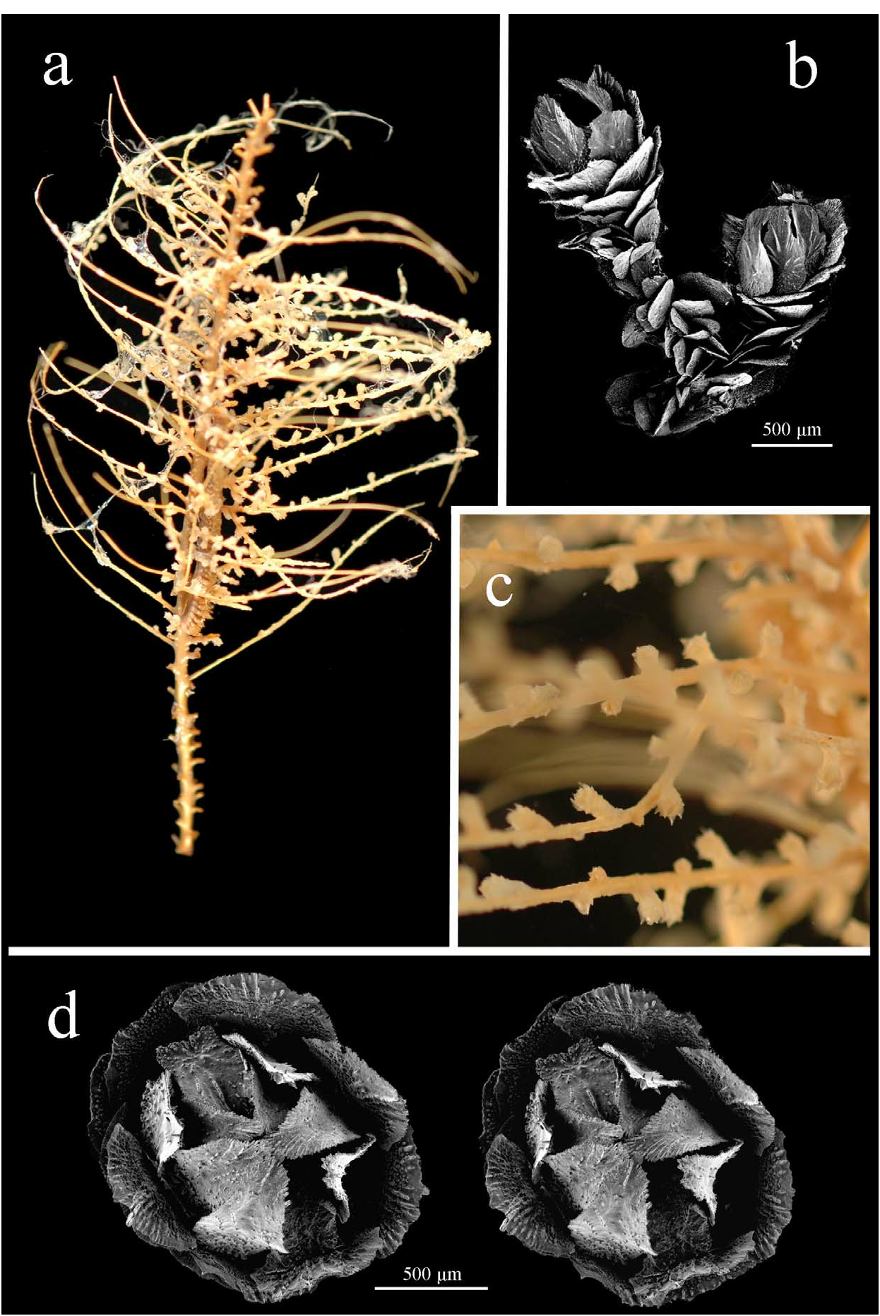
The colony is bottlebrush, ( Fig. 10a View FIGURE 10 ) but can appear bilateral. The axis is yellow, stiff, and brittle, although the branch apex is more flexible. The stem twists in a spiral from base to a quarter-length from the apex whereupon the next twist commences.
The branching of this species is dense, with only 1.5–2 mm between branchlets; becoming denser towards the apex. The branchlets are mostly simple, with some forking (dividing usually in basal region of branchlet), upwardly inclined 60–90˚, and up to 50 mm long. The branchlets are arranged in spirals from 3 sides of the main stem with 4 branchlets occurring within one spiral, but as the stem twists the spiral is difficult to follow.
The polyps are isolated, 1.1–2.1 mm high, at a density of 7–9 per cm (with denser placement at branchlet apex, which generally has a polyp at the tip). The polyps can be modestly flared but generally have a wide, rounded head extending from a slender polyp body making them clavate ( Fig. 10b,c; H View FIGURE 10 :W of 1.3–2.1, average 1.7). The polyps are arranged in short spirals of 3–4 and angled distally at 45–60˚. Each polyp has 8 longitudinal rows with 6–7 scales in each abaxial row and 5 adaxially.
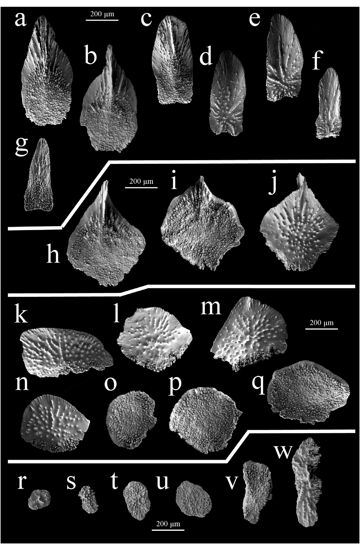
The tall rounded operculum rises above the marginal scales. The operculars are isosceles-triangle shaped ( Fig. 11a,b View FIGURE 11 ), tongue-shaped ( Fig. 11c–f View FIGURE 11 ), or lanceolate ( Fig. 11g View FIGURE 11 ), 540–780 µm high (average 610 µm) and 220–350 µm wide (average 280 µm), with an average H:W of 2.2 (range from 1.8–2.5). The outer scale surface is longitudinally concave with granules across the proximal area and occasional low striations spreading radially from centre towards distal edge ( Fig. 11d–f View FIGURE 11 ). The inner surface has a low, complex multi-keel ( Fig. 11a,b View FIGURE 11 ), or a dense area of low striations ( Fig. 11c View FIGURE 11 ); the proximal half of the scale is tuberculate.
The marginals are somewhat diamond-shaped ( Fig. 11h–j View FIGURE 11 ), 460–610 µm high (average 525 µm) and 460–640 µm wide, with an average H:W of 1 (ranges from 0.8–1.3). The inner surface has a smooth band along the distal edge which is broken by a small, simple keel ( Fig. 11h View FIGURE 11 ) with 2 or 3 adjacent striations whilst the remainder is tuberculate. The adaxial marginals frequently have no defined keel with just 4 or 5 short, sharp striations perpendicular to the distal edge ( Fig. 11i View FIGURE 11 ). The outer surface is mostly smooth with some granules that often form radial striations ( Fig. 11j View FIGURE 11 ).
The body-wall scales occur in a range of shapes from circular and irregular to elliptical ( Fig. 11k–q View FIGURE 11 ). They are generally broader than high, 350–490 µm high (average 430 µm), 380–780 µm wide (average 540 µm), with an average H:W of 0.8 (ranges from 0.6–1.1), and curved slightly away from the polyp body. Tubercles cover the inner scale surface. Granules occur sparsely on the outer surface and sometimes tubercles occur along the proximal edge. The distal edge of all sclerites of this species is finely serrate whilst the proximal edge is irregularly lobate.
There is a single layer of roughly circular ( Fig. 11 View FIGURE 11 r-u) or sometimes elongated coenenchymal scales ( Fig. 11v, w View FIGURE 11 ) that are 190–280 µm high (average 225 µm), 230–430 µm wide (average 300 µm), with an average H:W of 0.8 (range from 0.4–1.1). There are large prominent granules on the outer surface of the scales and fine tubercles on the inner surface.
Distribution
This species is known only from the type location Tristan de Cunha, and also Inacessible Island , mid-South Atlantic. The depth of observed occurrence is 91–800 m.
Remarks
The holotype material examined is damaged so the number of polyps per cm may be an underestimate.
Comparisons
Wright and Studer (1889) considered T. affinis to be very similar to T. antarctica and the polyps of these species do have a similar number of scales in the abaxial rows (6–7 T. affinis , 5–7 T. antarctica ). However, the latter has a more complexly structured marginal keel with large lateral projections compared to the modest keel found on marginals of the former.
The polyps of T. viridis are a similar shape and size to T. affinis and they also have similar shaped marginals, although those of T. viridis are slightly taller. However, the operculars of the polyps of T. viridis are pointed, while those of T. affinis are mostly tongue-shaped (with some triangular-shaped operculars with squared proximal edges). In addition, the body-wall scales of the former have more pronounced striations perpendicular to the distal edge (that are visible in lateral polyp view) than the latter.
The polyps of T. affinis are more rounded than those of T. brucei . The polyps of the latter also have fewer scales in the abaxial row and marginals that are narrower with a higher H:W ratio.
Polyps of T. koellikeri tend to be longer than those of T. affinis , with more scales in the abaxial row. Also, the former has pointed triangular operculars whereas those of the latter are mostly tongue-shaped.
| ZMH |
Zoologisches Museum Hamburg |
No known copyright restrictions apply. See Agosti, D., Egloff, W., 2009. Taxonomic information exchange and copyright: the Plazi approach. BMC Research Notes 2009, 2:53 for further explanation.
|
Kingdom |
|
|
Phylum |
|
|
Class |
|
|
Order |
|
|
Family |
|
|
Genus |
Thouarella affinis Wright and Studer, 1889
| TAYLOR, M. L., CAIRNS, S. D., AGNEW, D. J. & ROGERS, A. D. 2013 |
Thouarella (Thouarella) affinis
| Cairns, S. D. & Bayer, F. M. 2009: 27 |
Thouarella (Epithouarella) affinis Kükenthal 1915: 151
| Kukenthal, W. 1915: 151 |
Thouarella affinis
| Kukenthal, W. 1912: 302 |
| Thomson, J. A. & Henderson, W. D. 1906: 38 |
| Wright, E. P. & Studer, T. 1889: 68 |