
Thyropygus erectus, Pimvichai, Piyatida, Enghoff, Henrik & Panha, Somsak, 2009
|
publication ID |
https://doi.org/ 10.5281/zenodo.185971 |
|
DOI |
https://doi.org/10.5281/zenodo.6218638 |
|
persistent identifier |
https://treatment.plazi.org/id/1644D538-F459-FFD9-FF49-FF7A9C11FB83 |
|
treatment provided by |
Plazi |
|
scientific name |
Thyropygus erectus |
| status |
sp. nov. |
Thyropygus erectus View in CoL n. sp.
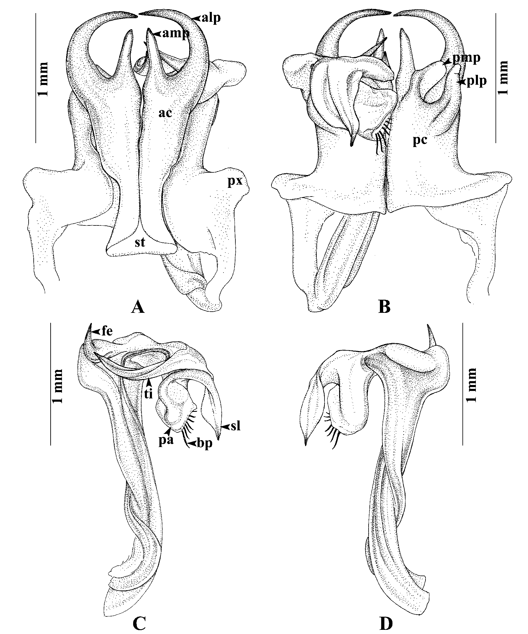
( Figs. 16 View FIGURE 16 A–D, 19C)
Material: HOLOTYPE male THAILAND, Satun Province, La-Ngu district, Koh Tarutao, 6° 49ˏ 36˝ N, 99° 38ˏ 30˝ E. 6 April 2008. P. Pimvichai, P. Prasankok, P. Tongkerd, R. Chanabun and Suwit Lhaokhum leg., ( CUMZ). – Paratypes: 10 females, same data as holotype ( CUMZ).
Etymology: The species epithet is a Latin adjective and refers to the erect femoral spine of the gonopod telopodite.
Diagnosis: A species of the opinatus subgroup. Spatulate lobe (sl) at the apical part of telopodite terminating in a sharp brown spine. Similar in this respect to T. opinatus , T. floweri and T. implicatus . Differs from these species by having the femoral spine (fe) directed distad and by having the mesal process of posterior coxal fold (pmp) flattened, curved distolaterad.
Description: Adult male with 56 podous rings, no apodous rings. Length ca. 7 cm, width ca. 4.3 mm. Adult females with 53–58 podous rings, no apodous rings. Length ca. 8–10 cm, width ca. 4.6–5.6 mm. Overall color of living animal ( Fig. 19 View FIGURE 19. A C) brown with a longitudinal orange band mid-dorsally on the body. Legs and antennae pale pink. Epiproct and margins of paraprocts yellow.
Gonopods ( Figs. 16 View FIGURE 16 A–D): Anterior coxal fold (ac) ( Fig. 16 View FIGURE 16 A): lateral process (alp) long, slender, regularly curved, tip close to tip of opposite alp, the two together forming a circle; mesal process (amp) almost as long as alp, straight, directed distad. Posterior coxal fold (pc) ( Fig. 16 View FIGURE 16 B) with lateral paracoxites (px) quite low, lateral process (plp) digitiform; mesal process (pmp) flattened, curved distolaterad. Telopodite ( Figs. 16 View FIGURE 16 C–D) leaving coxite between pmp and plp; femoral spine (fe) situated on the broad rounded lobe, directed distad, in situ resting close to amp; tibial spine (ti) very long, slender, curving in horizontal plane, its tip close to base of fe, in situ resting between amp and alp; apical part: spatulate lobe (sl) with a sharp dark brown spine at tip; palette (pa) simple, distally with about six brownish blepharochaetae (bp).
Distribution ( Fig. 20): Known only from the type locality.

| CUMZ |
Chulalongkorn University Museum of Natural History |
No known copyright restrictions apply. See Agosti, D., Egloff, W., 2009. Taxonomic information exchange and copyright: the Plazi approach. BMC Research Notes 2009, 2:53 for further explanation.