
Tinytrema sandy, PLATNICK, 2002
|
publication ID |
https://doi.org/ 10.1206/0003-0090(2002)271<0001:AROTAG>2.0.CO;2 |
|
persistent identifier |
https://treatment.plazi.org/id/03EAE52A-FFE2-A612-8225-245CDE724911 |
|
treatment provided by |
Felipe |
|
scientific name |
Tinytrema sandy |
| status |
sp. nov. |
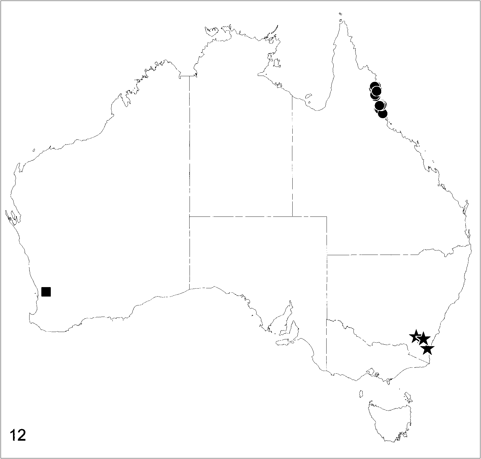
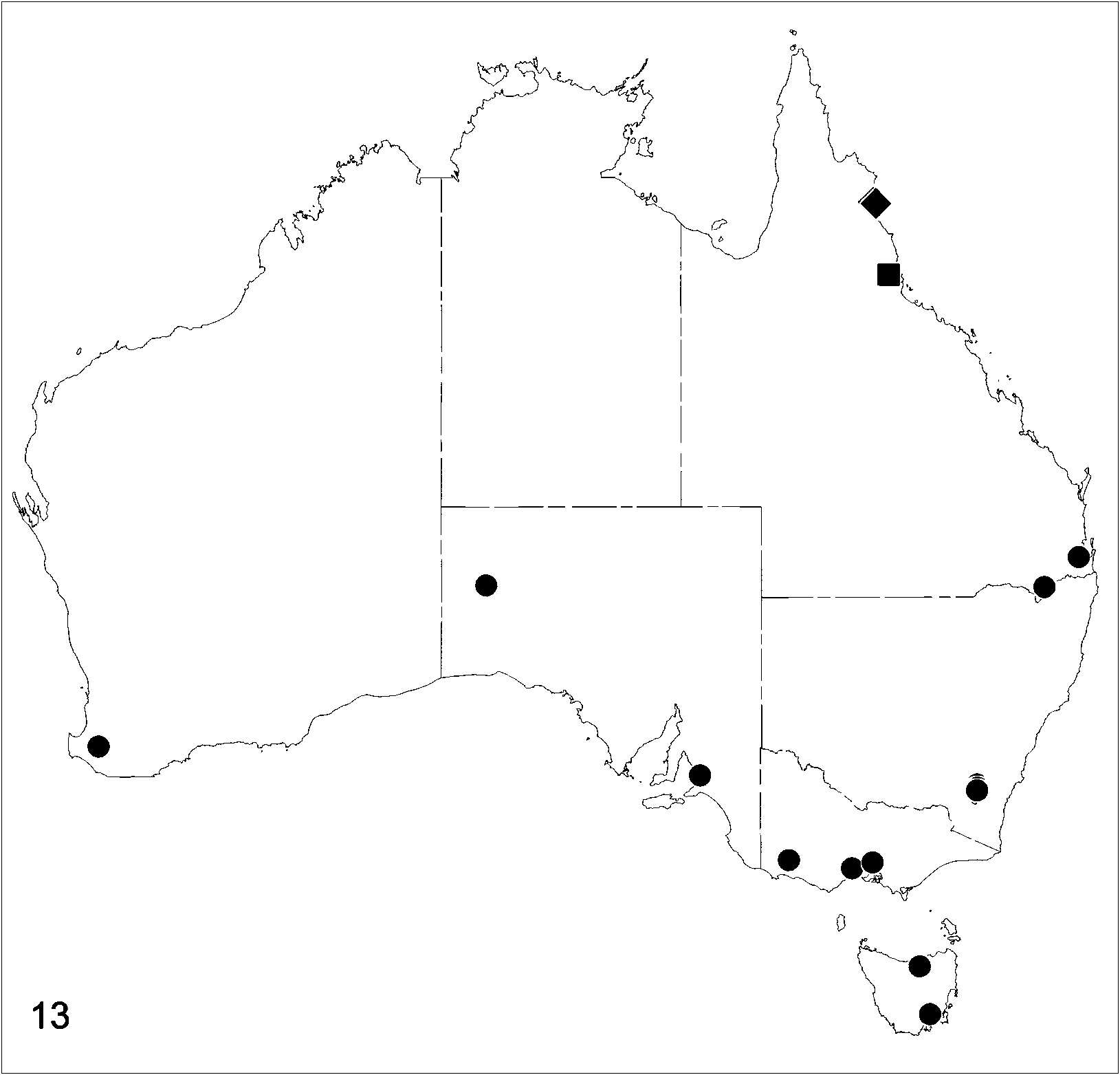
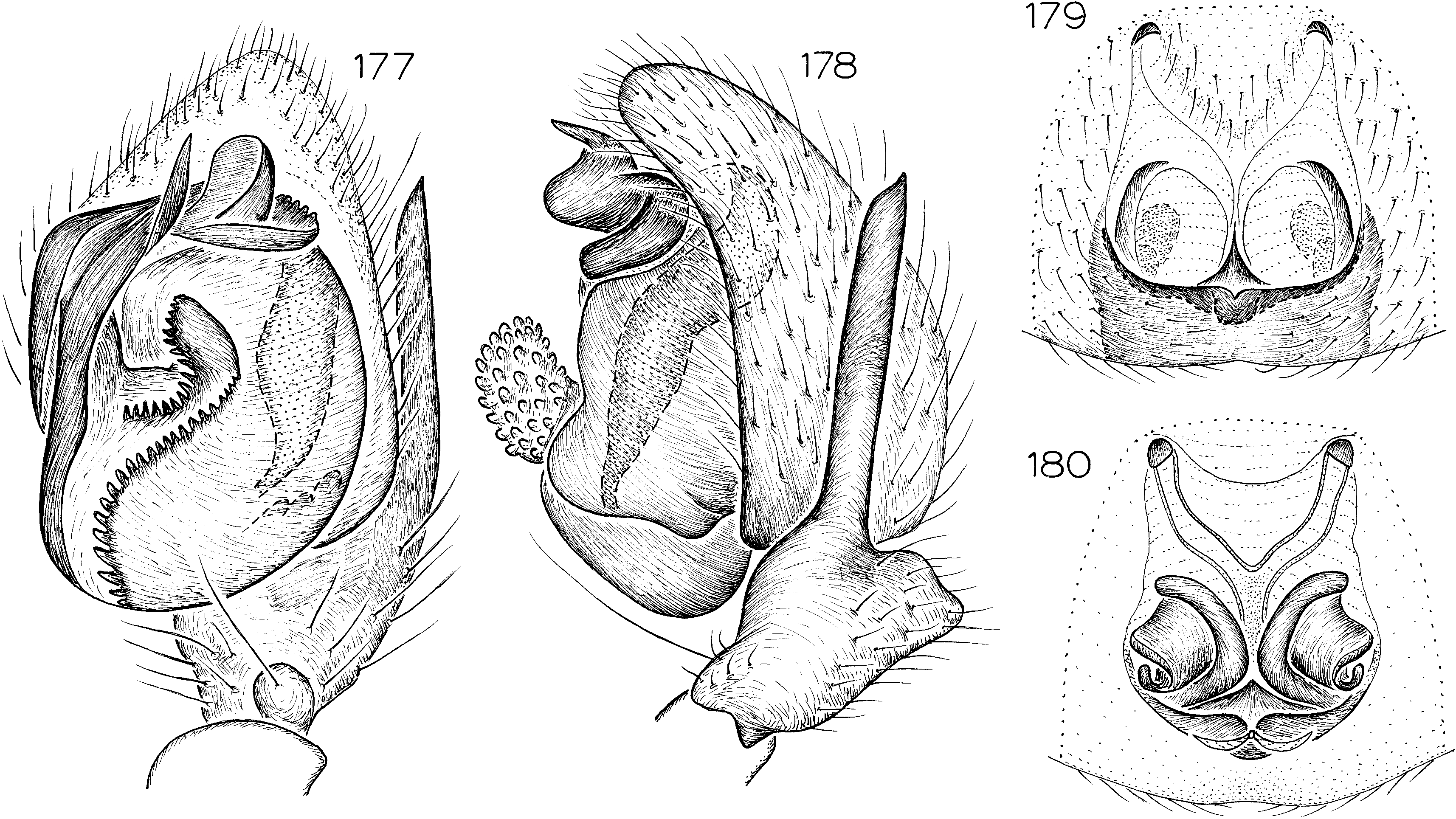
Tinytrema sandy , new species Figures 177–180 View Figs ; Map 13 View Map 13
TYPE: Male holotype taken in building at Sandy Bay , 42 ° 54 ̍ S, 147 ° 20 ̍ E, Tasmania (Mar. 28, 1967; J. Hickman), deposited in AMS (KS29305) .
ETYMOLOGY: The specific name is a noun in apposition taken from the type locality.
DIAGNOSIS: Males can easily be recognized by the long, narrow retrolateral tibial apophysis (fig. 178), females by the paired epigynal atria (fig. 179).
MALE: Total length 2.6. Carapace brown; abdominal dorsum gray, darkened along lateral margins, venter pale gray; anterior legs brown, posterior legs grayish brown. Legs spineless, but palpal femur with strong spine near dorsal apex. Retrolateral tibial apophysis extending almost entire length of cymbium (fig. 178), embolus situated prolaterally (fig. 177).
FEMALE: Total length 2.6. Coloration as in male except abdominal dorsum with pair of white marks at about onethird its length. Legs spineless. Epigynum with median septum originating from semicircular posterior ridge (fig. 179), ducts expanded, lobate laterally (fig. 180).
OTHER MATERIAL EXAMINED: Australian Capital Territory: Black Mountain , 35 ° 16 ̍ S, 149 ° 06 ̍ E, Dec, 29, 1984 (M. Harvey, R. Moran, A. Hastings, ANIC), 13 ; 23 Grylls Crescent, Cook , 35 ° 16 ̍ S, 149 ° 04 ̍ E, Nov. 10, 1984 (M. Harvey, ANIC), 13, Jan. 1, 1985 (M. Harvey, ANIC), 1♀ ; Kaleen , 35 ° 14 ̍ S, 149 ° 06 ̍ E, Apr. 23, 1994 (R. Leech, ANIC), 13 ; 14 Pera Place, Red Hill , 35 ° 20 ̍ S, 149 ° 07 ̍ E, Feb. 25, 1982, in house, on ceiling, daytime (M. Harvey, ANIC), 1♀, Nov. 8, 1982, dead on floor (M. Harvey, ANIC), 13 ; 42 Wheadon Street, Monash, 35 ° 25 ̍ S, 149 ° 06 ̍ E, Aug. 5–6, 1983 (R. Mor an, ANIC), 13. Queensland: Camira , 27 ° 38 ̍ S, 152 ° 55 ̍ E, Mar. 27, 1994 (R. Raven, QMB S31006 View Materials ), 13 ; 1 km along road 3.5 km E Pikedale, 28 ° 38 ̍ S, 151 ° 38 ̍ E, June 28, 1986 (M. Harvey, P. Vaughan, NMV Ent. 362), 1♀. South Australia: Amber Gully, Black Hill , Adelaide , 34 ° 55 ̍ S, 138 ° 42 ̍ E, July 14, 1984, under bark (D. Hirst, SAM N1999 View Materials /150), 1♀ ; 1 km W Vokes Hill corner, 28 ° 34 ̍ S, 130 ° 41 ̍ E, Apr. 14–19, 1994, pitfall (D. Hirst, SAM N1999 View Materials /149), 13. Tasmania: Exeter, 41 ° 18 ̍ S, 146 ° 56 ̍ E, Feb. 2, 1963, in house ( QVM 13 View Materials :24584), 1♀. Victoria: Hamilton , 37 ° 45 ̍ S, 142 ° 02 ̍ E, Aug. 29, 1948 (G. Stephens, NMV K3469 View Materials ), 13 ; Lara , 38 ° 01 ̍ S, 144 ° 24 ̍ E, Jan. 17, 1981, dead on ground (M. Harvey, ANIC), 13 ; Nunawading, 37 ° 49 ̍ S, 145 ° 10 ̍ E, Sept. 19, 1956 (Neboiss, NMV Ent. 362), 1♀. Western Australia: on Mockerdillup Road , 15 km SW Bridgetown, 33 ° 57 ̍ S, 116 ° 08 ̍ E, Nov. 28, 1986, found crawling on arm in garden (J. Waldock, WAM 99 View Materials /642), 13 .
DISTRIBUTION: Widespread in southern Australia, including Tasmania (map 13).
Tinytrema yarra , new species
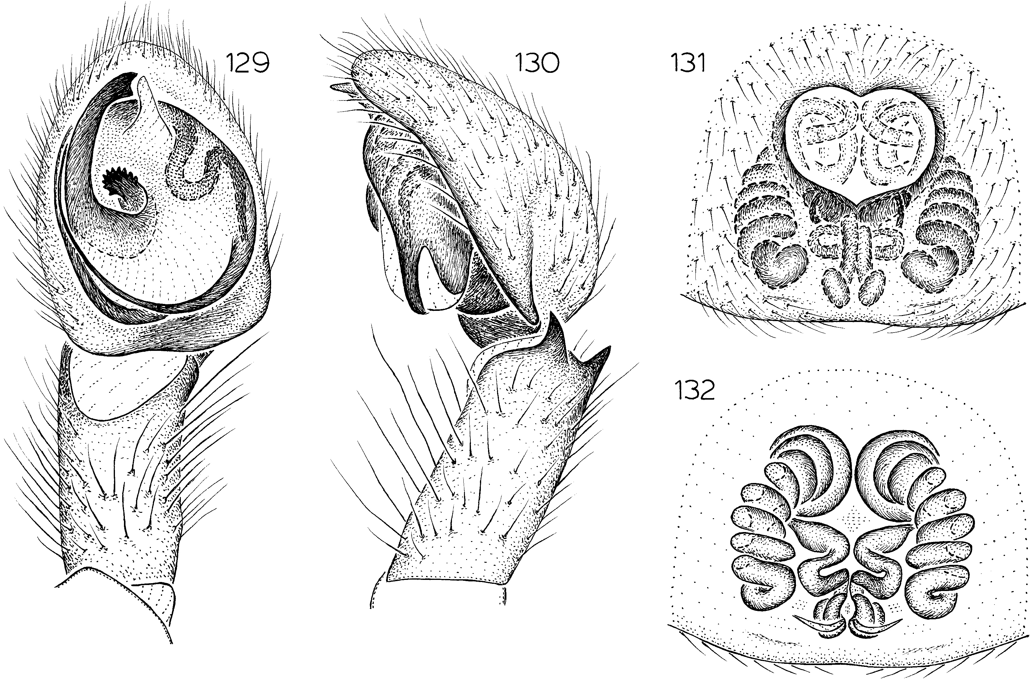
Figures 131, 132 View Figs ; Map 12 View Map 12
TYPE: Female holotype taken under the bark of a log at the junction of the Great Southern Highway and Yarra Road , 31 ° 51 ̍ S, 116 ° 28 ̍ E, Western Australia (Aug. 13, 1994; M. Harvey, M. Blosfelds), deposited in WAM (99/643) .
ETYMOLOGY: The specific name is a noun in apposition taken from the type locality.
DIAGNOSIS: Females can easily be recognized by the anteriorly situated epigynal atrium (fig. 131) and highly coiled epigynal ducts (fig. 132).
MALE: Unknown.
FEMALE: Total length 2.7. Coloration as in T. bondi except abdominal dorsum without white mark above spinnerets. Leg spination typical for genus. Epigynal atrium restricted to anterior half of epigynum (fig. 131); lateral ducts highly coiled (fig. 132).
OTHER MATERIAL EXAMINED: None.
DISTRIBUTION: Known only from the type locality in Western Australia (map 12).
Olin DeelemanReinhold, 2001: 565 (type species
by original designation O. platnicki Deeleman
Reinhold).
DIAGNOSIS: Members of this genus can be separated from those of Platyoides by the normal, rather than greatly elongated, fourth trochanters, from those of Tinytrema by the relatively unflattened body, and from those of Desognaphosa by the teeth on the tarsal claws, which resemble those of zodariids in being implanted on the inner sides of the claws.
DESCRIPTION: Mediumsized spiders, total length 5–7. Carapace only slightly flattened, without tubercles, with rebordered lateral margins and broadly reflexed posterior margin, setae few long, dark, weak, most abundant on ocular area and clypeus; thoracic groove long, longitudinal; cephalic groove obsolete. Eight or no eyes. Chelicerae porrect, divergent, with distinct oblique groove just below clypeus; anterior surface with few stiff setae; chilum wide, triangular, unipartite, entire, accompanied by second, elongated, posterior chilum (narrow, Tshaped sclerite separating bases of chelicerae posteriorly); chelicerae with distinct lateral boss, promargin with series of long setae originating in line along base of fang, those nearest base of fang not bent; promargin with four large, widely spaced teeth, second most distal smallest; retromargin with single low tooth near base of fang; cheliceral gland openings not obvious under light microscopy. Labium rectangular, flat, abruptly narrowed, deeply rebordered along posterior onefourth of length, anterior margin slightly invaginated near midline. Endites long, divergent, with oblique depression not restricted to their median edge; serrula present, sieve plate not conspicuous under light microscopy; anteromedian edges and apex bearing wide patch of long, stiff, dark setae. Sternum flat, with rebordered, slightly depressed lateral margins, not expanded anteriorly, with triangular extensions to and between coxae; surface smooth, with few long setae, posterior margin separating coxae IV, fused to ventral pedicel sclerite (at least in specimen discussed below). One weakly sclerotized epimeric sclerite on each side, not extending between coxae, not fused to carapace. Pedicel composed of two dorsal sclerites (anterior sclerite without deep posterior invagination, posteri or sclerite without beakshaped anterior extension) and strong, inverted yshaped ven tral sclerite with anteriorly expanded head surrounded by less heavily sclerotized cuticle fused with posterior sternal margin.
Abdomen reportedly with dorsal scutum in male; cuticle with sparse, erect setae; epigastric scutum weakly sclerotized, with wellmarked booklung openings at sides, with flat postepigastric sclerotizations, booklung covers slightly ridged; colulus represented only by scattered setae situated posterior of wide posterior spiracle. Anterior lateral spinnerets short, conical, separated by about their diameter at base, with two articles, distal article apparently with two major ampullate gland spigots and many small, unmodified piriform gland spigots; posterior median spinnerets of females with about seven mediumsized cylindrical gland spigots in two longitudinal rows; posterior lateral spinnerets with two articles, those of females with at least one cylindrical gland spigot.
Legs long, slightly laterigrade, leg formula 4123, with sparse setae and long spines; coxae and trochanters without dorsal tubercles, fourth trochanters only very slightly elongat ed; anterior coxae without protuberant posterolateral corners; trochanters not notched; metatarsal and tarsal scopula strong on anterior legs, weak on posteriors; posterior metatarsi with distal preening brushes; tarsi with two long claws bearing long rows of teeth implanted on inner sides of claw, teeth reduced in size but still present on tarsi IV (at least in female discussed below); weak claw tufts composed of rows of about six spatulate setae; tarsi without cuticular cracks, not shortened; morphologically dorsal surface with modified proximal margin consisting of patch of cuticle with vshaped sclerotization followed by strong cuticular ridge, that ridge opposing distinct distal extensions situated at distal edge of metatarsi; trichobothria present, in three rows on tarsi, two on metatarsi and tibiae. Female palpal femur with single dorsal spine near distal end, distal segments with long spines; female palpal tarsus with long, apparently smooth claw, without ventral scopula. Typical leg spination pattern (counts refer to morphological surfaces, only surfaces bearing spines listed): femora I–IV d100; tibiae: I, II v01r0; III d100, p011, v222, r011; IV d101, p011, v222, r011; metatarsi: I, II v20 0; III, IV p110, v222, r110.
Male palpal tibia with retrolateral apophysis, embolus and median apophysis short (per DeelemanReinhold, 2001). Epigynum with semicircular anterior margin, spermathecae short, with secondary bulbs.
Olin platnicki DeelemanReinhold
Figures 191, 192 View Figs Olin platnicki DeelemanReinhold, 2001: 565 , figs. 980–989 (holotype male and paratype female from Pulau Talata Koh, Togian Islands, Sulawesi, not examined).
DIAGNOSIS: With the characters of the genus. This species was originally described on the basis of a male and female taken in a cave on an island off the east coast of Central Sulawesi ; the female recorded below is also from the dark zone of a cave, on Christmas Island (an Australian territory off the southern coast of Java). The Christmas Island female differs obviously from the type specimens in lacking eyes (those of the type specimens were described as ‘‘possibly reduced in size as a consequence of the lightless environment of the cave’’). However , the details of the female epigynum (figs. 191, 192) match well with the drawings provided by DeelemanReinhold. It seems likely that these animals will also be found to occur in epigean sites on Indian Ocean islands, and the degree of eye reduction may well vary within and/or between cave populations. Until additional specimens become available, it seems best to regard the three specimens as conspecific .
MALE: See DeelemanReinhold (2001).
FEMALE: See DeelemanReinhold (2001).
MATERIAL EXAMINED: Christmas Island: Jedda Cave (CI5), Mar. 29, 1998, dark zone (S. Eberhard, WAM 98/1469), 1♀.
DISTRIBUTION: Known only from Christmas Island (near Java) and the Togian Islands off the east coast of Sulawesi.
Desognaphosa , new genus
TYPE SPECIES: Desognaphosa yabbra , new species.
ETYMOLOGY: The generic name is an arbitrary combination of letters taken from the informal, hybrid family name used for sort
ing these odd animals at the Queensland Museum, and is feminine in gender.
DIAGNOSIS: Members of this genus can be separated from those of Platyoides by the normal, rather than greatly elongated, fourth trochanters, from those of Tinytrema by the relatively unflattened body, and from those of Olin by the normal teeth on the tarsal claws, which are implanted ventrally rather than on the inner sides of the claws.
DESCRIPTION: Mediumsized spiders, total length of males 4–8.5, of females 3.5–9. Carapace only slightly flattened, without tubercles but with pits surrounding setal bases, with rebordered lateral margins and broadly reflexed posterior margin, setae long, dark, weak, arranged in rows radiating from thoracic groove; thoracic groove long, longitudinal; cephalic groove obsolete. Eight eyes in two rows; anterior medians smaller than other, subequal eyes, circular, dark, posterior medians irregularly oval, lenses flattened, canoeshaped tapetum present, laterals oval; from above, anterior eye row very slightly recurved, posterior row strongly recurved; from front, both rows almost straight; anterior medians separated by more than their diameter, much farther from anterior laterals; posterior medians separated by more than three times their diameter, farther from posterior laterals; anterior and posterior laterals separated by about twice their diameters; median ocular quadrangle much wider in back than in front or than long. Chelicerae porrect, divergent, with distinct oblique groove just below clypeus; anterior surface with few stiff setae; chilum wide, triangular, unipartite, entire, with projecting protrusion at midline, accompanied by second, elongated, posterior chilum (narrow, Ishaped sclerite separating bases of chelicerae posteriorly); chelicerae with distinct lateral boss, promargin with series of long setae originating in line along base of fang, those nearest base of fang not bent; promargin with five large, widely spaced teeth, second most distal smallest; retromargin with single low tooth near base of fang; cheliceral gland openings not obvious under light microscopy but retromarginal side of fang furrow distinctly ridged. Labium rectangular, flat, abruptly narrowed, deeply rebordered along posterior onefourth of length, anterior margin slightly invaginated near midline. Endites long, divergent, with oblique depression not restricted to their median edge; serrula present (fig. 152), sieve plate not conspicuous under light microscopy; anteromedian edges and apex bearing wide patch of long, stiff, dark setae. Sternum flat, with rebordered, deeply depressed lateral margins, not expanded anteriorly, with triangular extensions to and between coxae; surface smooth, with few long setae originating from pits, posterior margin separating coxae IV, not fused to ventral pedicel sclerite. One heavily sclerotized epimeric sclerite on each side, not extending between coxae, not fused to carapace. Pedicel composed of two dorsal sclerites (anterior sclerite without deep posterior invagination, posterior sclerite without beakshaped anterior extension) and strong, triangular ventral sclerite reaching almost to posterior sternal margin.
Abdomen without dorsal scutum; cuticle with sparse, erect setae; epigastric scutum heavily sclerotized, with wellmarked booklung openings at sides, with flat postepigastric sclerotizations, booklung covers slightly ridged; colulus represented only by scattered setae on rounded patch of sclerotized cuticle situated posterior of wide posterior spiracle. Six spinnerets (figs. 181–186), anterior laterals short, tubular, separated by about their diameter at base, with two articles, distal article with two major ampullate gland spigots and several small, unmodified piriform gland spigots; posterior medians of females with about six large cylindrical gland spigots in single longitudinal row; posterior laterals with two articles, those of females with single large cylindrical gland spigot.
Legs long, only slightly laterigrade, leg formula 4123, with sparse setae and few spines; coxae and trochanters without dorsal tubercles, fourth trochanters only very slightly elongated; anterior coxae without protuberant posterolateral corners; trochanters not notched; metatarsal and tarsal scopula weak on all legs; posterior metatarsi with weak distal preening brushes; tarsi with two long claws bearing long rows of teeth implanted ventrally on claw, teeth reduced in size but still present on tarsi IV; weak claw tufts composed of numerous, only slightly spatulate setae; tarsi without cuticular cracks, not shortened, higher subdistally than basally; morphologically dorsal surface with modified proximal margin consisting of patch of cuticle with ushaped sclerotization followed by strong cuticular ridge, that ridge opposing distinct distal extension situated at distal edge of metatarsi; trichobothria present, in three rows on tarsi, two on metatarsi and tibiae. Female palp with weak spines on tibiae and tarsi only; female palpal tarsus with long, dentate claw, with ventral scopula of stiff setae. Typical leg spination pattern (counts refer to morphological surfaces, only surfaces bearing spines listed): femora: III d001; IV d101; tibiae: III v1p02; IV v1p1p2; metatarsi: III v001p; IV 1p1p2.
Male palpal tibia with basal retrodorsal protrusion, retrolateral apophysis short, shift ed dorsally, opposite deeply invaginated retrolateral cymbial margin; embolus long, extending along prolateral side of bulb, terminating near sclerotized median apophysis and membranous conductor (separation of those sclerites sometimes indistinct). Epigynum relatively simple, with complexly arranged ducts.
SPECIES GROUPS: Two informal but putatively monophyletic species groups are recognized below, along with a small group of species of uncertain relationship. Within each group, the species are listed in geographic order, proceeding from south to north.
IDENTIFICATION: Because of the size of the genus, separate keys are presented for those species from northern Queensland (north of 20 ° S) and those from southern Queensland (south of 20 ° S) and New South Wales.
KEY TO SPECIES OF DESOGNAPHOSA KNOWN FROM SOUTHERN QUEENSLAND AND NEW SOUTH WALES
1. Males (those of bulburin , kroombit , funnel , and goonaneman unknown)......... 2
– Females........................... 4
2. Retrolateral tibial apophysis longer at middle than dorsally or ventrally (fig. 188)................................. yabbra
– Retrolateral tibial apophysis longer dorsally than at middle or ventrally (figs. 204, 208) ................................ 3
3. Embolus relatively short, originating near ventral edge of palpal bulb (fig. 203)............................... eungella
– Embolus relatively long, originating near re trolateral edge of palpal bulb (fig. 207)............................. dryander
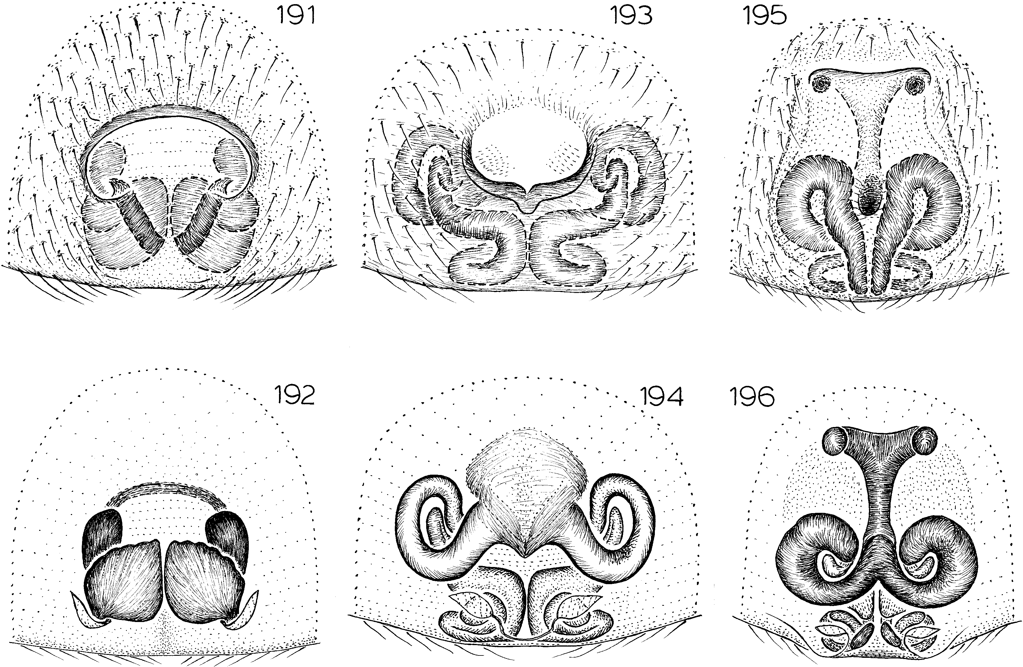
4. Epigynum without distinct anterior margin, with rounded atrium (fig. 193).................................. bulburin
– Epigynum with distinct anterior margin, without rounded atrium................ 5
5. Anterior epigynal margin distinctly bipartite (figs. 197, 205, 217)............... 6
– Anterior epigynal margin unipartite (figs. 189, 195, 209).................... 8
6. Spermathecae greatly reduced, far from anterior epigynal margin (figs. 217, 218)........................... goonaneman
– Spermathecae not reduced (figs. 198, 206) ................................. 7
7. Anterior epigynal ducts coiled (fig. 198)................................ funnel
– Anterior epigynal ducts not coiled (fig. 206).......................... eungella
8. Anterior epigynal margin relatively wide (fig. 195)...................... kroombit
– Anterior epigynal margin relatively narrow (figs. 189, 209)................... 9
9. Anterior epigynal margin relatively far from epigynal ducts (figs. 189, 190)................................... yabbra
– Anterior epigynal margin situated closer to epigynal ducts (figs. 209, 210).................................. dryander
No known copyright restrictions apply. See Agosti, D., Egloff, W., 2009. Taxonomic information exchange and copyright: the Plazi approach. BMC Research Notes 2009, 2:53 for further explanation.
|
Kingdom |
|
|
Phylum |
|
|
Class |
|
|
Order |
|
|
Family |
|
|
Genus |
Tinytrema sandy
| PLATNICK, NORMAN I. 2002 |
Olin DeelemanReinhold, 2001: 565
| Deeleman-Reinhold, C. L. 2001: 565 |