
Phyxioschema eripnastes, Schwendinger, Peter J., 2009
|
publication ID |
https://doi.org/10.5281/zenodo.188258 |
|
DOI |
https://doi.org/10.5281/zenodo.6214232 |
|
persistent identifier |
https://treatment.plazi.org/id/03A9B15A-FFB1-7E6C-FF06-B0F0FB53FF08 |
|
treatment provided by |
Plazi |
|
scientific name |
Phyxioschema eripnastes |
| status |
sp. nov. |
Phyxioschema eripnastes sp. n.
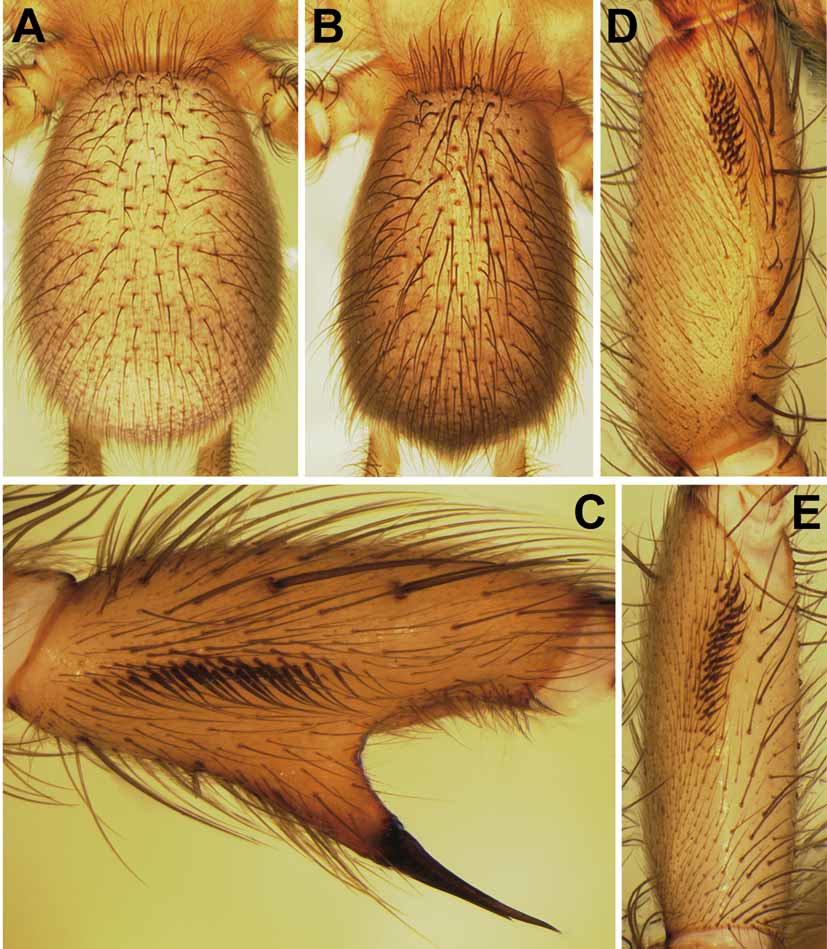
Figures 15–17 View FIGURE 15 A – E View FIGURE 16 A – N View FIGURE 17 A – D
Material: THAILAND (southern region): Phang Nga Province and District, limestone cliffs near Pha Phueng Cave ( 8°28’21”N, 98°32’12”E), 60 m, ca 3 km NE of Phang Nga city, male holotype (matured 30.IX.1997), 2 male paratypes (matured 1.X.1997, 8.X.1997) and 2 female paratypes, collected 29.IX.1997. From same locality: 1 male paratype (matured 14.I.2004) and 6 female paratypes, collected 17.V.2003 (sample TH-03/ 11). All specimens collected by P.J. Schwendinger and deposited in MHNG.
Etymology: Greek: "eripne" = precipice, cliff, "nastes" = inhabitant, dweller; noun in apposition.
Diagnosis: Medium-sized species, distinguished from the distinctly larger P. huberi sp. n. by: Males with more strongly curved embolus tip; leg tarsi with few or without spines; tibia I without spines in proximal third; tibia II with narrower prolateral band of elongated spinules, with a distally narrower and not bilobed ventral spur with a pair of megaspines of unequal lengths; metatarsus II with fewer spines, a more distally situated proventral keel and a more proximally situated retroventral keel. Females with much wider spermathecae carrying much shorter receptacles.
Description: MALE ( holotype). Colour in alcohol: Carapace, palps, booklung plates, genital region, anal mound and spinnerets light brown, legs and palpal bulbs slightly darker; eye mound black. Dorsal side of opisthosoma with very indistinct light transverse stripes forming incomplete chevrons; ventral side of opisthosoma mottled with dark spots; posterior lateral spinnerets with pattern of darker patches and light spots ventrally.
Body 8.9 long. Carapace 3.6 long, 3.3 wide, oval, almost flat, covered with light adpressed hairs interspersed by darker, more erect bristles (some wavy), these longest on carapace margin, in front, on and behind eye mound, and in front and behind fovea. Eyes on low mound; eye group ( Fig. 16B View FIGURE 16 A – N ) 0.41 long, anterior eye row slightly procurved, 0.75 wide, posterior eye row slightly recurved, 0.86 wide. Eye diameters and interdistances: AME 0.24, ALE 0.24, PME 0.16, PLE 0.21; AME–AME 0.04, AME–ALE 0.01, PME–PME 0.33, PME–PLE 0.01. MOQ 0.37 long, 0.43 wide anteriorly, 0.62 posteriorly. Fovea pit-like, with 3 long, thickened and slightly procurved foveal setae anterior to it.
Chelicerae weak, grooves with 9/10 prolateral teeth and 14/16 median proximal denticles. Maxillae ( Fig. 16C View FIGURE 16 A – N ) 1.0 long, 0.5 wide, with pallid prolateral zone; anterior lobe indistinct, with quite wide but fairly indistinct serrula on ridge. Labium ( Fig. 16C View FIGURE 16 A – N ) 0.3 long, 0.7 wide, anterior edge distinctly setose, followed by a pallid zone; posterior part pigmented, with few fine setae. Sternum ( Fig. 16C View FIGURE 16 A – N ) 1.9 long, 1.6 wide, cordate, with deeply excavated post-labial depression formed by fused anterior sigilla and labio-sternal groove, and with 3 pairs of indistinct marginal sigilla.
Palps ( Fig. 16D–E View FIGURE 16 A – N ) set with stiff bristles. 8+8 trichobothria in 2 rows on tibia, 8 in zig-zag row on tarsus. Palpal bulb with oval base and quite long embolus with distinctly curved tip.
Legs 2134. All tarsi pseudosegmented and without spines. Preening combs absent. I: tibia cylindrical, with strong spines prolaterally, ventrally and retrolaterally in distal two-thirds but without spines in proximal third ( Fig. 16G View FIGURE 16 A – N ); patella with row of 4 sigmoid spines retroventrally, without triangular projection on retrolateral margin ( Fig. 16F View FIGURE 16 A – N ); femur with short, fairly wide band of hooked spinules retrodorsally ( Fig. 15D View FIGURE 15 A – E ). II: metatarsus with only few spines, ventroproximally with 2 widely separated angular keels ( Fig. 16K View FIGURE 16 A – N ), proventral one ( Fig. 16J View FIGURE 16 A – N ) situated more distally than retroventral one ( Fig. 16K–L View FIGURE 16 A – N ), the latter only slightly projecting sideward ( Fig. 16K, M View FIGURE 16 A – N ); tibia moderately incrassate ( Fig. 16H View FIGURE 16 A – N ), band of elongated spinules on prolateral side straight, narrow, parallel to longitudinal axis of tibia, almost reaching height of distal side of ventral spur ( Fig. 15C View FIGURE 15 A – E ); ventral spur of tibia with apex not bilobed, pair of megaspines basally close to each other and distally slightly diverging, prolateral megaspine clearly longer than retrolateral one ( Fig. 16H View FIGURE 16 A – N ); femur with moderately long and relatively wide band of hooked spinules proventrally ( Fig. 15E View FIGURE 15 A – E ). Spination: I: patella d2, r4; tibia p11/12, r4, v4; metatarsus v 5. II: patella d2; tibia p2, v2 megaspines; metatarsus p2, v 4. III: patella d3; tibia d2, p2, r2, v5; metatarsus d8, v 8. IV: patella d3; tibia d1, p2, r2, v5; metatarsus d8, v7. Trichobothria: 2 rows of 7–9 each on tibiae, 13–16 in single row on metatarsi, 11–12 in single row on tarsi. Paired claws with 9–10 teeth in sigmoid row, unpaired claw with 5–7 sessile teeth.
Opisthosoma 4.2 long, 3.0 wide; dorsal side quite densely covered with fine light hairs, short dark hairs and long, strong dark bristles with darkened sockets ( Fig. 15B View FIGURE 15 A – E ); ventral side only with short dark and mediumsized dark hairs. Posterior median spinnerets 0.6 long, posterior laterals 6.0 long (proximal article 1.6, median article 1.7, pseudosegmented distal article 2.7).
FEMALE ("allotype"). As the male, except for: legs slightly lighter, chelicerae, labium, maxillae and tips of palpal tarsi darker; pale prolateral zone of maxillae and pale anterior zone of labium more distinct.
Body 14.4 long. Carapace 4.8 long, 4.2 wide. Eye group ( Fig. 16A View FIGURE 16 A – N ) 0.41 long, anterior eye row 0.86 wide, posterior eye row 0.96 wide. Eye diameters and interdistances: AME 0.24, ALE 0.25, PME 0.19, PLE 0.21; AME–AME 0.09, AME–ALE 0.04, PME–PME 0.38, PME–PLE 0.03. MOQ 0.38 long, 0.44 wide anteriorly, 0.67 posteriorly.
Chelicerae stronger than in male, grooves with 9 prolateral teeth and 17/18 median proximal denticles. Maxillae 1.5 long, 0.8 wide, serrula longer, wider and more pronounced than in male. Labium 0.5 long, 1.0 wide. Sternum 2.2 long, 2.0 wide.
Palps with 8+8 trichobothria on tibia and 15 on tarsus. Tarsal claws with 16/17 teeth.
Legs 1=234. All tarsi integral and armed with spines. Spination: I: patella d1; tibia p1, v3; metatarsus v7; tarsus p1, r 2. II: patella d2; tibia p2, v3; metatarsus p2, v7; tarsus p1, r 2. III: patella d3; tibia d2, p2, r2, v3; metatarsus d7, v7; tarsus p1, r 2. IV: patella d3; tibia d2, p2, r2, v3; metatarsus d7, v7; tarsus p1, r2. Trichobothria: 2 rows of 8–9 each on tibiae, 14–16 in single row on metatarsi, 12–13 in single row on tarsi. Paired claws with 12–16 teeth in sigmoid row, unpaired claw with 6–7 sessile teeth.
Opisthosoma ( Fig. 15A View FIGURE 15 A – E ) 7.5 long, 5.5 wide. Posterior median spinnerets 1.3 long; posterior lateral spinnerets 7.5 long (proximal article 2.2, median article 2.0, distal article 3.3).
Vulva ( Fig. 17A View FIGURE 17 A – D ) with 2 wide spermathecae, each carrying 4/5 short receptacles; lateral receptacle with short, wide and completely sclerotised base, and with small blister-like head carrying only few pores; median receptacle with fairly short stalk and globular head with pores; secondary receptacles similar to median receptacle, situated quite high up on the spermathecal trunk at almost the same level as the lateral receptacle.
Palp and leg measurements: See Table 1.
Variation: The number of foveal setae varies between two and four. Two males (including the holotype) have no spines on leg tarsi, a third male has only a single spine on one tarsus, and a fourth male has a single spine on each of tarsi II–IV. One male paratype has three retroventral spines on patella I of both sides, two others have three on one side and four on the other, the holotype has four on both sides. One male paratype possesses three megaspines on the ventral spur of its left tibia II ( Fig. 16I View FIGURE 16 A – N ). Another male paratype has a rather triangular retroventral proximal keel on metatarsus II ( Fig. 16N View FIGURE 16 A – N ). Usually there is only a single retroventral spine situated distally to the retroventral keel on metatarsus II ( Fig. 16K View FIGURE 16 A – N ), but two male paratypes additionally possess a proventral spine (in a more distal position than the retroventral one) on one side. The female genitalia examined (n=6) have 3–6 receptacles per spermathecal trunk; the base of the lateral receptacle is wide or slightly constricted to an indistinct neck ( Fig. 17A–D View FIGURE 17 A – D ). Measurements of males (n=4) (females in parentheses; n=8): body length 8.1–10.8 (11.5–16.0), carapace length 3.3–4.3 (4.1–5.4), width 3.0–4.0 (3.6–4.5). The smallest female produced eggs and was thus mature.
Relationships: Phyxioschema eripnastes sp. n. is most closely related to P. s p e l a e u m sp. n., both of them are very similar to P. sayamense sp. n. and P. huberi sp. n.
Distribution and habitat: This species is known only from near the Pha Phueng Cave close to Phang Nga city ( Fig. 1 View FIGURE 1 , locality 17). All spiders were collected from webs leading into crevices and holes of vertical limestone cliffs and into cracks between hardened mud and rock at the base of the cliffs. All were found in places shaded by remnants of an evergreen forest, not on the sun-exposed parts of the cliffs near the temple cave and near the huts of the resident monks.
Phenology ( Table 3): Three males collected in late September became mature soon afterwards ( 30 September, 1 October and 8 October); one immature male collected in May reached maturity in mid-January of the following year. The latter maturation date is presumably due to a delay in development caused by conditions in captivity. Mature males continued to feed for only about one month. No copulations were observed. Some females collected in mid-May had egg sacs suspended in their webs. One web was then seen with three egg sacs from two of which the spiderlings had already hatched, the third one contained 27 undeveloped, light yellow eggs. Two more egg sacs were later built in captivity by different females, one in early July ( 36 eggs), the other in early September ( 18 eggs). Either females are able to store sperm and produce eggs for almost a year, or there is a second mating period in April and/or May.


| MHNG |
Museum d'Histoire Naturelle |
No known copyright restrictions apply. See Agosti, D., Egloff, W., 2009. Taxonomic information exchange and copyright: the Plazi approach. BMC Research Notes 2009, 2:53 for further explanation.
|
Kingdom |
|
|
Phylum |
|
|
Class |
|
|
Order |
|
|
Family |
|
|
Genus |