
Phyxioschema sayamense, Schwendinger, Peter J., 2009
|
publication ID |
https://doi.org/ 10.5281/zenodo.188258 |
|
DOI |
https://doi.org/10.5281/zenodo.6214230 |
|
persistent identifier |
https://treatment.plazi.org/id/03A9B15A-FFB5-7E61-FF06-B311FD27FC06 |
|
treatment provided by |
Plazi |
|
scientific name |
Phyxioschema sayamense |
| status |
sp. nov. |
Phyxioschema sayamense sp. n.
Figures 12–14 View FIGURE 12 A – E View FIGURE 13 A – L View FIGURE 14 A – D
Material: THAILAND (southern region): Surat Thani Province, Phanom District, Khao Sok National Park, limestone hill near park headquarters (8°54’54”N, 98°31’39”E), 100 m, male holotype (matured 5.II.2004), 4 male paratypes (one collected mature, two matured 22.I.2005, fourth matured 18.III.2005) and 17 female paratypes. All specimens collected by P.J. Schwendinger on 14.V.2003 (sample TH-03/08) and deposited in MHNG.
Etymology: The species epithet is a latinized adjective of " Sayam " (English spelling: " Siam "), an old Chinese name for Thailand.
Diagnosis: Fairly large species, distinguished from all known congeners by retroventral spines on patella I of males relatively long and curved in lateral view (sigmoid shape only visible in ventral view), by tibia II of males longer than tibia I and by metatarsus II of males with strongly modified ventral keels (prolateral keel reduced to a small cone sitting on the much larger retrolateral keel, the latter projecting ventrally, not retroventrally as in other species). Different from the larger P. huberi sp. n. by: Males with more slender palpal bulb; palpal tibia more distinctly bulging on retrolateral side; femur I with longer and narrower band of spinules retrodorsally, femur II with narrower band proventrally; tibia I with only few proventral spines in a median position, retroventral and retrolateral spines lacking; tibia II more slender, with a narrower and shorter band of spinules prolaterally, and with more slender, not bilobed ventral spur with a pair of megaspines close to each other. Females without secondary spermathecal receptacles; median receptacle with distinctly longer stalk and larger head than lateral receptacle.
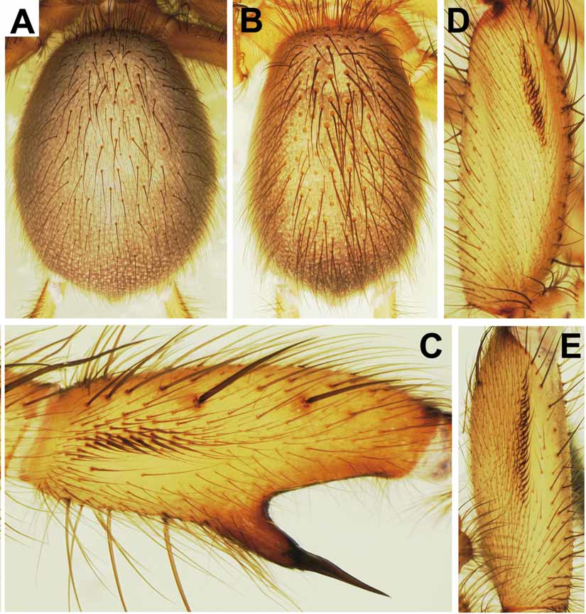
Description: MALE (holotype). Colour in alcohol: Body generally light reddish brown, ventral side lighter, eye mound black. Opisthosoma grey-brown, except for orange-brown bases of stiff dorsal bristles, very indistinct light transversal stripes forming incomplete chevrons in posterior half of dorsal side ( Fig. 12B View FIGURE 12 A – E ), light brown genital region and booklung plates, and light brown spinnerets with pattern of darker patches and light spots ventrally.
Body 11.9 long. Carapace 4.7 long, 4.2 wide, oval, almost flat, covered with light adpressed hairs interspersed by darker, more erect bristles (some wavy), these longest on carapace margin, in front, on and behind eye mound, and in front and behind fovea. Eyes on low mound; eye group ( Fig. 13B View FIGURE 13 A – L ) 0.45 long, anterior eye row slightly procurved, 0.89 wide, posterior eye row slightly recurved, 0.98 wide. Eye diameters and interdistances: AME 0.25, ALE 0.31, PME 0.17, PLE 0.22; AME–AME 0.07, AME–ALE 0.02, PME–PME 0.28, PME–PLE 0.02. MOQ 0.40 long, 0.48 wide anteriorly, 0.70 posteriorly. Fovea pit-like, with 2 long, thickened and slightly procurved foveal setae anterior to it.
Chelicerae weak, grooves with 10/11 prolateral teeth and 26/27 median proximal denticles. Maxillae ( Fig. 13C View FIGURE 13 A – L ) 1.1 long, 0.8 wide; prolateral zone pallid; anterior lobe indistinct, with quite wide but fairly indistinct serrula on ridge. Labium ( Fig. 13C View FIGURE 13 A – L ) 0.3 long, 0.7 wide, anterior edge distinctly setose, followed by pallid zone; posterior part pigmented, with few fine setae. Sternum ( Fig. 13C View FIGURE 13 A – L ) 2.4 long, 2.0 wide, cordate, with deeply excavated post-labial depression formed by fused anterior sigilla and labio-sternal groove, and with 3 pairs of indistinct marginal sigilla. Membrane close to posterior sternal exfoliations (between coxae IV) with dark pigment.
Palps ( Fig. 13D–E View FIGURE 13 A – L ) set with stiff bristles. 6+7 trichobothria in 2 rows on tibia, 10 in zig-zag row on tarsus. Palpal bulb relatively small (in comparison to tibia), with oval base and quite long, tapering, almost straight embolus with a slightly curved tip.
Legs 1234. All tarsi pseudosegmented and armed with spines. No preening combs. I: tibia cylindrical, with few prolateral and proventral spines in median third of tibia and with few ventral spines only in distal half, retroventral and retrolateral spines lacking ( Fig. 13G View FIGURE 13 A – L ); patella with a row of 3 quite long spines [curved in lateral view ( Fig. 13F View FIGURE 13 A – L ), slightly sigmoid in ventral view ( Fig. 13G View FIGURE 13 A – L )], without triangular projection on retrolateral margin; femur with relatively long and narrow band of hooked spinules retrodorsally ( Fig. 12D View FIGURE 12 A – E ). II: metatarsus ventroproximally with 2 keels close to each other and projecting ventrally (not sideward; Fig. 13J, L View FIGURE 13 A – L ), proventral keel reduced to small blunt cone ( Fig. 13I –J View FIGURE 13 A – L ) on the much larger, widely rounded retroventral keel with a distal angle ( Fig. 13K View FIGURE 13 A – L ); tibia II slightly incrassate, longer than tibia I (not so in males of other Phyxioschema species), band of elongated spinules on prolateral side straight, fairly narrow and exceptionally short, clearly not reaching height of proximal side of ventral spur, following longitudinal axis of tibia ( Fig. 12C View FIGURE 12 A – E ); ventral spur of tibia slender, its apex not bilobed, carrying 2 megaspines of equal length, prolateral one basally turning away from retrolateral one, distally both running more or less in parallel ( Fig. 13H View FIGURE 13 A – L ); femur II with long (slightly longer than on femur I) and narrow band of hooked spinules proventrally ( Fig. 12E View FIGURE 12 A – E ). Spination: I: patella p1, r3; tibia p7/8, v5; metatarsus v7; tarsus p 1. II: patella p1, r3; tibia p2, v2 megaspines; metatarsus p1/2, v4; tarsus r 2. III: patella p2/3, r1; tibia d1/2, p2/3, r2, v4; metatarsus d5, p1/2, v7; tarsus p1, r1/ 2. IV: patella p2, r1; tibia d2, p2, r2, v4; metatarsus d5, 2p, 1r, v7; tarsus r2. Trichobothria: 2 rows of 8–9 each on tibiae, 11–14 in single row on metatarsi, 10–11 in single row on tarsi. Paired claws with 11–13 teeth in sigmoid row, unpaired claw with 5–6 sessile teeth.
Opisthosoma 6.0 long, 3.9 wide; dorsal side quite densely covered with fine light hairs, stronger dark hairs and long, strong dark bristles with darkened sockets ( Fig. 12B View FIGURE 12 A – E ); ventral side only with short dark and medium-sized dark hairs. Posterior median spinnerets 0.8 long; posterior laterals 7.1 long (proximal article 2.1, median article 2.1, pseudosegmented distal article 2.9).
FEMALE ("allotype"). As male, except for: prosoma (especially chelicerae, maxillae and tips of palpal tarsi) darker; opisthosoma (especially ventral side) lighter; pale prolateral zone of maxillae and pale anterior zone of labium more distinct.
Body 16.4 long. Carapace 5.2 long, 4.4 wide. Eye group ( Fig. 13A View FIGURE 13 A – L ) 0.51 long, anterior eye row 1.13 wide, posterior eye row 1.19 wide. Eye diameters and interdistances: AME 0.24, ALE 0.36, PME 0.21, PLE 0.24; AME–AME 0.09, AME–ALE 0.02, PME–PME 0.43, PME–PLE 0.02. MOQ 0.45 long, 0.54 wide anteriorly, 0.83 posteriorly.
Chelicerae stronger than in male, grooves with 11/12 prolateral teeth and 24/25 median proximal denticles. Maxillae 1.5 long, 1.1 wide, serrula slightly wider than in male. Labium 0.4 long, 1.0 wide. Sternum 2.5 long, 2.2 wide.
Palps with 8+8 trichobothria on tibia and 10 on tarsus. Tarsal claw with 13/14 teeth.
Legs 2134. All tarsi integral. Tibia II shorter than tibia I. Spination: I: patella p1/2; tibia p1/2, v5 /6; metatarsus v9; tarsus p2, r 2. II: patella p2; tibia p1/2, v5; metatarsus p2, v7; tarsus p1, r1/ 2. III: patella p2/3, r1/2; tibia d1/2, p2/3, r2/3, v5; metatarsus d1, p4, r2/3, v8; tarsus p2, r2/ 4. IV: patella p2, r1; tibia d1/2, p2, r2, v5; metatarsus d5, p2, r2/4, v5; tarsus p1, r1/2. Trichobothria: 2 rows of 8–9 each on tibiae, 14–15 in single row on metatarsi, 12–15 in single row on tarsi. Paired claws with 11–13 teeth, unpaired claw with 4–5 sessile teeth.
Opisthosoma ( Fig. 12A View FIGURE 12 A – E ) 8.4 long, 6.4 wide. Posterior median spinnerets 0.9 long; posterior lateral spinnerets 8.3 long (proximal article 2.4, median article 2.3, distal article 3.6). Vulva ( Fig. 14A View FIGURE 14 A – D ) with 2 fairly narrow spermathecae, each carrying 2 receptacles; lateral receptacle with very short, sclerotised stalk and small head; median receptacle with much longer, slightly convoluted stalk and larger head.
Palp and leg measurements: See Table 1.
Variation: Measurements of males (n=5) (females with egg sacs in parentheses; n=7): body length 10.2–13.1 (11.7–19.9), carapace length 4.0–5.3 (4.2–5.2), width 3.5–4.5 (3.6–4.7). One male has its tibia II on one side shorter than tibia I, but on the other side it is the inverse (tibia II is always longer than tibia I in other males). One male has four retroventral spines on its right patella I, its left patella I has three spines (as in all other conspecific males). One female has three foveal setae (the unpaired one less pronounced than the paired ones), all other specimens examined have only two such setae. Variation in the shape of spermathecae (n=4), see Fig. 14A–D View FIGURE 14 A – D . One female possesses a vestigial secondary receptacle on one side ( Fig. 14C View FIGURE 14 A – D ). Very small lateral receptacular heads are without pores ( Fig. 14A View FIGURE 14 A – D ), larger ones with few pores ( Fig. 14D View FIGURE 14 A – D ).
Relationships: Judged by similarities in female genitalia, P. s a y a m e n s e sp. n. is closest to P. huberi sp. n.
Distribution and habitat: This species is known only from a small limestone hill, surrounded by remnants of semi-evergreen rain forest, in the Khao Sok N.P. ( Fig. 1 View FIGURE 1 , locality 16). All spiders examined were collected from quite large webs leading into crevices and holes in vertical rock walls.
Phenology ( Table 3): One mature male was collected at the type locality in mid-May. All other males were raised to maturity in captivity, either from specimens collected in the field, or from spiderlings that hatched in captivity. Maturations of males took place in late January to late March. No copulations were observed. Females which did not reproduce moulted in June to July and then again in January (n=6); females reproducing in May to July moulted in September to November and then again in January (n=4). In mid-May, three females were collected with egg sacs suspended in their webs. From two of them the spiderlings had already hatched, one contained second instar spiderlings. In two additional webs, third (or later) instar spiderlings were found at the entrance of the retreat. In captivity, other females laid eggs in June and July. Considering that male maturations in captivity were observed in January to March and that only a single mature male was collected in the field in mid-May, at the time when several females had already reproduced, the mating period was then obviously in its end phase. There is no indication for two mating periods per year. Male spiderlings which hatched and were raised in captivity reached maturity after 1.5 years. After the final moult they continued to feed for only a short time.


| MHNG |
Museum d'Histoire Naturelle |
No known copyright restrictions apply. See Agosti, D., Egloff, W., 2009. Taxonomic information exchange and copyright: the Plazi approach. BMC Research Notes 2009, 2:53 for further explanation.