
Paraulax queulensis Nieves-Aldrey & Liljeblad
|
publication ID |
https://doi.org/10.5281/zenodo.189597 |
|
DOI |
https://doi.org/10.5281/zenodo.5681154 |
|
persistent identifier |
https://treatment.plazi.org/id/03ADE415-FFC2-FF91-FF58-906CFBD4BAAE |
|
treatment provided by |
Plazi |
|
scientific name |
Paraulax queulensis Nieves-Aldrey & Liljeblad |
| status |
sp. nov. |
Paraulax queulensis Nieves-Aldrey & Liljeblad sp. nov.
( Figs. 4 View FIGURE 4 A–H & 10C–D)
Type material. Holotype Ƥ (in Museo Chileno de Historia Natural, Santiago de Chile, card mounted), CHILE, El Maule, VII Región, Cauquenes, Reserva Nacional Los Queules, 35º59´10´´S, 72º42´30´´O, 420 m; caught with a Malaise trap operating in a fragment of native forest , 21.ix/ 23.x.2005. J.L. Nieves-Aldrey & A. Grez leg. Paratypes: 13 same data as holotype, except collected 22/08-27/09 -2006. In MNCN; 13, Chile, Talca, Altos de Vilches. 18-25/X/1964, C. C. Porter leg. ( MCZC). 1Ƥ, Chile, Villarrica, Flor del Lago Ranch, canopy fogging of Nothofagus obliqua , 12/XII/2001, Arias et al. leg. ( UCRC).
Etymology. Named after the locality where it was collected, Reserva Nacional Los Queules.
Diagnosis. Closely allied to P. p e r p l e x a being similar in color, habitus and a majority of morphological characters. Differs by a more elongate body ( Figs. 4 View FIGURE 4 A & 10C), in the female 4 times longer than high; mesosoma 1.6 longer than high and metasoma 1.9 longer than high. Mesosoma also more dorsoventrally depressed ( Fig. 4 View FIGURE 4 A). Pronotum laterally 1.5 longer than high (only 1.1 in P. perplexa ). Longitudinal costulae running from lateral margin of pronotal plate to lateral surface of pronotum conspicuous and longer in P. queulensis ( Fig. 4 View FIGURE 4 B). Sculpture of mesoscutum coriaceous, striate ( Fig. 4 View FIGURE 4 C). Scutellar foveae more or less discernible even if shallow ( Fig. 4 View FIGURE 4 C). Mesopleural horizontal impression in lower part of mesopleuron meeting anterior margin of mesopleuron at a point further from meeting of anterior mesopleural margin and posterior pronotal margin ( Fig. 4 View FIGURE 4 B). Radial cell relatively long; 4.5 times longer than wide ( Fig. 4 View FIGURE 4 H). Antenna also differing; pedicel of female antenna 1.4 times longer than wide, as long as F1; F2 of male antenna not abruptly expanded towards apex, only excavated at base with F3 similar to F2 ( Fig. 4 View FIGURE 4 G) (these two segments not at all like in males of P. p e r p l e x a).
Description. Body length 2.1 mm (N = 1) for females; 2.5 mm (N = 1) for males. Color of body, coxae and first two antennomeres in both sexes black; antennal flagellum, tarsi, pro and mesotibia and apex of femora dark brown. Forewing hyaline, veins brown.
Female. Head, in dorsal view 1.9 times wider than long. Gena slightly expanded behind compound eye. POL 1.7 times longer than OOL, posterior ocellus separated from inner orbit of eye by about 2 times its diameter. Head in anterior view more or less oval. Face with sparse setation, denser in lower face; strong facial strigae radiating from clypeus, laterally reaching ventral margin of eye and centrally almost reaching ventral margin of toruli; vertical median carina absent. Upper face (frons) and vertex with shining coriaceous sculpture. Clypeus indistinct, more or less rectangular; ventral margin slightly projecting over mandibles. Subocular impression present though not well marked. About 10 regular vertical carinae present ventrolaterally in depression on gena ( Fig. 4 View FIGURE 4 B). Occiput dorsally pubescent with coriaceous-alutaceous sculpture.
Antenna ( Fig. 4 View FIGURE 4 F) 0.6 times length of body, with 12 antennomeres; flagellum slightly widened towards apex; antennal segments with coriaceous sculpture and setae not longer than width of a segment. Placodeal s en sil la v isi ble on ly on fla ge lla r se gm e nt s F7–F10. R a tio of a n ten na l se gm e nt len gt hs: 15:10:9:15:16:16:14:12:12:13:12:31; pedicel 1.4 times longer than wide; as long as F1; F1 2.2 times longer than wide. Apical flagellomere spindle-shaped, 3 times longer than wide, 1.4 times wider than penultimate, not truncate at apex.
Mesosoma. Pronotum, anterior view, almost glabrous in median area, strongly pubescent laterally. Pronotal plate distinct, dorsal part distinctly set off, anterolateral margins prominent and moderately projecting laterally. Pronotum in lateral view 1.5 times longer than high. Lateral surface of pronotum coriaceous, with a few strong, long rugae running horizontally from lateral margin of pronotal plate to posterior margin of pronotum ( Fig. 4 View FIGURE 4 B).
Mesonotum. Mesoscutum as wide as long; sculpture coriaceous-striate, more prominent on lateral lobe ( Fig. 4 View FIGURE 4 C). Median mesoscutal impression indicated only close to transscutal fissure. Notauli percurrent, straight and narrow, converging posteriorly.Separation of notauli posteriorly at transscutal fissure 1/3 of separation at anterior margin of mesoscutum. Anteroadmedian signa just visible. Scutellar foveae discernible ( Fig. 4 View FIGURE 4 C), shallow, with some rugae, confluent, indistinctly separated. Scutellum in dorsal view with strong transverse rugae, more irregular in posterior 1/5 of scutellum. Mesopleuron ( Fig. 4 View FIGURE 4 B) with a marked longitudinal impression, complete from anterior to posterior margins of mesopleuron. Some irregular longitudinal striae and area of coriaceous sculpture present above mesopleural impression. Smooth area ventral to mesopleural impression. Mesopleural triangle rhomboidal, distinctly impressed and densely pubescent; its dorsal margin diffuse at anterior end, not meeting area near prepectus but meeting posterolateral margin of pronotum well below prepectus.
Metanotum ( Fig. 4 View FIGURE 4 C). Metascutellum distinctly constricted in median area. Area posterior to median constriction of metascutellum not divided by a median vertical bar. Metascutellum as wide as a metanotal trough at center. Metanotal trough smooth, pubescent.
Lateral propodeal carinae narrow, parallel, subdivided into irregular carinae near nucha. Lateral and median propodeal areas smooth, pubescent. Nucha dorsally with strong, irregular longitudinal rugae.
Legs. Profemur with ventral swelling in basal third, with 4–5 rows of sharp, closely spaced, deep costulae. Metatarsal claws with basal acute lobe or tooth, about one fifth of length of apical tooth ( Fig. 4 View FIGURE 4 D).
Forewing ( Fig. 4 View FIGURE 4 H), slightly longer than body. Radial cell closed along anterior margin, 4.4 times longer than wide; R1 slightly depigmented along posterior one half of radial cell; radius (Rs) straight, reaching anterior margin of wing. Areolet absent; vein Rs+M and M invisible. Fringe of long setae along apical margin of wing.
Metasoma. Metasoma ( Fig. 4 View FIGURE 4 A) slightly shorter than head plus mesosoma; in lateral view 1.7 times longer than high; laterally compressed. Abdominal petiole dorsally smooth, ventrally with deep longitudinal grooves; about as long as high. T2 smooth and shining, covering about half of metasoma; anteromedian area of T2 with only 4–5 long setae. Projecting part of hypopygial spine 4 times longer than high; apical pubescence of hypopigial spine projecting beyond apex, subapical setae longer than apical ones, forming a small tuft ( Fig. 4 View FIGURE 4 E).
Male. Similar to female except for the following: Antenna ( Fig. 4 View FIGURE 4 G) with 15 antennomeres. Flagellum not widened towards apex. F1 cylindrical, 1.6 longer than pedicel; F2 and F3 excavated and curved in basal third; not expanded towards apex; outer apical margin of flagellum straight; about 2.5 times longer than pedicel. Relative length of antennomeres: 15:10:16:24:24:20:18:17:16:15:16:15:14:13:15. Placodeal sensillae present on all flagellomeres except F1, arranged in a row of 4–5 sensillae on each flagellomere.
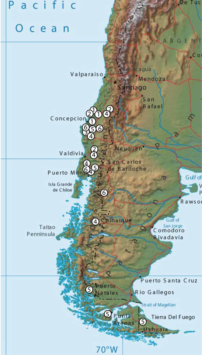
Distribution. Chile, From Talca, in the north, to Villarrica ( Fig. 15 View FIGURE 15 ). As P. p e r p l e x a, it seems to be associated with Nothofagus obliqua forests.
Biology. Unknown. Likely associated with galls induced by Espinosa on N. obliqua .
Flight period is late winter and early spring like P. p e r p l e x a (viii, ix and x).

No known copyright restrictions apply. See Agosti, D., Egloff, W., 2009. Taxonomic information exchange and copyright: the Plazi approach. BMC Research Notes 2009, 2:53 for further explanation.
|
Kingdom |
|
|
Phylum |
|
|
Class |
|
|
Order |
|
|
Family |
|
|
Genus |