
Paraulax Kieffer, 1904
|
publication ID |
https://doi.org/10.5281/zenodo.189597 |
|
DOI |
https://doi.org/10.5281/zenodo.5681149 |
|
persistent identifier |
https://treatment.plazi.org/id/03ADE415-FFC8-FF99-FF58-9586FE12BBC5 |
|
treatment provided by |
Plazi |
|
scientific name |
Paraulax Kieffer, 1904 |
| status |
|
Paraulax Kieffer, 1904: 59 . Type species: Paraulax perplexa Kieffer, 1904: 60 , by original designation. Note. As was pointed out by Rohwer & Fagan (1919), the description of Paraulax was duplicated in Kieffer, 1904b: 43, being impossible to determine which has priority. However Neave (1940): 605, gave the first reference above as valid that is here accepted.
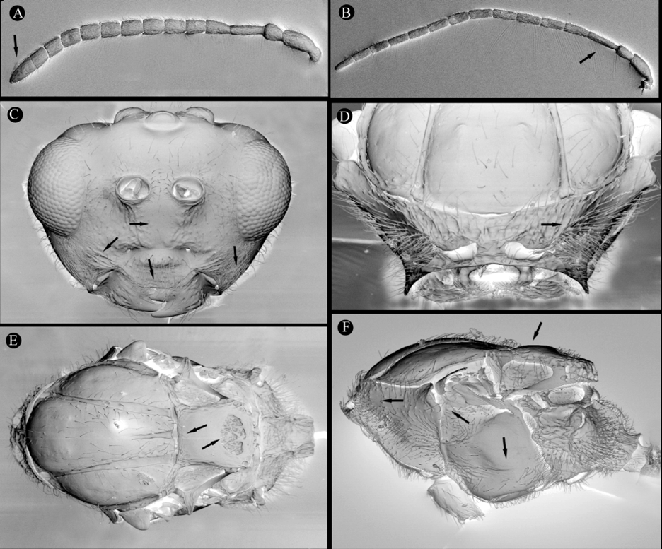
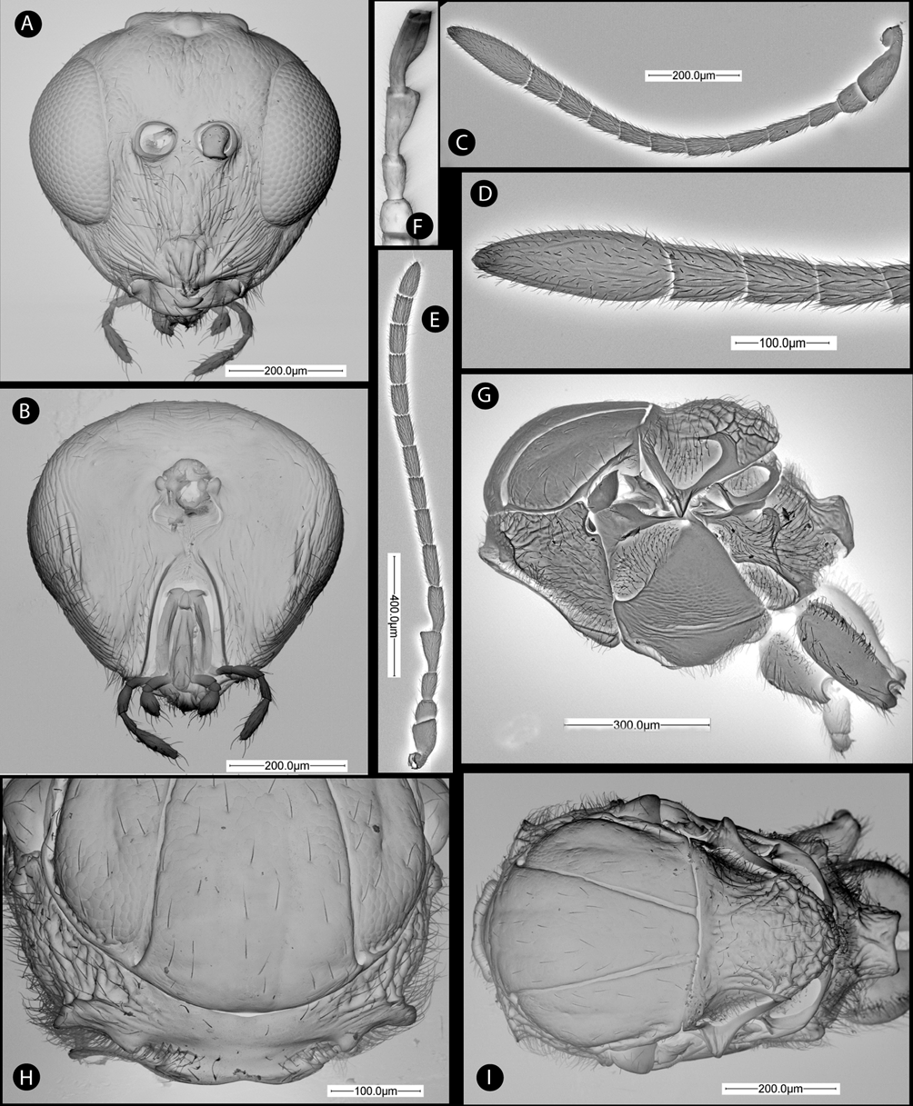
Diagnosis. Similar to Cecinothofagus in general appearance, but separated by the following character states: face without a distinct median vertical carina ( Fig. 2 View FIGURE 2 A); facial strigae radiating from clypeus laterally present also medially, reaching ventral margin of antennal socket ( Fig. 2 View FIGURE 2 A). Ventral part of clypeus slightly projecting over mandibles ( Fig. 2 View FIGURE 2 A). Last flagellomere of the female antenna 3 times longer than wide ( Fig. 2 View FIGURE 2 D); more than 2.5 times as long as penultimate. F2 and F3 of male antenna modified ( Fig. 2 View FIGURE 2 E). Dorsolateral margin of pronotal plate strongly projecting laterally ( Fig. 2 View FIGURE 2 H). Notauli straight, strongly converging and close posteriorly, almost as wide as anteriorly ( Fig. 2 View FIGURE 2 I). Separation of notauli at the meeting of the transcutal fissure relatively short 0.2–0.3 times compared to separation at anterior margin of mesoscutum. Scutellar foveae sometimes indicated, albeit shallow ( Fig. 2 View FIGURE 2 I). Sculpture present dorsal to mesopleural impression ( Fig. 2 View FIGURE 2 G). Claws with an acute basal lobe or tooth; about 1/3 to ¼ length of apical tooth ( Fig. 3 View FIGURE 3 D). Projecting part of hypopygial spine relatively long, 4 times longer than wide ( Fig. 3 View FIGURE 3 G).
Redescription. Head. Some scattered setae along face, gena and occiput dorsally. Gena not expanded behind compound eye. Vertical median carina absent; strong facial strigae radiating from clypeus, laterally reaching ventral margin of eye, medially almost reaching torulus; Frons and vertex with coriaceous sculpture. Clypeus indistinct, ventral margin slightly projecting over mandibles. Subocular impression present though not well marked ( Fig. 1 View FIGURE 1 A). gena with 5–7 regular vertical carinae present ventrolaterally ( Fig. 4 View FIGURE 4 B). Anterior tentorial pits visible; epistomal sulcus and clypeo-pleurostomal lines indistinct. Occiput without dorsal occipital carina ( Fig. 2 View FIGURE 2 B); some strong longitudinal rugae present on lateral margin of head, but without a distinct genal carina. Hypostomal sulci meeting slightly before hypostoma.
Antenna. Female with 10 flagellomeres; flagellum widening towards apex ( Fig. 2 View FIGURE 2 C); Placodeal sensilla visible on F7–F10 ( Fig. 2 View FIGURE 2 D). Apical flagellomere spindle-shaped, not apically truncate. Male with 13 flagellomeres. Flagellum not widening towards apex. F1 cylindrical, F2 and F3 excavated and curved in basal third ( Fig. 2 View FIGURE 2 F). Placodeal sensilla present on all flagellomeres except F1.
Pronotum. Pronotal plate distinct, dorsal part distinctly set off, with anterolateral margins marked and moderately projecting laterally. Admedian pronotal depressions widely separated ( Fig. 2 View FIGURE 2 H). Lateral surface of pronotum coriaceous, some strong, short rugae running from the lateral margin of pronotal plate ( Fig. 2 View FIGURE 2 G).
Mesoscutum with weak coriaceous sculpture, more marked on lateral lobes. Mesoscutal pubescence comprised of some sparse setae. Median mesoscutal impression absent. Notauli complete, straight and narrow, converging posteriorly ( Figs. 2 View FIGURE 2 I & 4C). Anteroadmedian signa visible. Mesoscutum and mesoscutellum separated by a narrow transscutal fissure. Scutellar foveae indistinct, visible only as a shallow depression with some rugae ( Figs. 2 View FIGURE 2 I & 4C). Scutellum, in dorsal view with strong rugae. Posterodorsal and posterior margins of axillula distinct. Mesopleuron ventrally of mesopleural triangle with a marked longitudinal mesopleural impression, more or less complete, ending at margin of mesopleural triangle ( Fig. 2 View FIGURE 2 G). Above furrow with some irregular longitudinal striae and coriaceous sculpture ( Figs. 2 View FIGURE 2 G & 4B). Mesopleuron smooth below mesopleural impression. Metascutellum distinctly constricted medially.
Metapectal-propodeal complex. Metapleural sulcus meeting posterior margin of mesopectus at about mid height of metapectal-propodeal complex ( Fig. 2 View FIGURE 2 G). Lateral propodeal carinae narrow, parallel ( Fig. 3 View FIGURE 3 A). Lateral and median propodeal areas smooth, pubescent. Nucha dorsally with some irregular longitudinal rugae.
Legs. Profemur with a ventral swelling in basal third, with 4–5 rows of sharp closely spaced, deep costulae ( Figs. 3 View FIGURE 3 B & 3C). Metatarsal claws with a basal acute lobe or tooth ( Figs. 3 View FIGURE 3 D & 4D).
Forewing. Radial cell closed along anterior margin; R1 slightly depigmented along radial cell ( Figs. 3 View FIGURE 3 E & 4H); areolet absent; vein Rs+M and M almost invisible, directed towards lower half of median vein. Fringe of long setae along apical margin of wing.
Female metasoma laterally compressed ( Fig. 2 View FIGURE 2 F). Abdominal petiole smooth dorsally, ventrally with deep longitudinal grooves, about as long as high. T2 smooth and shining, covering about 2/3 of metasoma; anteromedian area of T2 with only 4–5 long setae ( Figs. 2 View FIGURE 2 F & 4A). Projecting part of hypopygial spine 4 times as long as high; apical pubescence projecting beyond apex, subapical setae longer than apical ones, forming a small tuft ( Figs. 3 View FIGURE 3 G & 4E).
Included species
Paraulax perplexa Kieffer, 1904 . Types lost. A neotype here designated.
P. queulensis sp. n.
P. ronquisti sp. n.
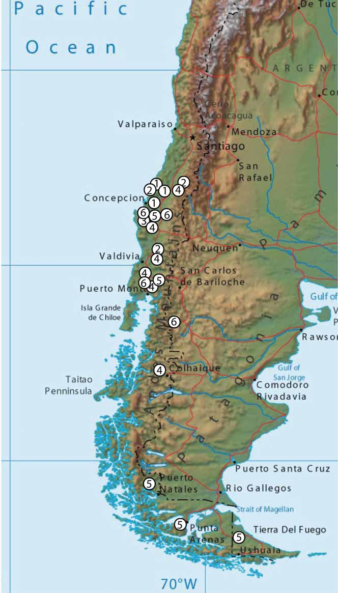
Distribution. Chile, as far as we know, and as here defined, the species of Paraulax occur in the VI to X regions of Chile, in Nothofagus forest habitats, mainly N. obliqua , roughly extending from Santiago to Villarrica ( Fig. 15 View FIGURE 15 ).
Biology. Unknown. Adults were captured in Nothofagus forests, probably associated with galls induced by species of Aditrochus (Pteromalidae) on Nothofagus obliqua .
Remarks. Closely related to Cecinothofagus , the sister genus within the Paraulacini . Although many characters are shared with Cecinothofagus , the two genera are readily separated as detailed in the diagnosis and the key for identification.


No known copyright restrictions apply. See Agosti, D., Egloff, W., 2009. Taxonomic information exchange and copyright: the Plazi approach. BMC Research Notes 2009, 2:53 for further explanation.
|
Kingdom |
|
|
Phylum |
|
|
Class |
|
|
Order |
|
|
Family |
Paraulax Kieffer, 1904
| Nieves-Aldrey, José Luis, Liljeblad, Johan, Nieves, María Hernández, Grez, Audrey & Nylander, Johan A. A. 2009 |
Paraulax
| Kieffer 1904: 59 |
Paraulax perplexa
| Kieffer 1904: 60 |