
Daiphron bipartitus Pic, 1934
|
publication ID |
https://doi.org/ 10.2478/aemnp-2019-0012 |
|
publication LSID |
lsid:zoobank.org:pub:BA2B16BE-CE3D-4746-94DD-4597E515E640 |
|
DOI |
https://doi.org/10.5281/zenodo.5062400 |
|
persistent identifier |
https://treatment.plazi.org/id/03AE466E-FFF9-1025-0EEA-A8B5EE1BC558 |
|
treatment provided by |
Felipe |
|
scientific name |
Daiphron bipartitus Pic, 1934 |
| status |
|
( Figs 1–45 View Figs 1–3 View Figs 4–7 View Figs 8–17 View Figs 18–25 View Figs 26–28 View Figs 29–38 View Figs 39–45 )
Daiphron atripenne Pic, 1934: 130 ; DELKESKAMP (1939):309; BLACKWELDER (1945): 374.
Daiphron (Championellum) proteum var. atripennis: DELKESKAMP (1977) : 463 [altripennis]; DELKESKAMP (1978): 491; BIFFI (2019): 32 (syn.) [junior homonym of Daiphron polemioides var. atripennis Pic, 1927 ].
Daiphron atripenne var. bipartitum Pic, 1934: 130 ; DELKESKAMP (1939): 309; BLACKWELDER (1945): 374.
Daiphron (Championellum) proteum var. bipartita: DELKESKAMP (1977) : 463.
Daiphron bipartitus: BIFFI (2019) : 32 View Cited Treatment (replacement name).
Type material examined. Daiphron atripenne Pic, 1934 : LECTOTYPE: ♀, Brazil, ʻMinasʼ [= Brazil: Minas Gerais] ( MNHN). PARALECTOTYPES: 2 ♀♀ 1 ♂, ʻMinasʼ [= Brazil: Minas Gerais] ( MNHN).
Daiphron atripenne var. bipartitum Pic, 1934 : LECTOTYPE: ♀, ʻMinasʼ [= Brazil: Minas Gerais] ( MNHN). PARALECTOTYPES: 1 ♀ 1 ♀, ʻMinasʼ [= Brazil: Minas Gerais] ( MNHN).
Additional material examined. Adults. BRAZIL: MINAS GERAIS: Vila Monte Verde , ix.1967, J. Halik col.(1♂ 1♀, MZSP 31007 – 31008 ); idem, 17.ix.1971, J. Halik col. (1 ♀, MZSP 31009 ); Itajubá ( Serra dos Toledos ), S.P. Rosa & D.S. Dias col. (1 ♂, MZSP 31010 – spirit collection). SÃO PAULO: Serra da Bocaina , 1500 m, xi.1965 (4 ♂♂ 1 ♀, NHMB); 1600 m, São José do Barreiro, xi.1967, Alvarenga & Seabra cols. (4 ♀♀, DZUP 273492 – 273495 ); São José do Barreiro, xi.1969, Alvarenga col. (2 ♀♀, MZSP 31011 – 31012 ); Campos do Jordão, xi.1936, J. Lane col. (1 ♀, MZSP 31013 ); Campos do Jordão (lot. Quebradas ), 1–28.x.2017, Malaise, Rosa col. (1 ♂, MZSP 31014 – spirit collection).
Immature stages. BRAZIL: SÃO PAULO: Campos do Jordão ( lot. Quebradas ), 22°40′50.1″S 45°35′35.0″W, 25.vi.2017, inside bromeliad, Rosa col. (1 larva reared to adult: larval and pupal exuviae, male adult, MZSP 10357 ); GoogleMaps same locality, 27.viii.2017, in the soil (1 larva reared to adult:larval and pupal exuviae, male adult, MZSP 10358 ); GoogleMaps same locality, 5.v.2018, inside bromeliad (1 larva reared to pupa: larval exuvia and pupa, MZSP 10359 ); GoogleMaps same locality, 5.v.2018, inside bromeliad (1 larva reared to adult:larval and pupal exuviae, male adult, MZSP 10360 ); GoogleMaps same locality, ix.2017 (1 male and 1 female in copula, MZSP 10361 ); GoogleMaps same locality, 11.viii.2018, inside bromeliad (1 larva, MZSP 10362 ); GoogleMaps idem, 8.xii.2018, inside bromeliad (1 larva, MZSP 10363 ). GoogleMaps
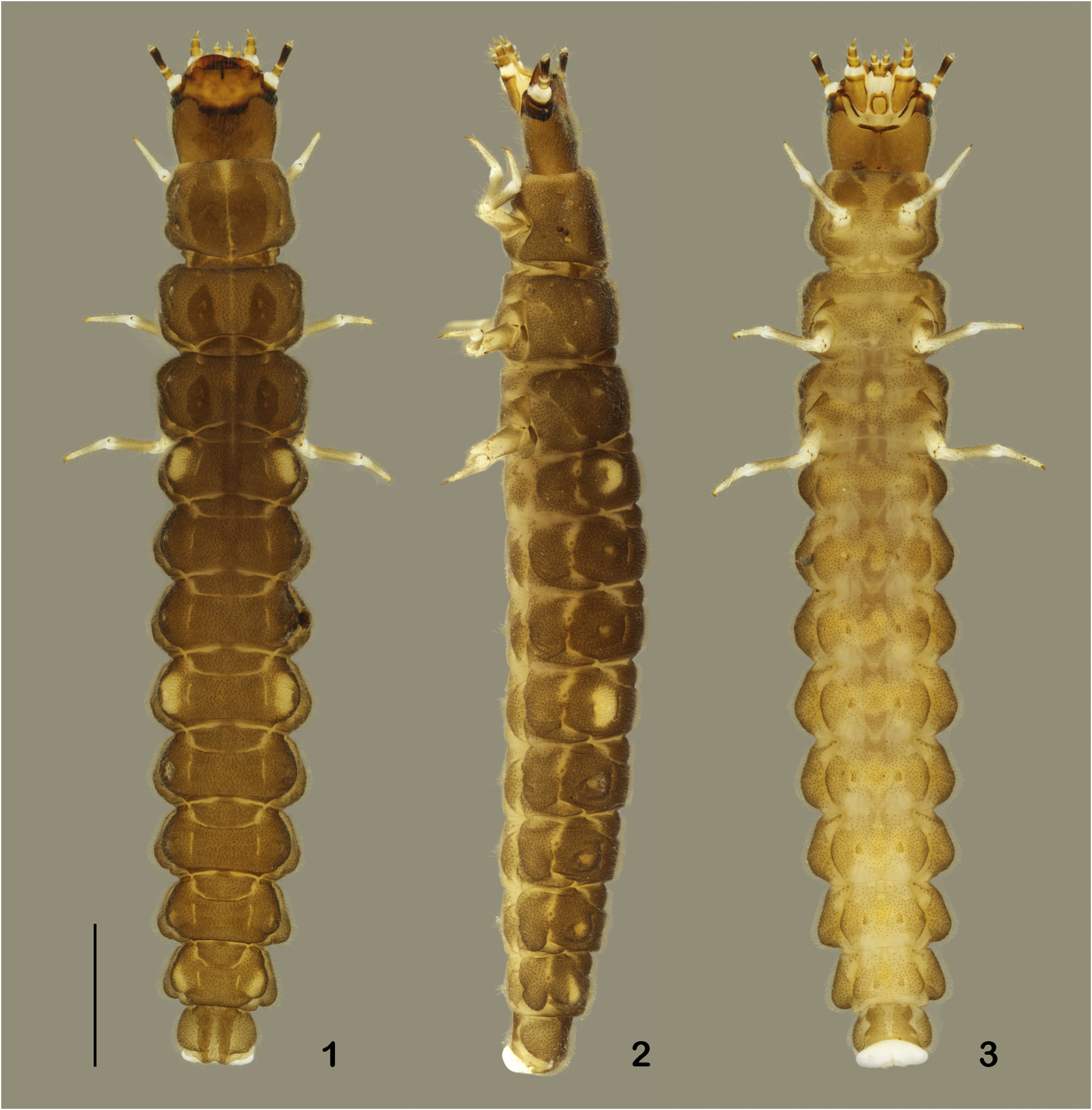
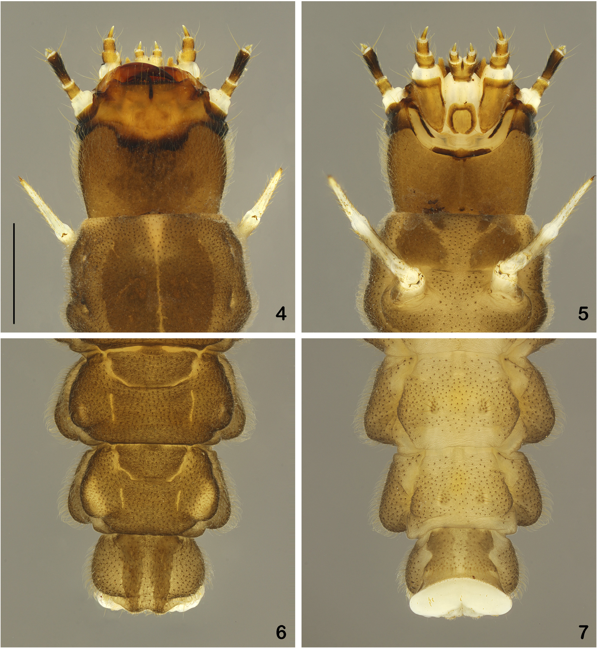
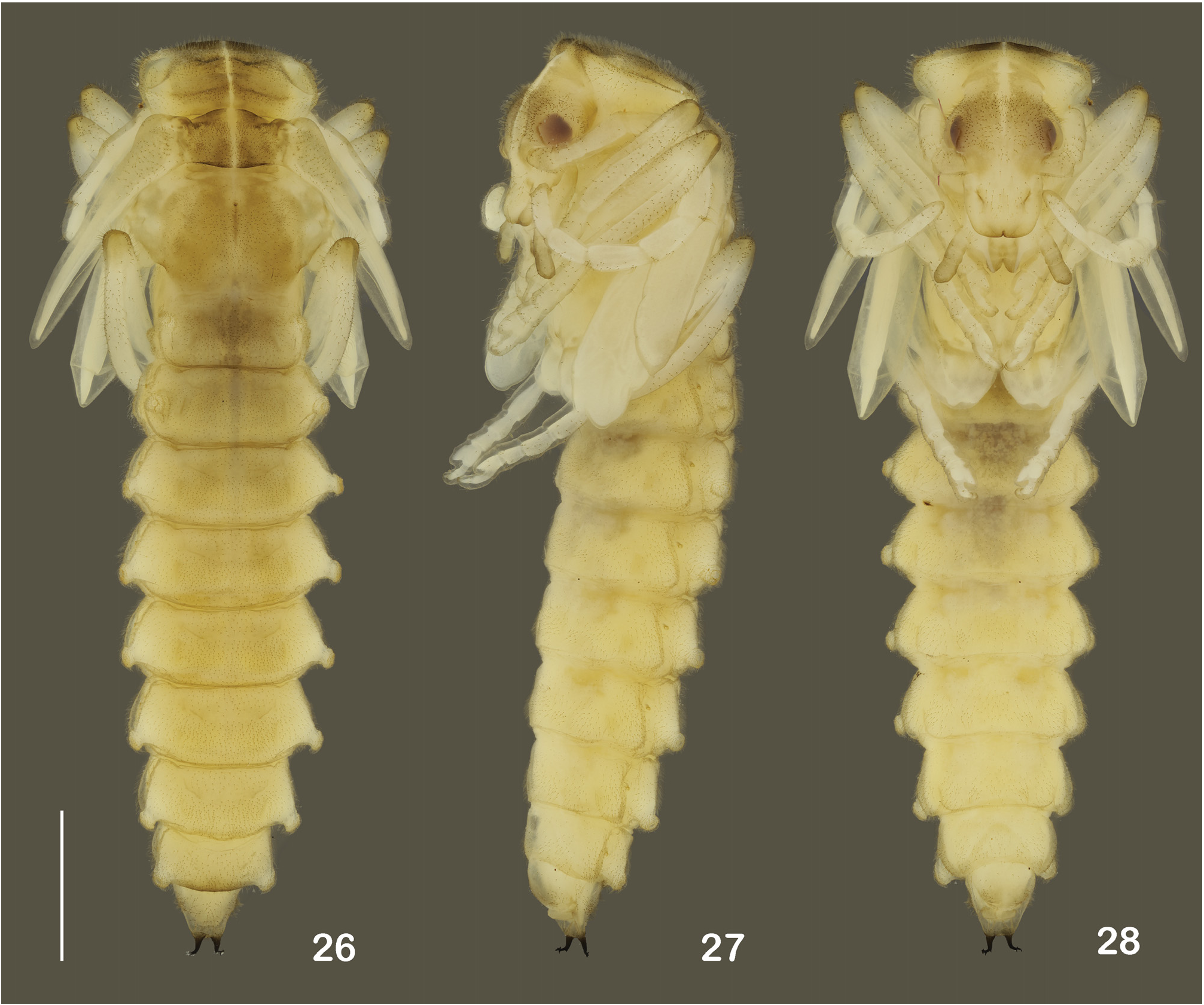
Description of the last instar larva ( Figs 1–24 View Figs 1–3 View Figs 4–7 View Figs 8–17 View Figs 18–25 , 49–50 View Figs 46–50 ). Colouration. In fixed specimens, head ( Figs 4–5 View Figs 4–7 , 8 View Figs 8–17 ) dark brown to black, dull in basal two thirds; lighter and bright in apical third; limits between dull and bright areas with a sinuous and darker stripe towards base of stemmata. Mandibles ( Figs 11–14 View Figs 8–17 ) dark brown in apical half and brown in basal half. Basal and distal antennomeres light brown, median antennomere dark brown; maxillae and labium light brown, except for palpigers and distal half of postmentum, translucent. Body ( Figs 1–7 View Figs 1–3 View Figs 4–7 ): integument mostly dark greyish brown dorsally, pale yellow along ecdysial line of thorax and anterior part of abdominal segment I; thorax with one pair of longitudinal darker spots: almost contiguous, occupying median two thirds of prothorax, separated on meso- and metathorax, with one third of width of each lateral half; abdomen with pale yellow spots on apex of dorsal ampulla of segments I, IV and VIII, and dark brown lateral stripes on segment IX; ventral surface of prothorax and abdominal segment IX ( Figs 4–6 View Figs 4–7 ) brown with paired darker spots. When alive larval tegument is more greyish dark brown. Legs light brown, translucent on articulating areas; pretarsi brown.
Pubescence moderately long and dense; setae light brown, inserted in dark punctures; abdominal segments II–VIII with paired patches of longer setae.
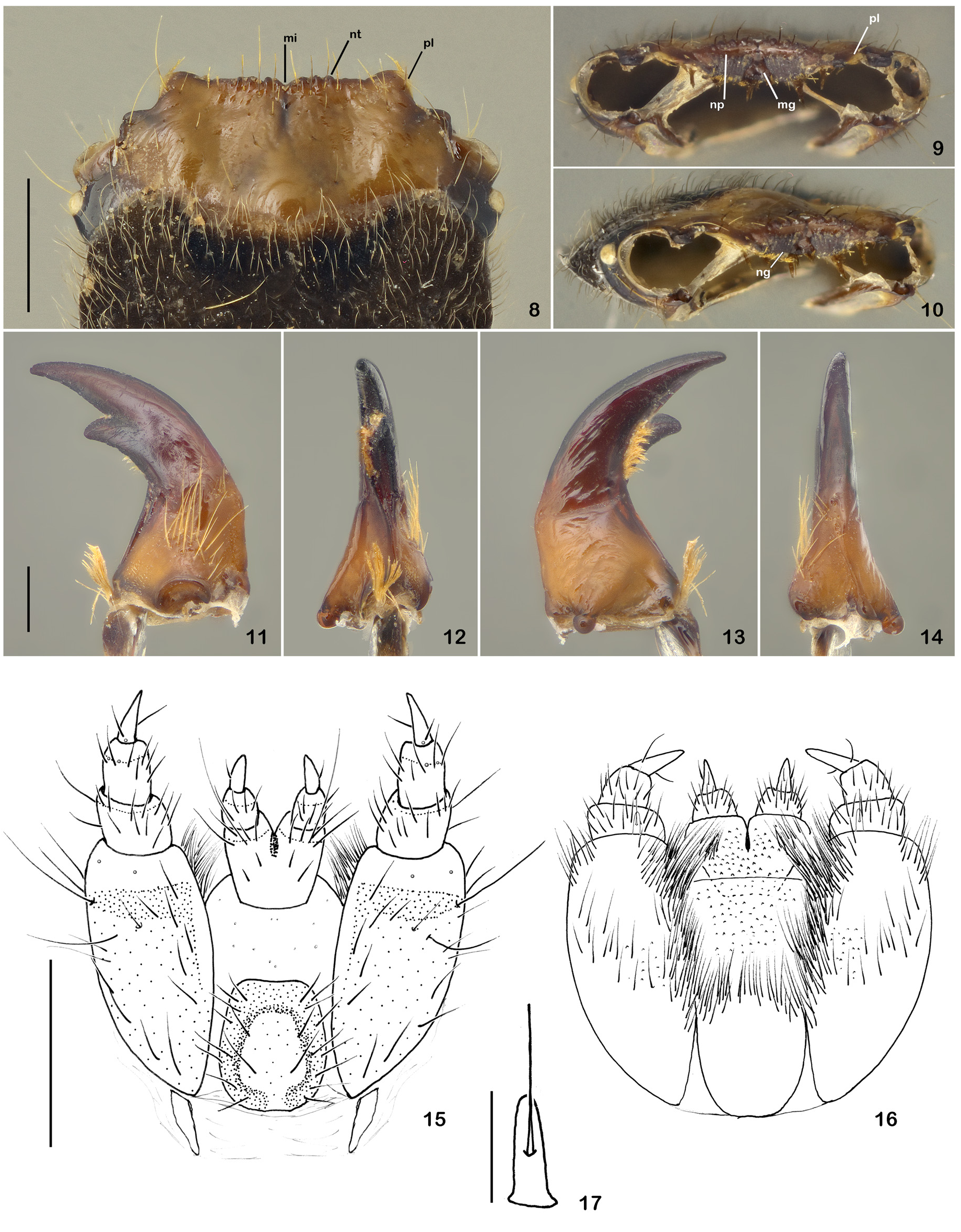
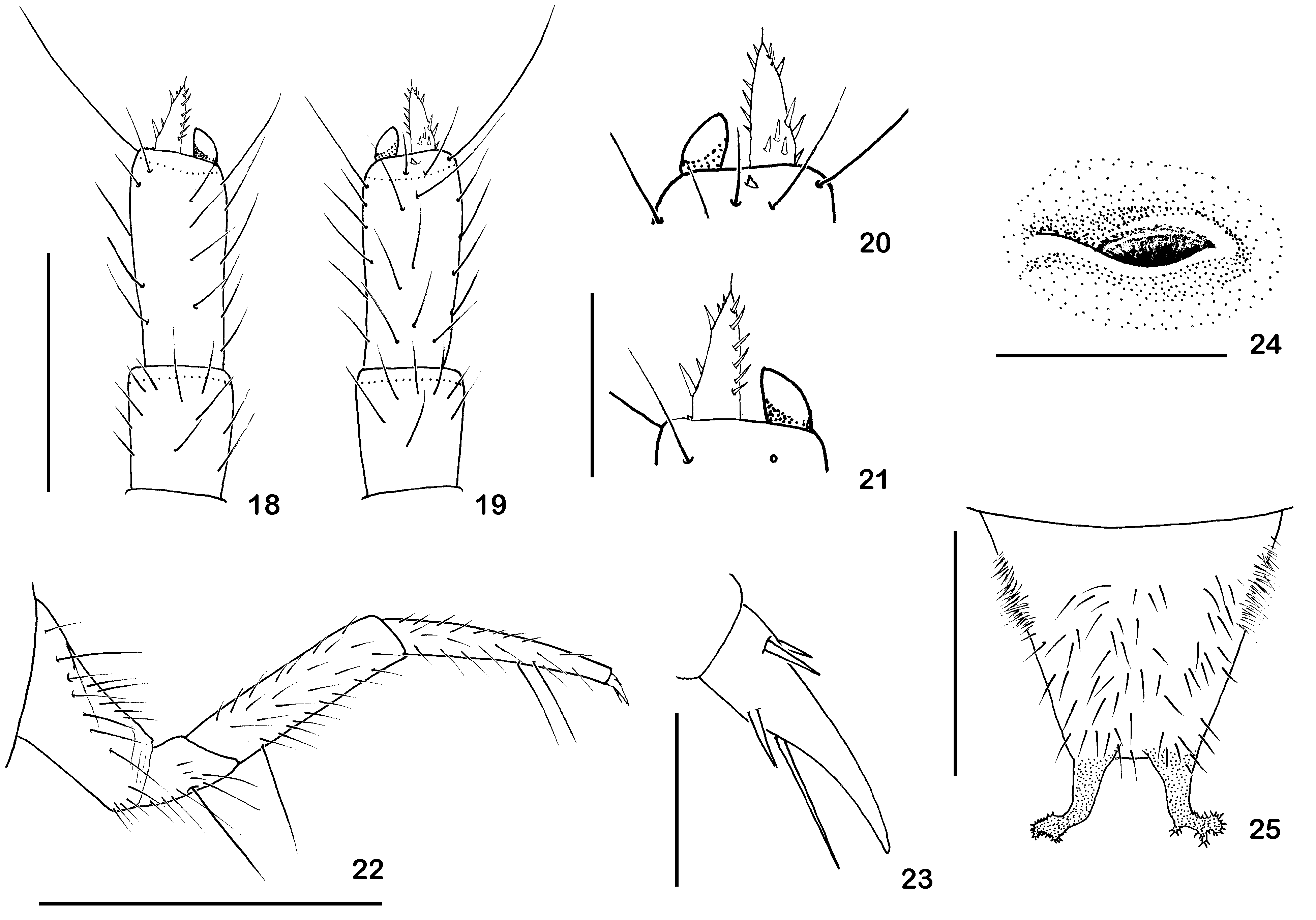
Structure. Head ( Figs 2 View Figs 1–3 , 4–5 View Figs 4–7 ) with lateral margins slightly rounded; partially retracted into prothorax; prognathous and flattened dorso-ventrally; densely covered with fine and long yellow setae on basal third, with a velvety aspect; almost smooth anteriorly; epicranial suture absent; gular suture obsolete. One large stemma on each side, behind antenna. Nasale ( Figs 8–10 View Figs 8–17 ) slightly shorter than paranasal lobes, 5 prominent acute teeth on each side of median incision dorsally; transverse, oblique, sclerotized plate ventrally, with deep irregular grooves on each side; median groove very wide with a small rounded central protuberance; fringe of long setae, longer laterally, appearing dorsally below paranasal lobes, near sclerotized plate. Paranasal lobes longer than nasale, inner margins strongly declivous from apex to nasale; outer angles rounded.Antennae ( Figs 18–21 View Figs 18–25 ) densely setose, with three antennomeres; setae long, longer near apex of median antennomere; basal antennomere elongate, 0.6 times as long as median antennomere; median antennomere cylindrical, apex truncate bearing terminal membranous sensorium on outer margin; sensorium elongate with an sclerotized basal band; median antennomere with two dorsal campaniform sensilla and one tiny wart-like sensorium near apex, between sensorium and distal antennomere; distal antennomere elongate and narrow apically, with one spine-like seta at apex; one third of width of median antennomere, inserted latero-internally at apex of median antennomere, next to sensorium; 6 latero-internal, 6 latero-external, and 3 dorsal setae near base. Mandibles ( Figs 11–14 View Figs 8–17 ) symmetrical, slightly arcuate, incisor acute; retinaculum dorsal, apex round; mesal channel well-developed, extended ventrally at apex, almost straight, slightly directed dorsally near base; elongate, meso-dorsal, weakly striate concavity below retinaculum; dense fringe of fine yellow setae on ventral margin of distal third of longitudinal channel; penicillus formed by tuft of long yellow setae; a few long yellow setae forming irregular row in basal half, near acetabulum, and a few shorter setae near middle laterally. Maxillae ( Figs 15–16 View Figs 8–17 ). Cardo, elongate, tapering towards apex, weakly sclerotized. Stipes elongate, narrow basally, ventral sclerotized surface with darker transverse band at base of palpigers and numerous long setae along anterior, inner and lateral margins; ventral surface of stipes with microspines near middle and long setae on inner margin and anterior corner, intermingled among long, fine and ramified setae dorsally. Galea ( Fig. 17 View Figs 8–17 ) minute, elongate and narrow, one-segmented, located dorsally near apex of stipes, bearing one long seta near middle. Lacinia formed by fringe of short and long, fine and ramified setae densely distributed near inner margin. Maxillary palpi with three palpomeres; densely setose, except for distal palpomere, which bears two setae; setae long, denser dorsally; basal palpomere wider than long, with transverse sclerotized band at base; median palpomere longer than wide with two ventral campaniform sensilla near apex; distal palpomere elongate, tapering apicad with one lateral seta ventrally and one inner seta dorsally, and campaniform sensillum ventrally at base. Labium ( Figs 15–16 View Figs 8–17 ). Postmentum elongate, translucent with circular brownish sclerotized area at base, with numerous long setae along lateral edges, dorsal area covered with microspines distally, without ramified setae. Prementum translucent with elongate sclerite between palpigers; numerous long setae near anterior margin, and one longer seta on each side; hypopharynx (dorsal area) covered with microspines and long setae. Palpi with two palpomeres; basal palpomere longer than wide, setose; setae long, denser on ventral surface; distal palpomere elongate, tapering towards apex, with one lateral seta, one ventral seta, and one campaniform sensillum ventrally at base. Legs ( Fig. 22 View Figs 18–25 ) long, increasing in size from anterior to posterior; densely setose; setae yellow, a few longer setae on ventral margin of trochanter, femur and tibia; coxae inserted distant one from the other; femora as wide as trochanter, as long as tibiae; tibiae tapering apicad; pretarsi ( Fig. 23 View Figs 18–25 ) with four stout setae; one basal seta straight, reaching or surpassing apex of pretarsus. Thorax and abdomen. Pro-, meso- and metathorax and abdominal segments I–IX with one pair of latero-dorsal glandular pores each. Meso- and metathoracic and abdominal glandular pores I–VIII on latero-dorsal ampullae ( Fig. 1 View Figs 1–3 ). Mesothorax with one pair of C-shaped spiracles located ventro-anteriorly ( Fig. 24 View Figs 18–25 ); abdominal segments I–IX with one pair of dorsolateral spiracles each, smaller than thoracic ones, located latero- anteriorly between dorsal and lateral ampullae.Abdominal segment I–V subequal in width, VI–IX gradually narrowed apicad; IX weakly widened posterad and bilobed at apex. Metathorax and abdominal segments I–VIII with median anterior transverse ampulla. Abdominal sternites I–VIII with long and thick setae arranged in one pair of tufts per segment ( Fig. 7 View Figs 4–7 ); sternite IX with long setae, denser near apex. Segment X reduced, membranous and bilobed.
Measurements of one fixed larva. Body length approx. 14 mm; head 1.1× as long as wide, 0.8× narrower than prothorax; prothorax 1.3× longer than wide.
Pupa ( Figs 25–28 View Figs 18–25 View Figs 26–28 ). Pubescence long, dense, bristly and light brown, inserted in dark punctures. Pronotum with pair of glandular pores, near fore angles, smaller than abdominal ones. Abdominal segments I–VIII with one pair of glandular pores each; each glandular pore at apex of tubular projection. One pair of lateral round spiracles on mesothorax and on abdominal segments I–VIII; last spiracle smaller. Palpi slightly sclerotized; abdominal segment IX ( Fig. 25 View Figs 18–25 ) densely setose, denser laterally at base, with pair of strongly sclerotized urogomphi; apices of urogomphi curved, with 4–5 projections with short and sharpened spines.
Measurements. Body length 12 mm; head (up to base of mandibles) as long as wide; pronotum 1.3× wider than long, 1.4× wider than head width.
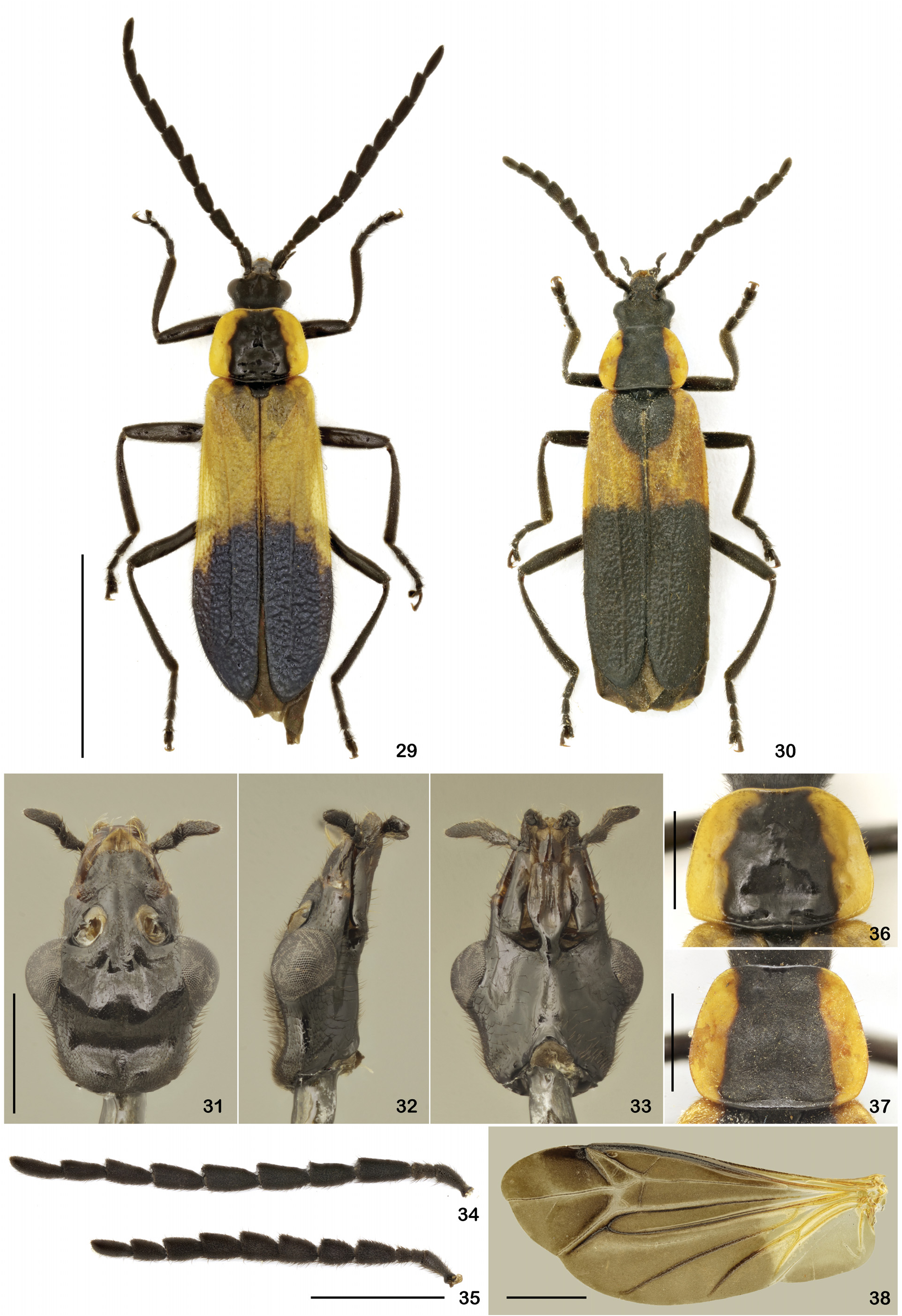
Redescription of the adult ( Figs 29–45 View Figs 29–38 View Figs 39–45 ). Colouration. Head, antennae, palpi, thorax, legs and abdomen dark brown to pitch-black, sometimes clearer in front of eyes and lateral margin of clypeus; pronotum orangish yellow with broad black band extending longitudinally from anterior to posterior margin; scutellum black; elytral background orangish-yellow, black in distal two-thirds, sometimes with broad round black spot around scutellum, which may be merged with distal black marks, leaving two narrow humeral yellow bands; wings yellow on basal third and light brown in distal two thirds. Body densely covered with short pubescence and sparse longer setae, more concentrated on tibiae and tarsi.
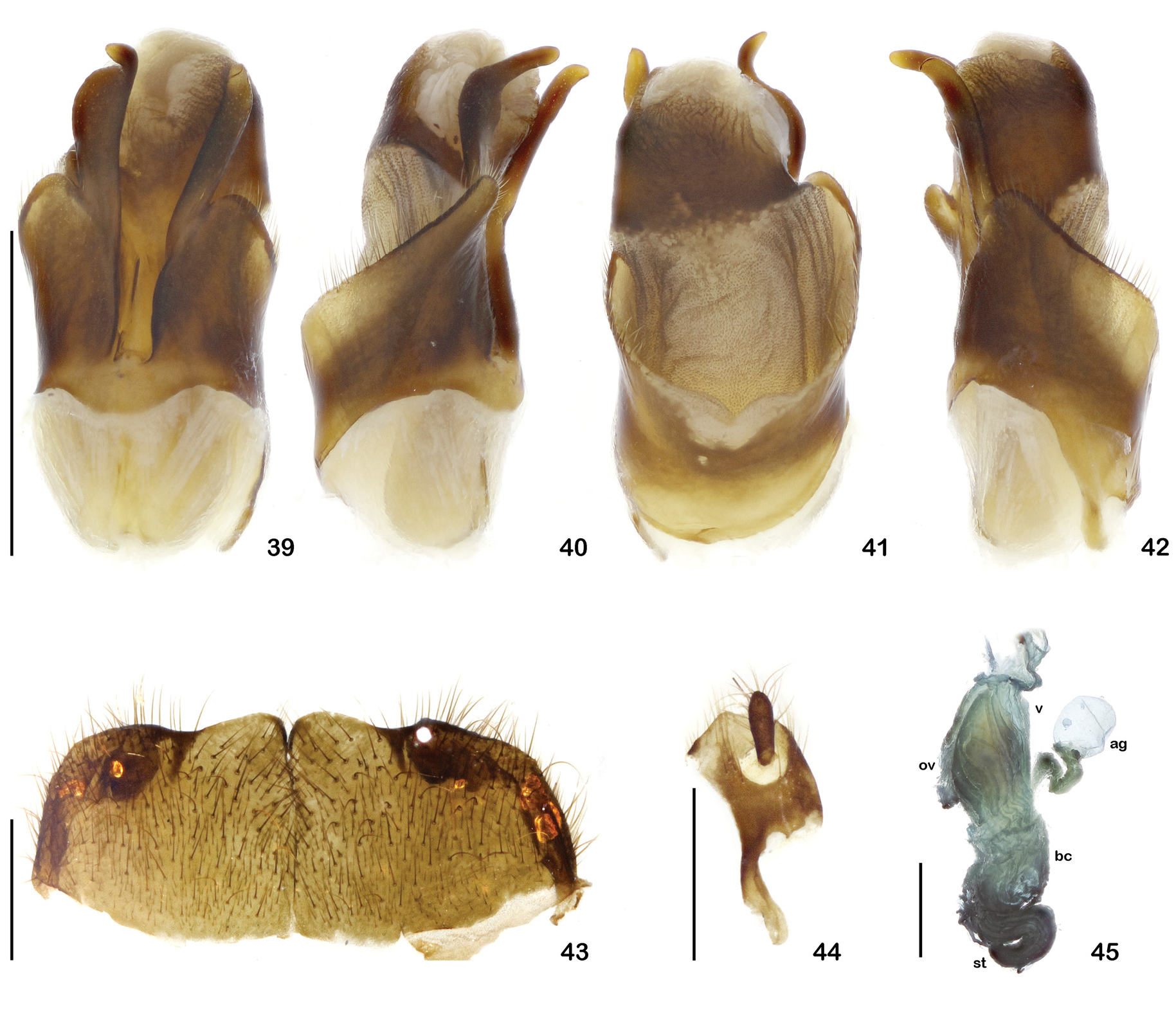
Structure. Male ( Fig. 29 View Figs 29–38 ). Head ( Figs 31–33 View Figs 29–38 ) elongate, narrow, slightly compressed dorsoventrally; 1.7× longer than wide behind eyes; eyes large, very prominent, 1.3× wider than head width; head narrow and elongate in front of eyes, sides nearly parallel, slightly convergent; clypeus long and broad; fronto-clypeal suture broadly arched, distal margin slightly emarginated. Maxillae and labium long and narrow; maxillary palpi long, fourth palpomere 2.7× longer than wide.Antennae ( Fig. 34 View Figs 29–38 ) robust; antennomere I almost cylindrical, slightly swollen apically; antennomeres II and III very short, subequal in length, each 3.2× shorter than antennomere I; antennomeres IV–X subserrate, broad, slightly compressed dorsoventrally; antennomere XI narrow, cylindrical, elongate. Pronotum ( Fig. 36 View Figs 29–38 ) subtrapezoidal, 1.3× wider than long, wider posteriorly, lateral margins arched, convergent anteriorly, forming flattened flaps directed upwards; anterior margin and angles broadly arched; a rounded elevation on the disk posteriorly with a central impression. Elytra ( Fig. 29 View Figs 29–38 ) long, 3.1× longer than humeral width; sides nearly parallel, slightly wider posteriorly; apices rounded; dorsal surface rough, sometimes lustrous; each elytron with one distinct longitudinal costa fading near the apex. Wings venation ( Fig. 38 View Figs 29–38 ): radial cell 2R1 closed; Rr extending up until reaching r-m; Cu curved, running from anterior third of M3+4; Mr and Ax2 sclerotized; transverse cu-a absent; margin of anal area slightly sinuous. Legs long and slender, increasing in size posteriorly; tarsal claws simple. Abdomen weakly sclerotized; ventrites I– VI wide, entire, not indented; distal margin of ventrite VII strongly notched, U-shaped; ventrite VIII oblong and strongly convex. Abdominal glandular pores broad. Aedeagus ( Figs 39–42 View Figs 39–45 ) asymmetrical. Right paramere flat, entire, bent to the right, apex rounded with a small tip directed outwards. Left paramere broad and flat, turned to the left; apex with a long projection curved to the left. Median lobe short and broad, slightly twisted in its longitudinal axis to the left; ventral face with short well-marked longitudinal keel between parameres; dorsal surface short, rough; apex broad, weakly sclerotized.
Female ( Fig. 30 View Figs 29–38 ). Usually slightly larger than males; eyes less prominent; antennae ( Fig. 35 View Figs 29–38 ) more robust, antennomeres I–III similar to males, IV–XI shorter. Pronotum ( Fig. 37 View Figs 29–38 ) less wide than in males; lateral margins broadly arched posteriorly; anterior angles less arched; posterior elevation and central impression less pronounced than in males. Seventh abdominal ventrite ( Fig. 43 View Figs 39–45 ) wider than long, distal margin broadly sinuate; two broad rounded lobes meeting medially, forming a weak internal longitudinal keel. Coxites ( Fig. 44 View Figs 39–45 ) elongate, slender anteriorly and short and broad posteriorly; styles very short, slightly wider apically; long setae concentrated on apex of style and on distal surface of coxite, around style. Genitalia ( Fig. 45 View Figs 39–45 ): vagina membranous, sacciform; oviduct joining posterior region of vagina; bursa copulatrix short, globose; spermatheca consisting of dense tuft of long filaments joining anterior region of bursa copulatrix; accessory gland long, sinuous, globose distally, in dorsal region of bursa copulatrix.
Measurements. Body length: 11.0–13.0 mm.
Remarks. PIC (1934) described the species as Daiphron atripenne based on a specimen with elytra nearly entirely black, and the variety D. atripenne var. bipartitum , with anterior half of elytra orangish yellow and posterior half black. Both phenotypes are just colour variations of the same species Daiphron bipartitus ( BIFFI 2019) , the latter being the most frequent amongst examined specimens.
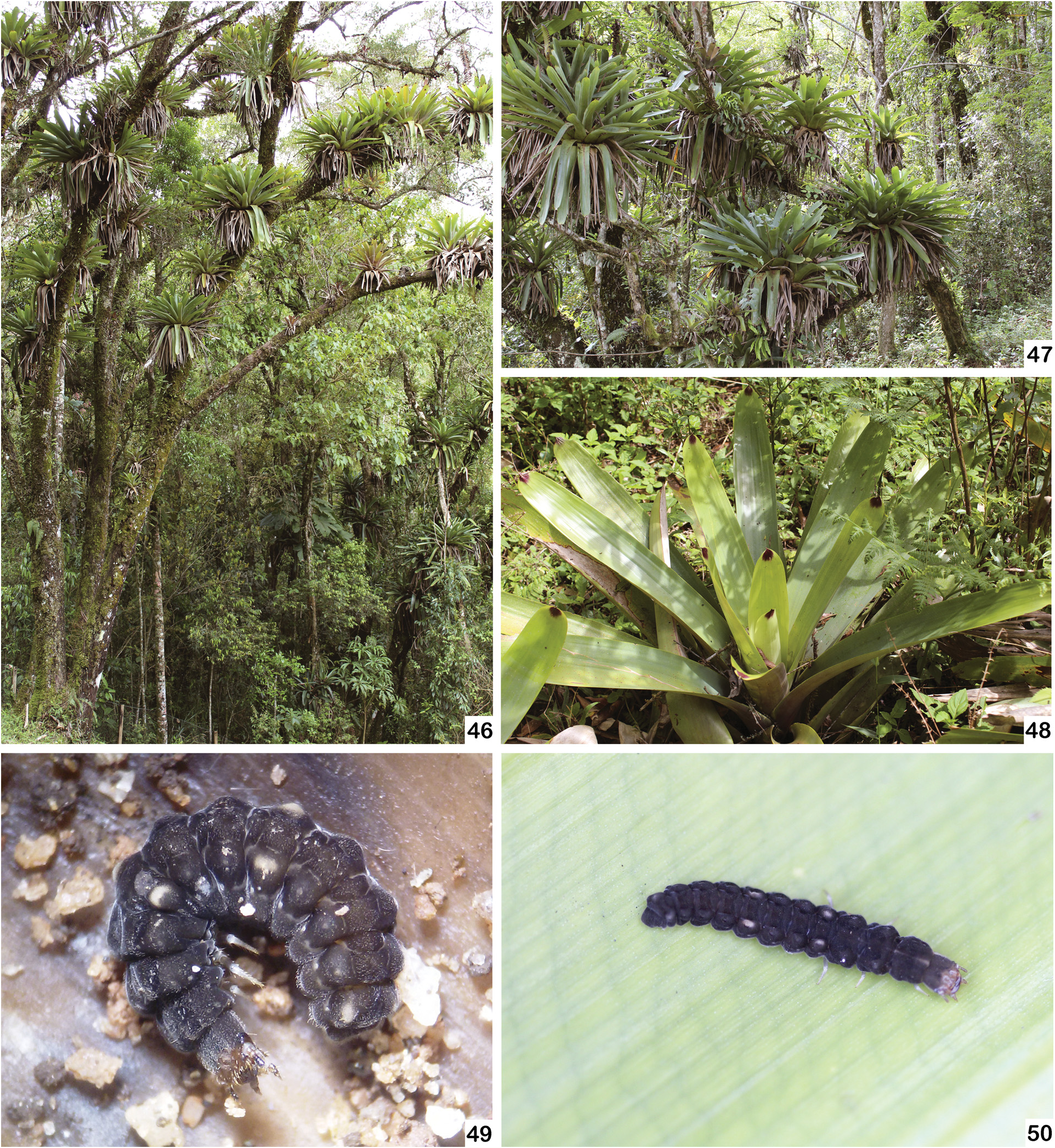
Biological notes. One larva of Daiphron bipartitus was collected in the soil while six larvae were found amongst leaves of fallen bromeliads Vriesea bituminosa Wawra ( Bromeliaceae : Tillandsioideae ). Most of the larvae were found amongst the innermost, humid leaf sheaths of fallen bromeliads ( Figs 46–48 View Figs 46–50 ) during the winter, the dry season. In the laboratory, larvae were observed always inactive inside a chamber in the soil at the bottom of the container. However, they seemed to feed, since the offered termites disappeared every week. Larvae were reared for up to 36 days until they reached the pupal stage. The pupal period of three specimens lasted 6, 7, and 15 days. All the specimens of D. bipartitus known so far, including immatures and adults, were collected in nearby localities along Serra da Mantiqueira, at altitudes ranging from 1,400 m to 1,600 m.
No known copyright restrictions apply. See Agosti, D., Egloff, W., 2009. Taxonomic information exchange and copyright: the Plazi approach. BMC Research Notes 2009, 2:53 for further explanation.
|
Kingdom |
|
|
Phylum |
|
|
Class |
|
|
Order |
|
|
Family |
|
|
Genus |
Daiphron bipartitus Pic, 1934
| Biffi, Gabriel & Rosa, Simone Policena 2019 |
Daiphron (Championellum) proteum var. atripennis:
| BIFFI G. 2019: 32 |
| DELKESKAMP K. 1978: 491 |
| DELKESKAMP K. 1977: 463 |
Daiphron (Championellum) proteum var. bipartita:
| DELKESKAMP K. 1977: 463 |
Daiphron atripenne
| BLACKWELDER R. E. 1945: 374 |
| DELKESKAMP K. 1939: 309 |
| PIC M. 1934: 130 |
Daiphron atripenne var. bipartitum
| BLACKWELDER R. E. 1945: 374 |
| DELKESKAMP K. 1939: 309 |
| PIC M. 1934: 130 |