
Ramazzottius littoreus, Fontoura, Paulo, Rubal, Marcos & Veiga, Puri, 2017
|
publication ID |
https://doi.org/10.11646/zootaxa.4263.3.2 |
|
publication LSID |
lsid:zoobank.org:pub:093CCBCF-E709-4C9D-8697-BF18BFC635F1 |
|
DOI |
https://doi.org/10.5281/zenodo.6015868 |
|
persistent identifier |
https://treatment.plazi.org/id/03BF87BA-0C5F-756B-B7C9-A9CC328BFE63 |
|
treatment provided by |
Plazi |
|
scientific name |
Ramazzottius littoreus |
| status |
sp. nov. |
Ramazzottius littoreus sp. nov.
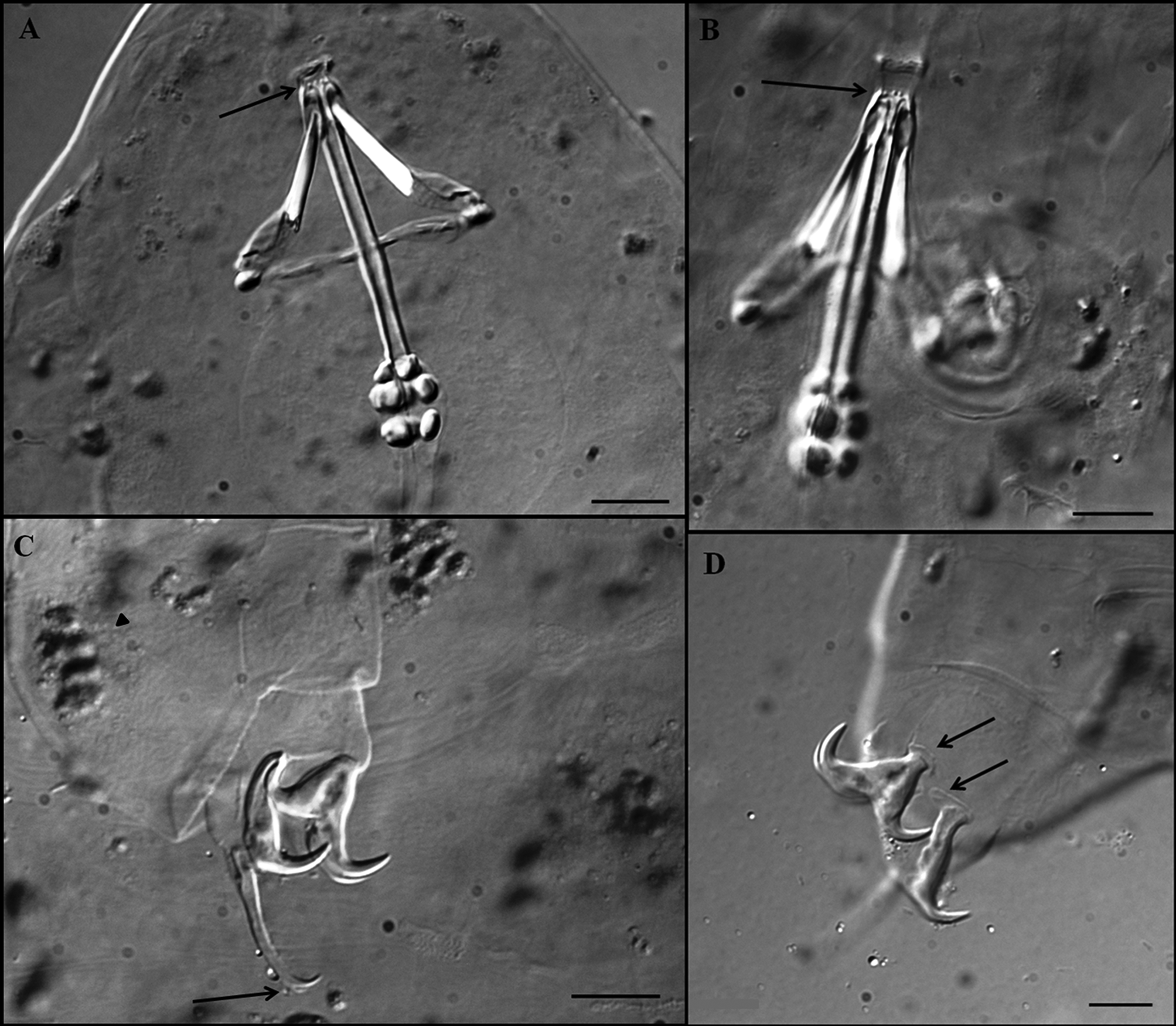
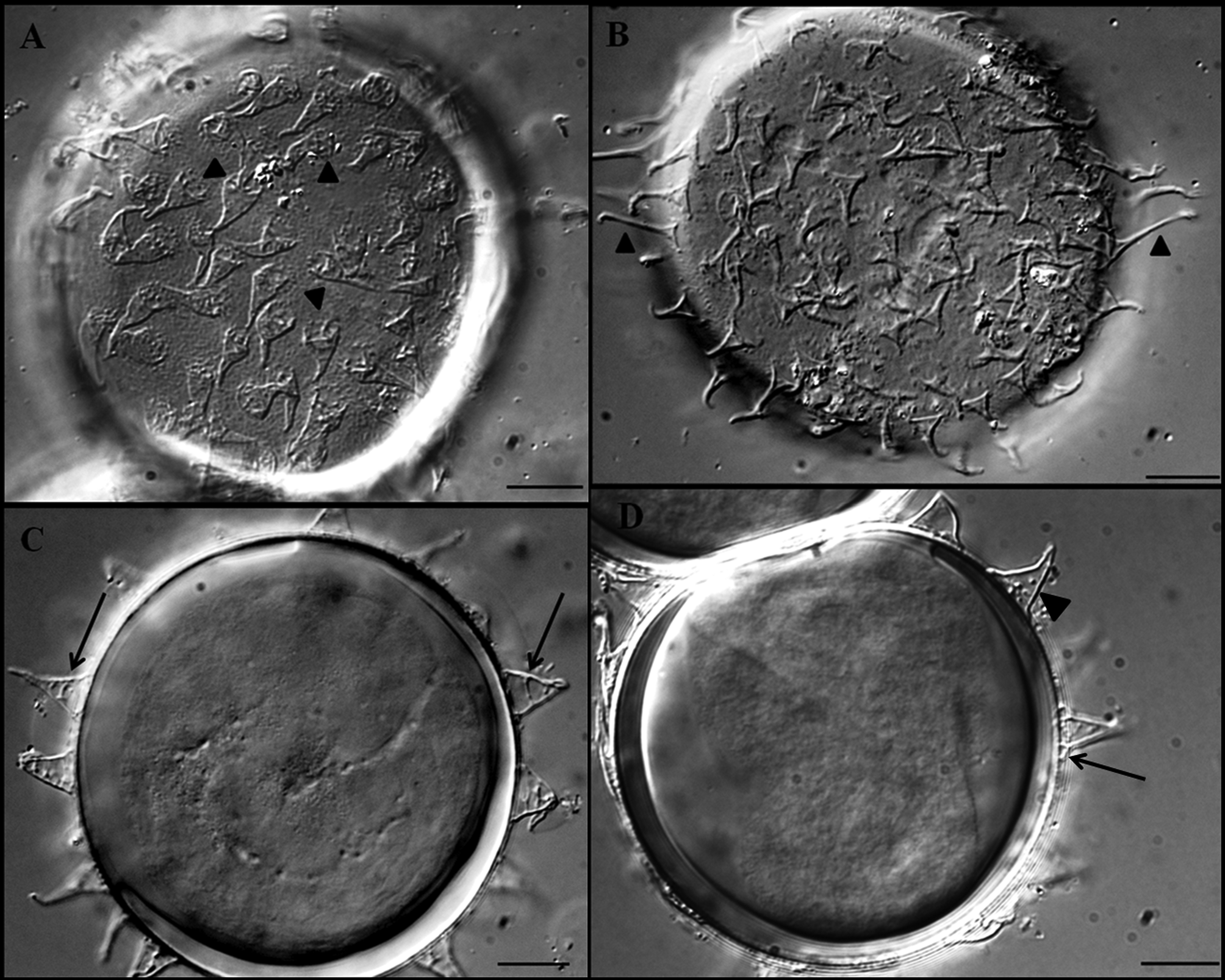
( Figures 1 View FIGURE 1 , 2 View FIGURE 2 A, B, 3, 4; Tables 1, 2)
Material examined. Holotype (slide C. VII-60); 62 paratypes (49 animals, slides C. VII-60 to C. VII-74 and C. VII- 76 to C. VII-78, and 13 eggs, of which three were embryonate, slides C. VII-71 to C. VII-78).
Type repository. Holotype and paratypes are deposited in the Department of Biology of the Faculty of Sciences, University of Porto, Portugal (collection P. Fontoura).
Type locality. 43°41'37.77''N; 7°26'13.24''W, San Cibrao , Lugo, Galicia, Spain .
Specific diagnosis. Ramazzottius with granulated cuticle. Cuticular granules also present on legs. Eyes always absent. Elliptical cephalic organs present. Five longitudinal and nine transversal pinkish/brownish bands. Lateral gibbosities on hind legs. Claws of Ramazzottius type with accessory points on main branches and lunules on hind legs. Posterior part of mouth cavity with four dorsal and four ventral teeth. Anterior portion of the buccal tube bent ventrally. Pharyngeal bulb with two macroplacoids, first macroplacoid slightly longer than the second (1>2). Free laid eggs, with cone-shaped processes of variable size with reticulated bulbous bases. Surface between processes finely dotted.
CHARACTERS Holotype Mean ± SD (Range); (N) Mean ± SD (Range); (N) Description of the holotype: Body length 397 µm (table 1). Before being mounted on a slide five longitudinal and nine transversal pinkish/brownish bands separated by unpigmented areas are visible. Small and sparse clumps of dark pigment are present ( Figs 1 View FIGURE 1 A, C). Eyes absent. Elliptical cephalic organs present ( Fig. 1 View FIGURE 1 B, asterisk). Cuticular ornamentation consisting of granules (polygonal tubercles) extending dorso-laterally from the head to the caudal region; where it is more visible. In the posterior part of the body tubercles have a diameter of about 2.0–3.0 µm ( Figs 1 View FIGURE 1 B, C, arrowhead). External side of legs I–III with small, difficult to see, granules ( Fig. 3 View FIGURE 3 C, arrowhead). Dorsal side of legs IV sclerotised and also covered with obvious granules. Lateral gibbosities present on hind legs ( Figs 1 View FIGURE 1 A, C, arrows). Ventral cuticle smooth. Granular nodes corresponding to muscle attachment points are only clearly seen under DIC, particularly in segmental folds deprived of tubercles ( Fig. 1 View FIGURE 1 B, arrows). They are arranged in ten ventral and eight dorsal rows ( Figs 2 View FIGURE 2 A, B).
Bucco-pharyngeal apparatus of Ramazzottius type, with a rigid buccal tube without ventral lamina ( Fig. 3 View FIGURE 3 A); asymmetrical dorsal and ventral blunt hook shaped apophyses for the insertion of stylet muscles. Antero-ventral mouth opening, without peribuccal papulae and lamellae. Oral cavity with an anterior band of teeth poorly visible and a posterior band with four evident dorsal and four ventral teeth ( Figs 3 View FIGURE 3 A, B, arrows). Buccal tube with thick wall, particularly posterior to the insertion of stylet supports ( Fig. 3 View FIGURE 3 A). Stylet supports inserted on the buccal tube at 60.8% of its length. Ovoid pharyngeal bulb with large apophyses and two grain-like macroplacoids without constrictions. First macroplacoid slightly longer than the second (respectively: 3.4 and 3.3 µm long). Microplacoid and septulum absent ( Fig. 3 View FIGURE 3 A). Placoid row, 8.1 µm long.
Claws of Ramazzottius type, well developed ( pt of posterior claw IV ca. 85%), long main branches on external claws with robust accessory points ( Fig. 3 View FIGURE 3 C, arrow). Poorly visible lunules on legs I–III and well developed lunules on the hind legs ( Fig. 3 View FIGURE 3 D, arrows). Other cuticular thickenings on legs absent.
Eggs ( Figs 4 View FIGURE 4 A, D; table 2): free laid, whitish, spherical (diameter 51.3–68.2 and 61.3–84.1µm without and with egg processes respectively). Egg processes are well spaced (24–28 processes on the circumference) and coneshaped with a bulbous base and a thinner apical point. The size and shape of the egg processes are variable between eggs and, sometimes also within the same egg (some egg processes have a longer, thin apical point, Fig. 4 View FIGURE 4 A arrowheads, and a few are funnel-shaped with enlarged tips, Fig. 4 View FIGURE 4 B arrowheads). Length of egg processes 5.4–16.3 µm and with base diameter 1.8–6.2 µm. The bulbous base of egg processes is clearly reticulated ( Figs 4 View FIGURE 4 A, arrowheads, C, arrows). However, the egg process coverage of this reticulation is variable, and can be restricted to the base ( Fig. 4 View FIGURE 4 D, arrow), in some egg processes is not visible ( Fig. 4 View FIGURE 4 D, arrowhead), or can cover almost the entire processes, tip excluded ( Fig. 4 View FIGURE 4 C, arrows). The inter-process egg surface is faintly dotted/granulated (in some eggs the inter-process surface appears smooth when observed with PCM but it is dotted/granulated when observed with DIC).
Characteristics of the egg Mean ± SD (Range; N) Diameter without processes 59.7± 4.8 (51.3–68.2; 9) Diameter with processes 74.2 ± 6.7 (61.3–84.1; 9) Number processes on circumference 24.9 ± 1.5 (24–28; 9)
Processes base 4.2 ± 1.1 (1.8–6.2; 29) Processes length 9.9 ± 2.9 (5.4–16.3; 26) Remarks. Measurements and statistics of structures obtained for specimens of R. littoreus sp. no v. are provided in table 1, and raw data as Supplementary Files.
Although not visible in the holotype, in paratypes mounted in lateral position the anterior portion of the buccal tube is bent ventrally. A very slight second bend is also visible in the posterior part of the buccal tube, posterior to the insertion of stylet supports.
According to Baumman (1966), Biserov (1985) and Rebecchi & Bertolani (1988), some Ramazzottius species can have lateral gibbosities on hind legs that are flattened in males and roundish or absent in females. Sex dimorphism in Ramazzottius species can indicate an amphimictic mode of reproduction (Rebecchi & Bertolani 1994; Dastych 2011). In the new species, lateral gibbosities on hind legs were exhibited by all examined specimens. However, distinctly formed gonads with male germ cells were not observed in the examined aceticorcein stained specimens of R. littoreus sp. nov. We were, therefore, unable to determine gender of specimens of the new species and thus the reproductive mode is inconclusive.
In the same sample R. littoreus sp. nov. was found jointly with Milnesium tardigradum .
Etymology. The name littoreus refers to the environment where the species was found, the littoral, a derivation from the Latin noun litus (genitive litoris) meaning the shore; littoreus = inhabiting near the shoreline.
Differential diagnosis. Currently, 27 Ramazzottius species have been described. Some are very similar and can only be distinguished by a different egg morphology.
Of these 27, there are seven Ramazzottius sp. that share the R. littoreus sp. nov. characters of: absence of eyes, narrow buccal tube (less than 2.0 µm inner diameter), the dorsal cuticle without dorsal gibbosities and entirely sculptured with small tubercles (diameter about 2–3 µm), and by having medium sized claws ( pt posterior claw IV <100) with accessory points on main branches.
Of these seven, three species are clearly differentiate from those, like R. littoreus sp. nov., that produce eggs with well separated, conical processes. Ramazzottius oberhaeuseri and R. lybicus Pilato, D’Urso & Lisi, 2013 , produce eggs with hemispheric processes, and R. bunikowskae Kaczmarek, Michalczyk & Diduszko, 2006 has egg processes with low enlarged bases (almost in contact) and long slender tips.
The remaining four species have eggs with similar shaped egg processes but, contrary to the new species, R. tribulosus Bertolani & Rebecchi, 1988 , R. andreevi Biserov, 1997 /98 and R. rupeus Biserov, 1999 , lay eggs with smooth egg shells. Only R. horningi Binda & Pilato, 1994 lay eggs with dotted shells but, unlike the new species, the base of the egg processes is not sculptured. Adult specimens of R. horningi differs from R. littoreus sp nov. in having unsculptured bands on dorsal side of the body, the cuticular granules are smaller (the largest have a diameter less than 1.9 µm; but up to 3.0 µm in the new species), and the placoid row and macroplacoids, particularly the first, are longer ( pt of placoid row and first macroplacoid 23.5 and 12.9 respectively in a 233 µm specimen of R. horningi ; pt 19.1–23.5 and 8.3–11.9 in the new species).
As the dotted/granulated egg surface is faint and appears smooth in some R. littoreus sp. nov. eggs, we think opportune to distinguish the new species from R. tribulosus . The presence of conical egg processes with bases sculptured with a reticular pattern, puts R. tribulosus as the most similar species to R. littoreus sp. nov. This character was not referred to in the original description of R. tribulosus , despite being obvious in the figure (see fig. 4, page 368, in Bertolani & Rebecchi 1988). Furthermore, Dastych (1993), noted in his description of the R. tribulosus egg processes as having “irregular sculptures internally”. However, adult specimens of the two species can be distinguished by the different buccal armature (posterior band with four ventral and four dorsal teeth in the new species, and six ventral and six dorsal teeth in R. tribulosus ), and by the unconstricted first macroplacoids and undivided elliptical organs in the new species ( R. tribulosus has macroplacoids with constriction and elliptical organs divided by a longitudinal sulcus).
Superfamily: Macrobiotoidea Thulin, 1928 in Marley et al. 2011 Family: Macrobiotidae Thulin, 1928


No known copyright restrictions apply. See Agosti, D., Egloff, W., 2009. Taxonomic information exchange and copyright: the Plazi approach. BMC Research Notes 2009, 2:53 for further explanation.
|
Kingdom |
|
|
Phylum |
|
|
Class |
|
|
Order |
|
|
Family |
|
|
Genus |