
Dentirotacorimus reticulatus Yoshida & Hirowatari
|
publication ID |
https://doi.org/10.11646/zootaxa.4258.4.4 |
|
publication LSID |
lsid:zoobank.org:pub:4114BB44-D3E9-4B9A-A64C-04E5BE3E1011 |
|
DOI |
https://doi.org/10.5281/zenodo.6033636 |
|
persistent identifier |
https://treatment.plazi.org/id/03F587E1-FFBA-FFB8-FF42-FE6BFE79FCE1 |
|
treatment provided by |
Plazi |
|
scientific name |
Dentirotacorimus reticulatus Yoshida & Hirowatari |
| status |
sp. nov. |
Dentirotacorimus reticulatus Yoshida & Hirowatari , sp. nov.
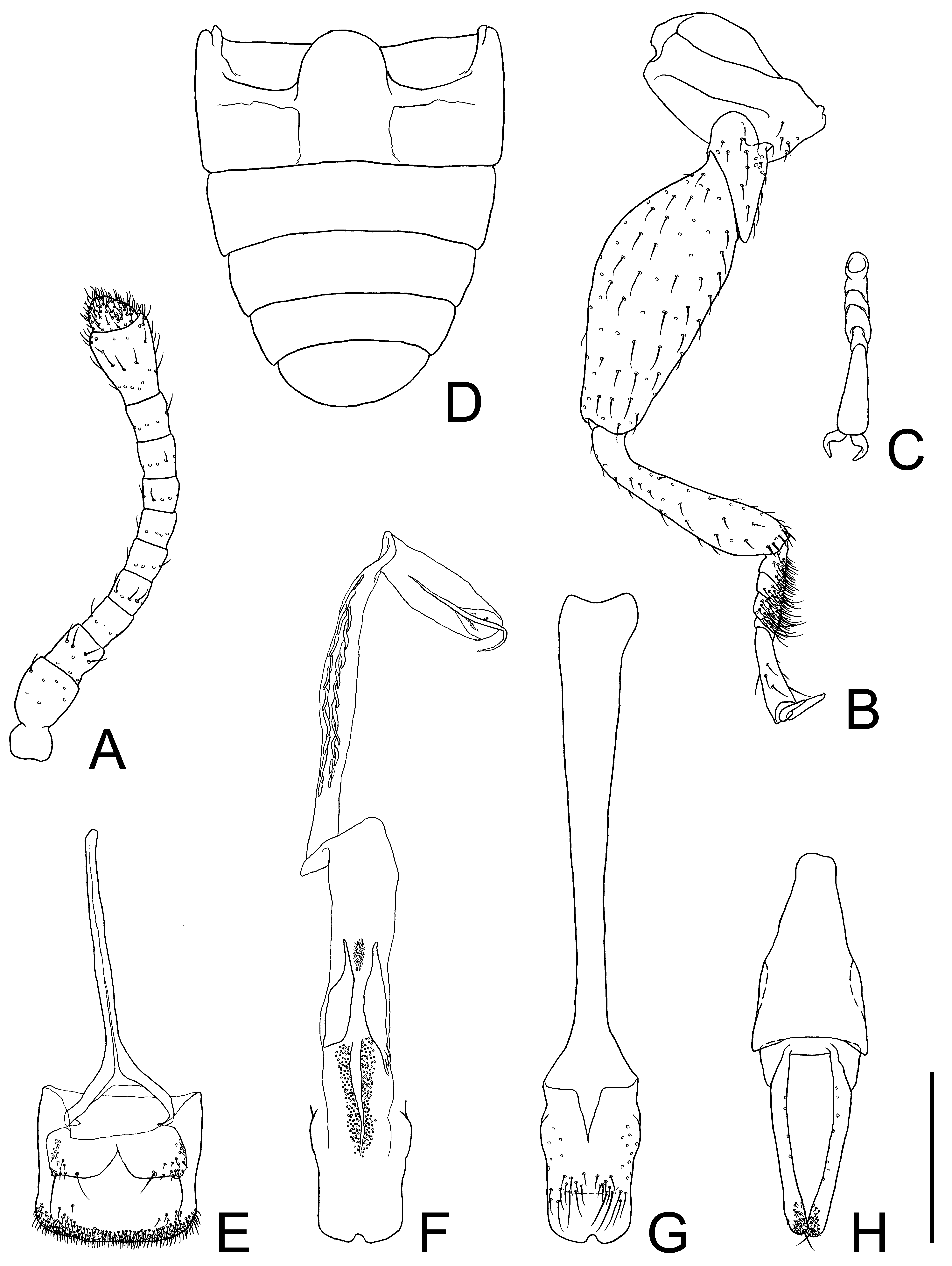
( Figs 1 View FIGURE 1 A,B, 2, 3)
Diagnosis. This species differs from the following species by the shape of the lateral pronotal teeth, the reticulate punctation on the pronotal disk and the explanate lateral margins of the elytra being narrowed towards the humeri, the longer and narrower 5th tarsomere and the femoral line curved posteriorly at the level of the metacoxal process.
Body ( Figs 1 View FIGURE 1 A,B). Body length from anterior margin of clypeus to apex of elytra measured along the median line: 2.33–2.60 mm (n=4). Surface dark brown; sides of elytra and legs somewhat lighter.
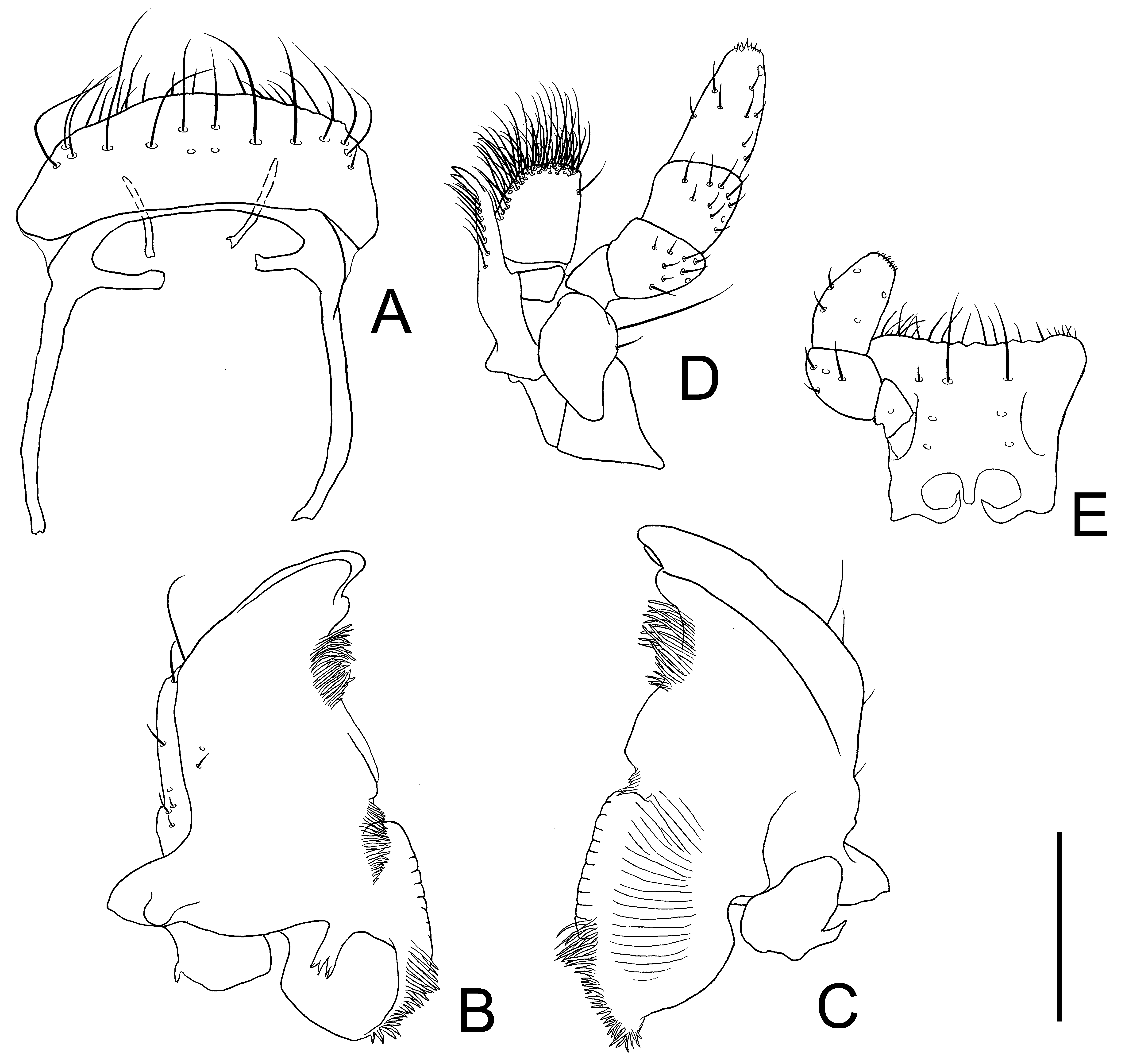
Head ( Figs 1 View FIGURE 1 A,B, 2A, 3). Subquadrate, maximum width across eyes 0.48–0.54 mm (n=4); clypeal margin slightly reflexed; genal region prominent (front of eye) laterally but not exceeding eye in width. Eye small, strongly protruding, as long as combined lengths of antennomeres 3 and 4. Punctation coarse and strong on posterior half; ventral surface with sparser punctation than dorsum. Pubescence inconspicuous; dorsally composed of very short and thick setae; ventrally composed of finer setae. Labrum ( Fig. 3 View FIGURE 3 A) oblong, ventrally with many setae along anterior margin, dorsally with some long setae and a few short setae. Antenna ( Fig. 2 View FIGURE 2 A) 0.56–0.60 mm long (n=4), relatively thick; antennomere 10 about twice as long as 9; covered with fine and relatively short setae; approximate ratios of antennomere lengths of holotype as follows: 2.0: 1.5: 1.4: 1.3: 1.2: 1.1: 1.3: 1.3: 1.3: 2.5: 1.0. Mandible ( Figs 3 View FIGURE 3 B,C) densely, finely pubescent on about anterior 1/4 and posterior 2/5 of inner margin, dorsally with long oblique furrow on distal half; lateral region ventrally with one long and a few short setae at each anterolateral angle and a few short setae; mola widely extended posteriorly, with some teeth, with dense pubescence on posterior half of inner margin; outer posterior angles strongly extended laterally. Maxilla ( Fig. 3 View FIGURE 3 D) with lacinia and galea; lacinia with two long apical teeth, dorsally with some long setae in a longitudinal row along inner margin; galea broad, divided into basigalea and distigalea, distigalea with dense long setae along anterior and inner margins, with a long seta on anterior outer margin, connected to basigalea by a membrane; palpifer with some short to medium length setae; palpomere 1 small, curved outwards; palpomere 2 strongly widening distally, covered with setae of various length except on inner area; palpomere 3 cylindrical, gradually widening distally, covered with setae of various length except on inner area; palpomere 4 long and conical, relatively densely covered with short spines on apex, sparsely covered with setae of various length, with a large puncture around apex; stipes protruding posteriorly at basal angle, ventrally with a very long seta and a few long setae. Labium ( Fig. 3 View FIGURE 3 E) dorsally covered with many setae of various lengths, ventrally with paired long and a few short setae, with two paired punctures around palpomeres, with some medium length setae along anterior margin; palpomere 1 small and stout, incised at middle of outer portion, with a puncture; palpomere 2 widening anteriorly, sparsely covered with short setae except on inner area; palpomere 3 conical, longer than 2, with some short setae, with many minute spines along distal margin.
Thorax and abdomen ( Figs 1 View FIGURE 1 A,B, 2B–D). Pronotum subquadrate, wider than long, length along median line 0.56–0.70 mm (n=4), maximum width including lateral teeth 0.85–0.98 mm (n=4), with paired shallow depressions on posterior half; six long lateral teeth, rounded at apices; anterior angle (1st tooth) broader than others, directed anterolaterally; 6th tooth extended in posterolateral direction; interstices of 2nd to 6th teeth as wide as teeth, wider than interstice of 1st and 2nd teeth; punctation reticulate and dense; small setiferous punctures inserted within ridge of reticulum; setae very short and thick. Thoracic ventrites with strong punctation; punctation on mesoventrite denser than on other ventrites; covered with coarse transverse microsculpture laterally; setae very short and fine; intercoxal process of procoxae parallel-sided; mesocoxal process narrowed posteriorly, widened around apex. Scutellar shield small, transverse, twice as wide as long, width a little shorter than eye length, with a transverse furrow at middle, partly concealed by elytral base. Legs ( Fig. 2 View FIGURE 2 B) covered with setae longer than those of other parts; pro- and mesocoxae rounded; metacoxa oblong, with transverse ridge, with some setae on medial areas; trochanters with well-extended inner distal angles; femora inflated but metafemur less so than others; tibiae widening distally, with some conical setae around apices; tarsomere 5 long, more than 3× as long as 4 ( Fig. 2 View FIGURE 2 C); claws simple. Abdomen more than 4/5 as wide as long; intercoxal process wide and rounded, with rim attaining near basal margin of 1st abdominal ventrite; with a pair of femoral lines running along metacoxal cavities and curved posteriorly at level of metacoxal process ( Fig. 2 View FIGURE 2 D); setae short and fine.
Elytra ( Fig. 1 View FIGURE 1 A). Oval, length along median line 1.40–1.62 mm (n=4), maximum combined width 0.93–1.06 mm (n=4) just behind middle, with distinct humeral carinae; apices sharply protruding. Rows of punctures almost as wide as the space between rows, with very short and thick setae on anterior margins of the punctures. Lateral margins widely flattened; flattened areas gradually narrowed towards humeri, widened apically; many denticles with short setae on lateral margins, sparser and smaller posteriorly.
Male genitalia ( Figs 2 View FIGURE 2 E–H). Tergite VIII square, wider than long, with many short setae along posterior margin; sternite VIII somewhat protruding at posterior angles, with a very long seta at posterior angle, a seta of medium length near each posterior angle, and some short setae near lateral margins and posterior angles; spiculum gastrale Y-shaped, thin and long ( Fig. 2 View FIGURE 2 E). Median lobe ( Fig. 2 View FIGURE 2 G) connected to long median strut by deeply incised membrane, median strut 3× as long as median lobe, incised at middle of posterior margin, with ostium opening dorsally at posterior 2/5, sparsely punctate in anterior half, with short to long setae on about posterior 2/5. Parameres ( Fig. 2 View FIGURE 2 H) fused with phallobase, a little widened around apices, sparsely punctate on inner areas, densely covered with short setae around apices, with a few setae of medium length near apex. Phallobase ( Fig. 2 View FIGURE 2 H) triangular; tegminal strut gradually narrowed anteriorly; lateral margins curled inwards for about half the length; basal piece narrow; posterior angles protruding posteriorly along basal parameres. Internal sac ( Fig. 2 View FIGURE 2 F) long, with paired sclerotized longitudinal portions with dense punctation around base and paired longitudinal plates without rods next to the sclerotized portions, with long armature around apex, and thin strut at apex.
Type series. Holotype: male, Ulu Gombak , Setapak Subdistrict, Gombak District, Selangor State, Malaysia, 6‒16. V. 2016, FIT with light, T. Yoshida leg. ( ELKU) . Paratypes: 1 female and 1 ex., same data as the holotype ( ELKU) ; 1 male, same locality ( 250 m), 13‒21. XI. 2009, LT, Maruyama & Tanaka Y. leg. ( ELKU) .
Distribution. Malaysia.
Etymology. The specific name means “reticulate”. The punctation on the pronotal disc of this new species is reticulate.
Remarks. The internal spherical space in the base of the mandible was observed under a compound microscope. It appears to be a mycangium, but it is difficult to regard it as such, since the opening of the structure could not be observed.

No known copyright restrictions apply. See Agosti, D., Egloff, W., 2009. Taxonomic information exchange and copyright: the Plazi approach. BMC Research Notes 2009, 2:53 for further explanation.
|
Kingdom |
|
|
Phylum |
|
|
Class |
|
|
Order |
|
|
Family |
|
|
Genus |