Glomerida
Chang, Hsueh-Wen, 2010, Pill-millipedes (Glomerida, Diplopoda) in Taiwan, Zootaxa 2477, pp. 1-20 : 18
publication ID |
https://doi.org/10.5281/zenodo.195313 |
DOI |
https://doi.org/10.5281/zenodo.6212127 |
persistent identifier |
https://treatment.plazi.org/id/332987AD-F56D-FFD2-38E9-86846CCB22AE |
treatment provided by |
Plazi (2016-04-09 18:17:13, last updated 2024-11-27 02:51:58) |
scientific name |
Glomerida |
status |
|
Key to the known species of Taiwanese Glomerida View in CoL
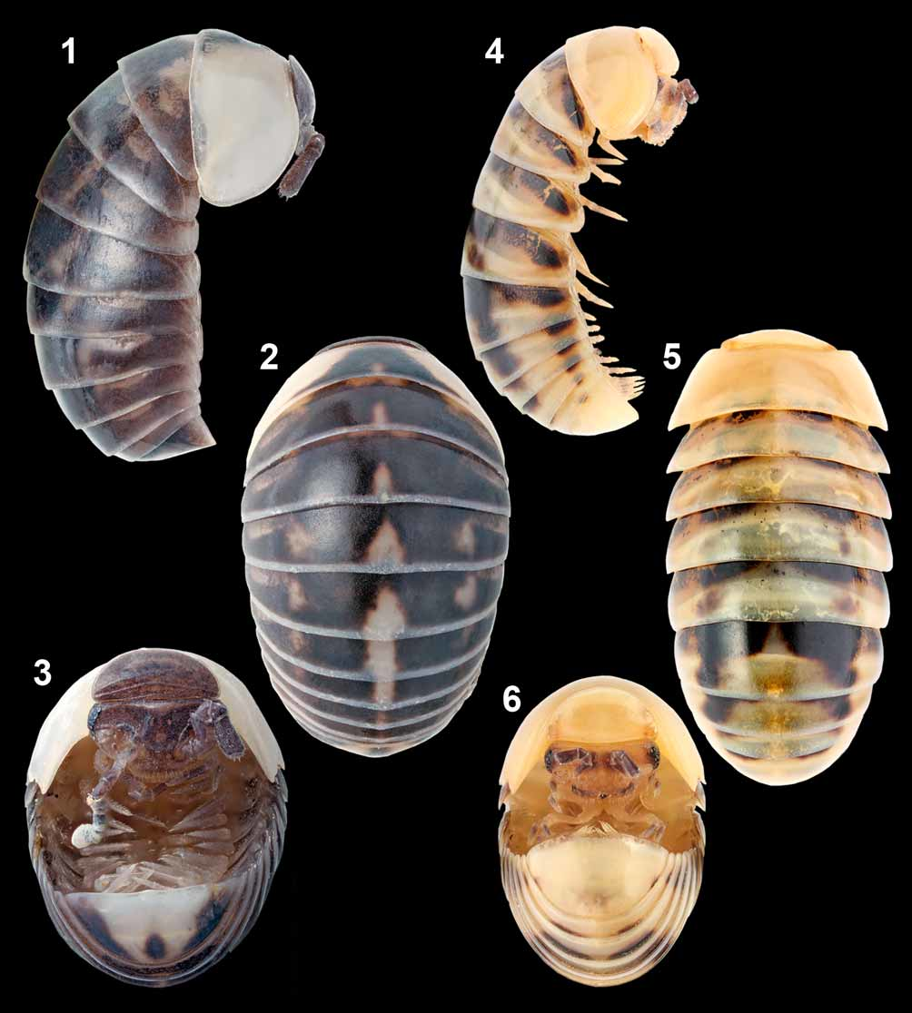
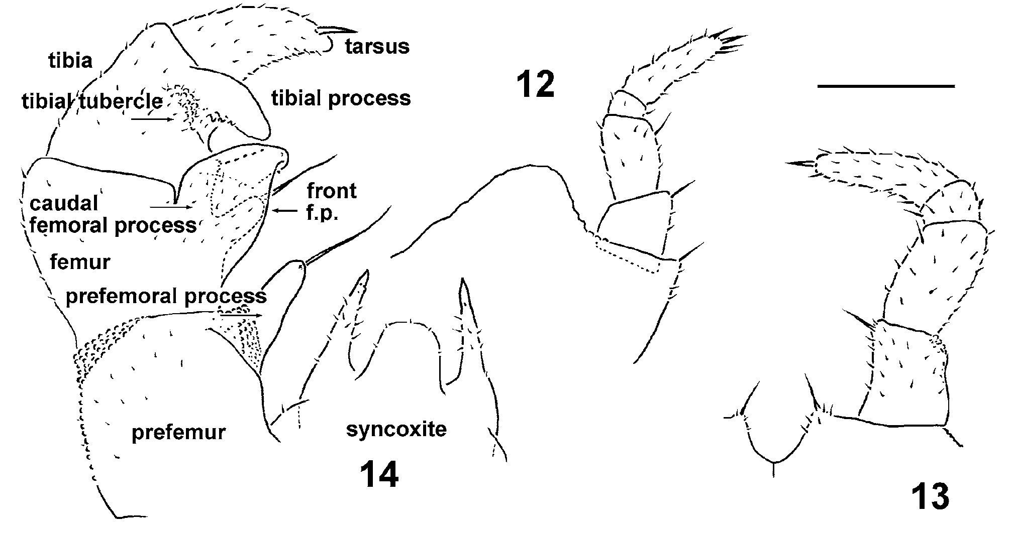
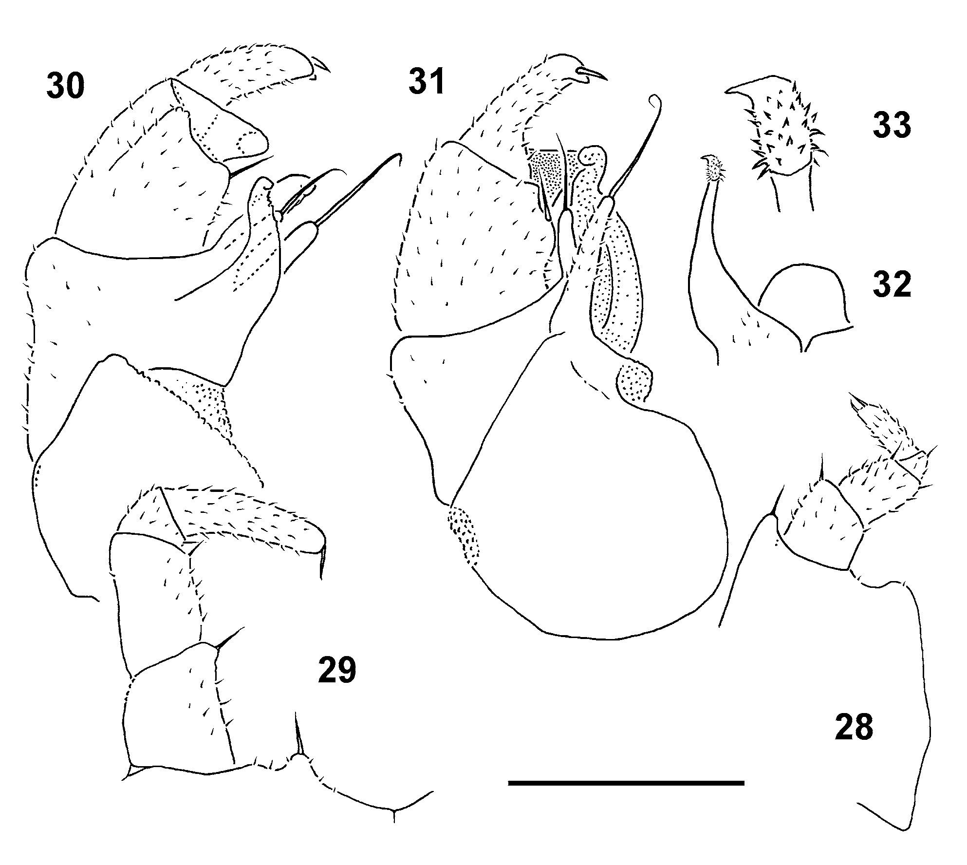
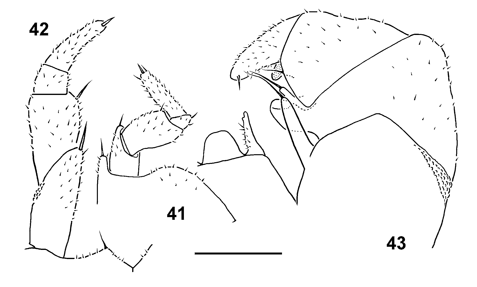
1. Male pygidium with a medial lobule at caudal edge ( Fig. 3 View FIGURES 1 – 6 ). Telopods extremely short and stout, without long setae on fingers ( Figs 10 and 11 View FIGURES 7 – 11 ) ............................................................................................. Mauriesia splendida View in CoL sp. nov.
– Male pygidium either (nearly) regularly convex or at least faintly concave medially at caudal edge ( Fig. 50 View FIGURES 44 – 50 ). Telopods much longer, both prefemur and femur with a long seta borne on a finger ( Figs 14 View FIGURES 12 – 14 , 31 View FIGURES 28 – 33 , 36, 39 View FIGURES 34 – 40 and 43 View FIGURES 41 – 43 ) .............. ................................................................................................................................................ genus Hyleoglomeris View in CoL , 2.
FIGURES 1 – 6. Mauriesia splendida sp. nov., male holotype (1 – 3), and Hyleoglomeris aurata sp. nov., female paratype (4 – 6). Habitus in lateral, dorsal, ventral, lateral, dorsal and ventral views, respectively. Photographed not to scale.
FIGURES 7 – 11. Mauriesia splendida sp. nov., male paratype. 7, sketches of Tőmősváry’s organ (left) and caudomedian lobule on pygidium (right); 8, leg 17; 9, leg 18; 10 and 11, telopod (front and caudal views, respectively). Scale bar: 0.2 mm; figure 7 drawn not to scale. Special designations: k, distomedian knob on femur; p, tibial process; s, setose bulge on femur; f, distal finger on femur; a, caudomedian apophysis on femur.
FIGURES 44 – 50. Hyleoglomeris vittata Verhoeff, 1929, male syntype (44 and 45) and males from Taroko National Park (46 – 50). Habitus in ventrolateral, dorsolateral, lateral, dorsal, ventral, lateral and ventral views, respectively. Photographed not to scale.
FIGURES 12 – 14. Hyleoglomeris aurata sp. nov., male holotype. 12, leg 17; 13, leg 18; 14, telopod (caudal view). Scale bar: 0.2 mm. Special designation: front f. p., front femoral process (hidden behind caudal femoral process).
FIGURES 28 – 33. Hyleoglomeris sinuata sp. nov., male paratype from Shangping Workstation. 28, leg 17; 29, leg 18; 30 and 31, telopod (caudal and front views, respectively). 32, telopod syncoxite (caudal view). 33, tip of telopod syncoxital horn. Scale bar: 0.1 mm; figure 33 drawn not to scale.
FIGURES 34 – 40. Hyleoglomeris proximata sp. nov., male paratype, and Hyleoglomeris vittata Verhoeff, 1929, male from Taroko National Park. 34 and 37, leg 17; 35 and 38, leg 18; 36, 39 and 40, telopod (caudal, caudal and front views, respectively). Scale bar: 0.2 (34 – 36) and 0.5 mm (37 – 40).
No known copyright restrictions apply. See Agosti, D., Egloff, W., 2009. Taxonomic information exchange and copyright: the Plazi approach. BMC Research Notes 2009, 2:53 for further explanation.
Kingdom |
|
Phylum |
|
Class |
|
Order |
|
Genus |
1 (by plazi, 2016-04-09 18:17:13)
2 (by ImsDioSync, 2016-12-07 15:39:05)
3 (by ImsDioSync, 2016-12-07 15:40:21)
4 (by ImsDioSync, 2017-06-26 03:01:50)
5 (by ImsDioSync, 2017-06-26 03:45:35)
6 (by ImsDioSync, 2019-03-29 22:35:20)
7 (by ExternalLinkService, 2019-09-26 21:15:31)
8 (by ExternalLinkService, 2022-01-30 17:23:52)
9 (by ExternalLinkService, 2022-02-21 22:24:06)
10 (by plazi, 2023-10-25 12:51:25)