
Achelidelphys steinitzi Lafargue and Laubier, 1977
|
publication ID |
https://doi.org/ 10.5281/zenodo.176361 |
|
DOI |
https://doi.org/10.5281/zenodo.5661737 |
|
persistent identifier |
https://treatment.plazi.org/id/C03D8785-0459-FFA0-FF2D-FCE4FED7FE9C |
|
treatment provided by |
Plazi |
|
scientific name |
Achelidelphys steinitzi Lafargue and Laubier, 1977 |
| status |
|
Achelidelphys steinitzi Lafargue and Laubier, 1977
Material examined: Holotype, reg. no. ZMA CO. 102.628. Four females from Monniot collection, from Didemnum sp. collected intertidally on shore at Mont Dore, New Caledonia. Three females in alcohol reg. nos MNHN-Cp2320. One female in alcohol, reg. no. BMNH 2006.1189.
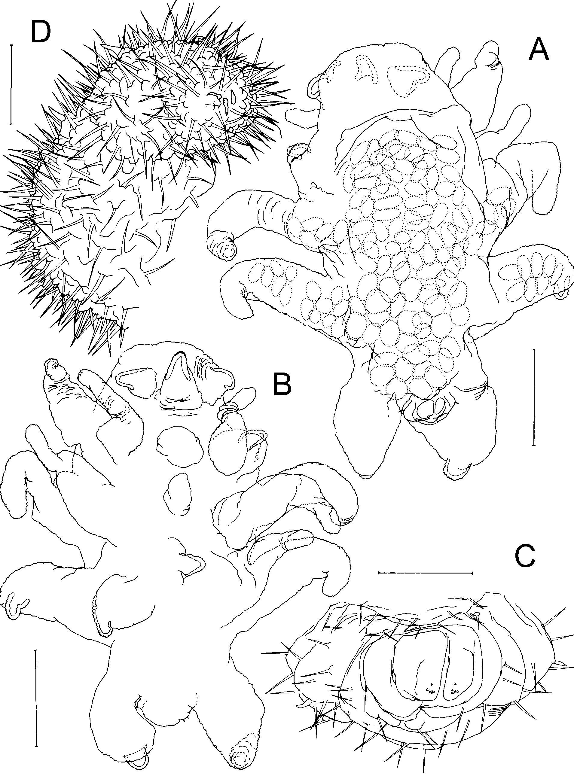
Differential Diagnosis: Body highly transformed, stellate ( Figs 6 View FIGURE 6 A–D, 7A–B); segmentation indistinct with segmental boundaries marked by superficial folds. Cephalosome with frontal margin merging into tapering, laterally-directed antennulary lobes. Rostrum anteroventrally-directed; elongate lobe, without accessory median lobe. Post-rostral median lobe absent. Labrum forming rounded hemispherical lobe. Lateral margin of cephalosome not produced into ridge-like swellings. Antenno-medial processes absent. Metasome truncated, with urosome hardly extending posterior to origin of leg 4. Legs 1–4 transformed, originating laterally, each occupying entire margin of somite; produced laterally giving body a stellate appearance. Mid-ventral metasomal processes present between legs 2 and 3 ( Figs 6 View FIGURE 6 A, C; 7B). Urosome vestigial, located terminally; bearing partly incorporated caudal rami. Surface of body, rostrum, labrum, cephalosomic processes, and legs densely ornamented with surface setules.
Antennules forming tapering lobe on either side of frontal margin of cephalosome. Antenna to maxilliped lacking. Legs 1–3 biramous; rami represented by unsegmented, tapering lobes; exopodal lobe laterallydirected, with broad base, carrying smaller endopodal lobe ventrally. Leg 4 uniramous, comprising short, posterolaterally-directed lobe representing exopod. Exopodal lobes of legs 2 and 3 each housing internal expansion of uterus, containing eggs visible through body wall ( Fig. 7 View FIGURE 7 A). Leg 5 absent.
Body length of female 1.40–2.50 mm. Male unknown.
Remarks: The three available, undamaged females of A. steinitzi from New Caledonia were compared in an attempt to assess the variability of different characters and their relative merit for species discrimination. These three specimens share with the holotype the number and arrangement of body processes, in particular they all possess median ventral processes between legs 2 and 3, the tapering conical rostrum, the hemispherical labrum, and the surface ornamentation of long setules. The new material also falls within the range of body lengths given for the type material ( Lafargue & Laubier 1977). One of the new females contains eggs in the uterus, lobes of which extend into the exopodal lobes of legs 2 and 3, as in the holotype. There are differences in the degree of development of the cephalosomal, metasomal and leg processes but given the variability in specimens collected from the same host individual (cf. Figs 6 View FIGURE 6 A, C and 7B), we do not regard them as sufficient to justify the establishment of a new species. All specimens also display some asymmetry in the development of processes, but this asymmetry varies individually and may possibly be due to the position within the host.

| ZMA |
Universiteit van Amsterdam, Zoologisch Museum |
No known copyright restrictions apply. See Agosti, D., Egloff, W., 2009. Taxonomic information exchange and copyright: the Plazi approach. BMC Research Notes 2009, 2:53 for further explanation.