
Ancyrona diversa Pic, 1921
|
publication ID |
https://doi.org/ 10.5281/zenodo.195206 |
|
DOI |
https://doi.org/10.5281/zenodo.6201466 |
|
persistent identifier |
https://treatment.plazi.org/id/19682A00-AB5D-FF92-5880-FEFADF25F967 |
|
treatment provided by |
Plazi |
|
scientific name |
Ancyrona diversa Pic, 1921 |
| status |
|
Larva of Ancyrona diversa Pic, 1921
Specimens examined: Five larval specimens: “Primorskyi krai, Lazovskyi zapovednik; Under the bark of Quercus mongolica ; 43°15'17"N, 134°07'59"E; A. Zaitsev; 08.VIII.2007 ”. Adult specimen: “Primorskyi krai, Lazovskyi zapovednik; on the bark of Quercus ; 43°00'34"N, 134°07'43"E; K. Makarov; 9.VII.2005 ”.
Description: Measurements (larger specimen): Body length (incl. mandibles and antennae) 7.3 mm, cranium length (without mandibles and antennae) 0.6 mm, cranium width 0.7 mm, thorax length 1.2 mm, abdomen length (incl. urogomphi) 5.2 mm, urogomphi length c. 0.2 mm.
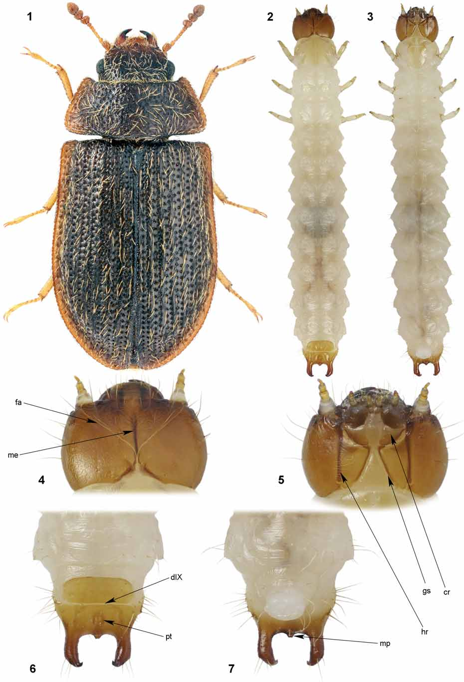
Coloration and pubescence ( Figs 2–7 View FIGURES 1 – 7 ): Head, urogomphi and dorsal part of abdominal segment IX heavily sclerotized, brown; prosternum, protergum and mesotergum with weakly sclerotized yellow-brown plates; metathorax and abdomen up to segment VIII yellowish white. Cranium with long sparse pubescence (c. 24 setae) situated laterally and along anterior margin; thorax with 2 long setae at each side and 2–5 short setae situated nearby; legs with few long setae; abdominal segments I–VIII with about 5 long setae situated dorsolaterally at each side; abdominal segments IX–X at sides densely and longly pubescent, urogomphi also with several setae.
Head ( Figs 4, 5 View FIGURES 1 – 7 ): Cranium wider than long, lateral sides rounded (convex); two stemmata occur at each side; frontal arms V-shaped, nearly straight; epicranial stem minute (nearly absent); median endocarina present, extending to 3/4 of cranium length; frontoclypeal suture absent; gular sutures widely separated at base, strongly convergent, extending to approximately midpoint of cranium; hypostomal rods distinct, nearly reaching base of cranium; paragular sclerites absent.
Antennae ( Figs 9, 10 View FIGURES 8 – 13 ): 3-segmented, 1st antennomere longer than 2nd, the latter as long as 3rd; sensory appendix relatively large, extending approximately to midpoint of 3rd antennomere.
Mandible ( Figs 8, 9 View FIGURES 8 – 13 ): with two sharp apical teeth situated side by side (in horizontal axis); two large blunt medial teeth present; mola absent; prostheca (lacinia mandibulae) plumose, long.
Maxilla ( Figs 13 View FIGURES 8 – 13 , 14 View FIGURES 14 – 16 ): mala small, blunt, with approximately 5 clavate setae along outer margin and numerous hair-like setae in centre; pedunculate seta present but small and pale; small pigmented area situated in inner basal corner; palpi 3-segmented; cardo and stipes distinctly separated; cardo nearly as large as stipes.
Labium ( Fig. 14 View FIGURES 14 – 16 ): ligula indistinct – small, pale, membranous, finely pubescent; palpi 2-segmented; prementum longitudinaly divided into two parts; postmentum somewhat shorter than stipes.
Labrum ( Figs 12 View FIGURES 8 – 13 , 15 View FIGURES 14 – 16 ): free; epipharynx with about 8 clavate setae along anterior margin, ciliate along lateral margins; labrum dorsally with 4 long hair-like setae at disc; rigid pigmented tormal plate situated between pair of short simple tormal sclerites.
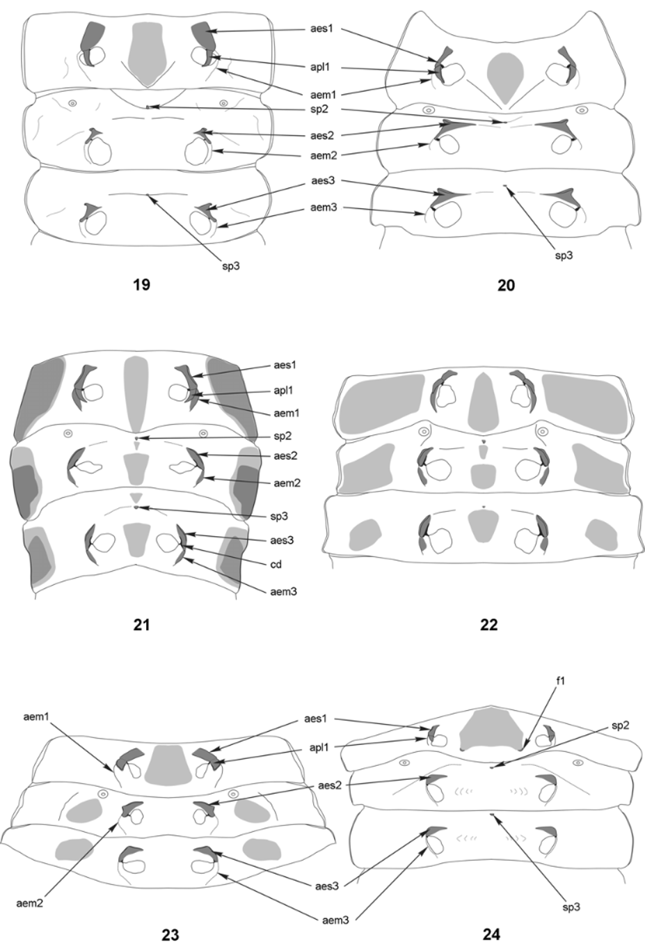
Thorax ( Figs 2, 3 View FIGURES 1 – 7 , 19 View FIGURES 19 – 24 ): protergum weakly sclerotized and pigmented, tergal plate longitudinaly divided by medial suture; mesotergum with two small hardly visible softly sclerotized plates in posterior half; metatergum without sclerites. Prosternum with distinct elongate sclerotized plate; meso- and metasterna without visible sclerites.
Thoracic endoskeleton ( Fig. 19 View FIGURES 19 – 24 ): Prothorax with pleural apodemes triangular, elongated, sharpened apically; epimeral and episternal apodemes poorly developed. Meso- and metathorax: episternal apodemes well developed, distinctly extended; epimeral apodemes as on prothorax; pleural apodemes absent. Mesothoracic spina trapeziform, well developed; metathoracic spina much smaller, oval.
Legs ( Fig. 11 View FIGURES 8 – 13 ): Coxae projecting; trochanteri oblong (not triangular); femora shorter than tibiae; tarsunguli with single seta.
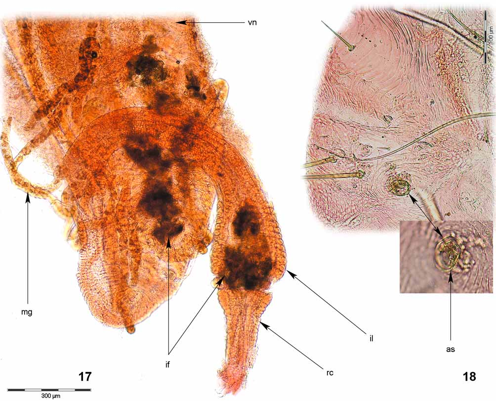
Abdomen ( Figs 6, 7 View FIGURES 1 – 7 , 18 View FIGURES 17 – 18 ): segments I–VIII membranous, terga without sclerites or pigmented plates; spiracles annular-biforous; segment IX with tergum transversely divided into basal disciform sclerite and apical sclerite bearing pair of hooked urogomphi; distinct median process with glandular opening present between urogomphi (no other glandular openings observed in abdominal segments); large shallow pit occurs in centre of apical sclerite; urogomphi smooth, turned upwards, well-developed.
Alimentary canal ( Figs 16 View FIGURES 14 – 16 , 17 View FIGURES 17 – 18 ): Ventriculus with micropapillae, its surface not smooth and not with netlike structure; ileum distinctly curved; four Malpighian tubules observed, probably all of the same length.
Biology: Larva found under oak bark ( Quercus mongolica ). Predatory – distinct remnant of arthropod antenna or palpus were observed inside ventriculus together with crushed remnants of arthropod cuticle inside ventriculus and ileum. Adult ( Fig. 1 View FIGURES 1 – 7 ) collected on dry oak log at night, they are probably predatory in the same way as other species of Ancyrona ( Kolibáč 2005, 2007).


No known copyright restrictions apply. See Agosti, D., Egloff, W., 2009. Taxonomic information exchange and copyright: the Plazi approach. BMC Research Notes 2009, 2:53 for further explanation.