
Astrodendrum spinulosum, Okanishi & Fujita, 2018
|
publication ID |
https://doi.org/ 10.11646/zootaxa.4392.2.4 |
|
publication LSID |
lsid:zoobank.org:pub:C1A8F758-D41A-404C-A5C4-6CBC476EA324 |
|
DOI |
https://doi.org/10.5281/zenodo.5998498 |
|
persistent identifier |
https://treatment.plazi.org/id/2526AA33-FF9A-417B-D9C2-EFB8FDDCD6BB |
|
treatment provided by |
Plazi |
|
scientific name |
Astrodendrum spinulosum |
| status |
sp. nov. |
Astrodendrum spinulosum View in CoL sp. nov.
( Figs 2–7 View FIGURE 2 View FIGURE 3 View FIGURE 4 View FIGURE 5 View FIGURE 6 View FIGURE 7 )
[Japanese Name: Toge-Tsuruboso-Tezurumozuru]
Astrodendrum sagaminum View in CoL — Irimura, 1982. 79, text fig. 4, pl. 2, figs 4–5. (Non Astrodendrum sagaminum Döderlein, 1911 View in CoL . 38– 39, pl. 2 figs 3–5, pl. 7, fig. 8)
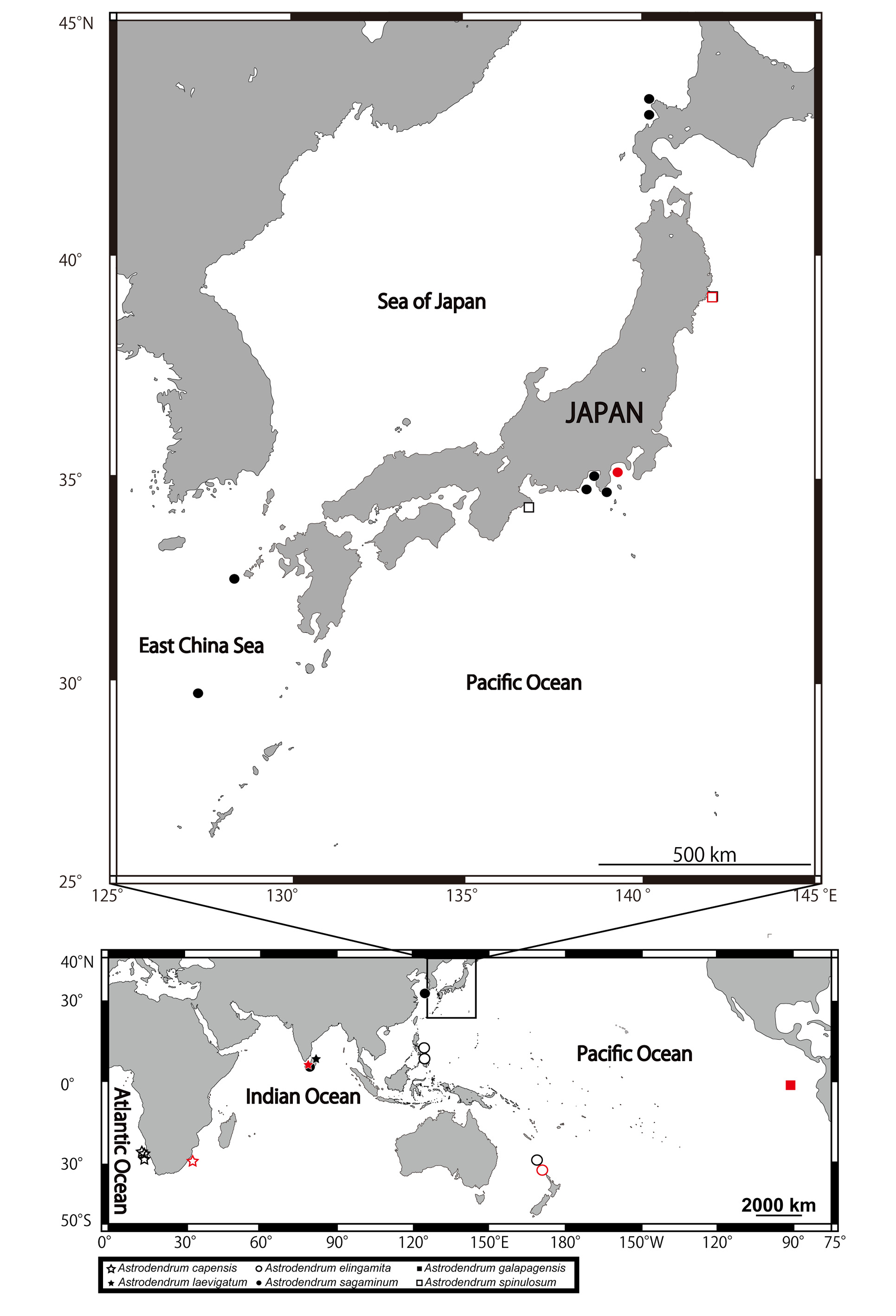
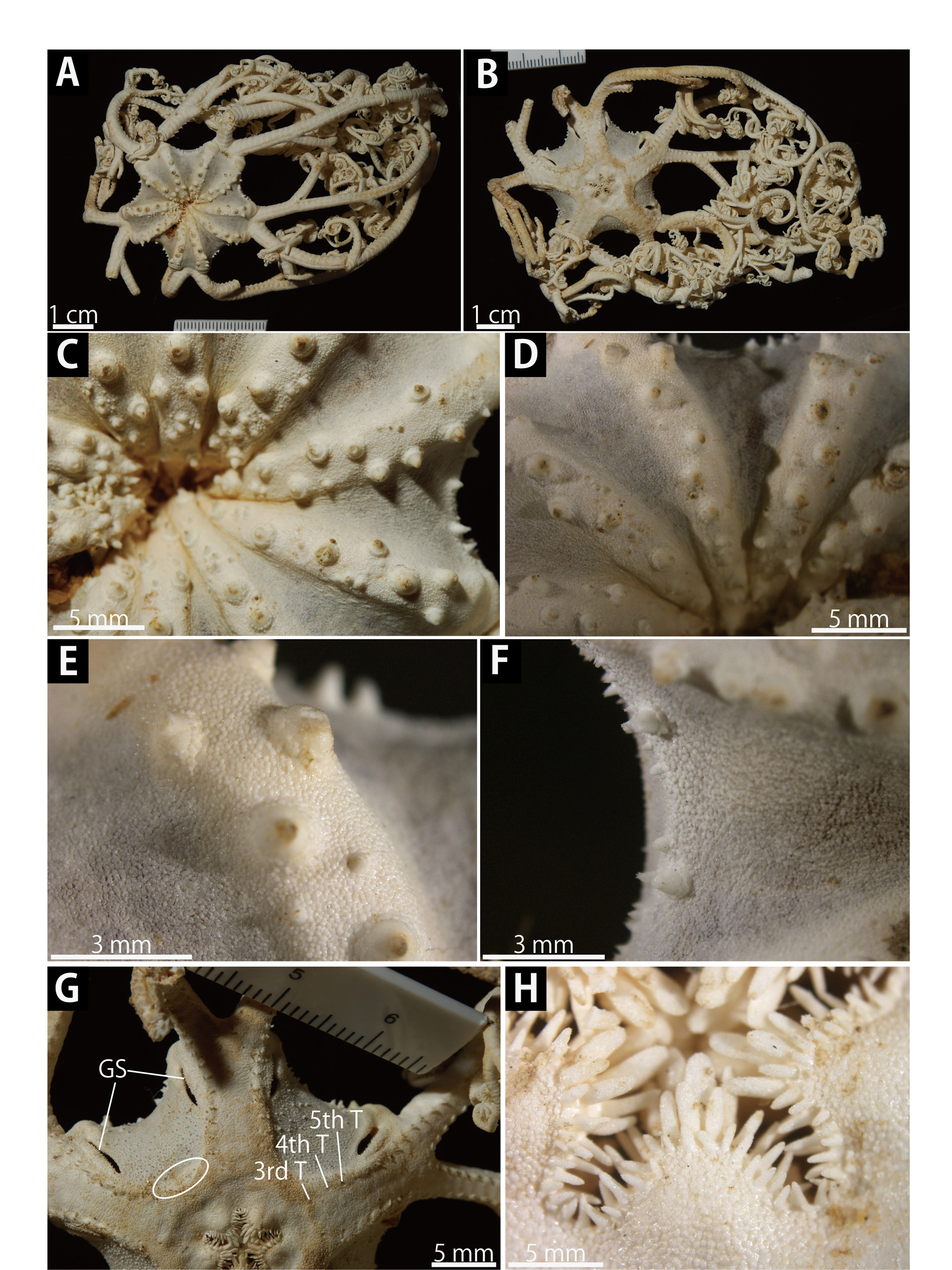
Type materials. NSMT E-6237, holotype, collected by fishery boat Taku-Maru , off Ohakozaki, Otsuchi, Iwate Prefecture, northeastern Japan, 39˚21.N, 142˚00.E, 75 m depth, 9 May 2008 (Type locality) . NSMT E-10715, one paratype, collected by T/ S Seisui-Maru, off Shima Peninsula, Mie Prefecture, central Japan, 34˚11.42’N, 136˚41.54’E, 111 m depth, 12 October 2016 . NSMT E-6905, one paratype, collected by R/ V Yayoi, off Ohakozaki, off Otsuchi, Iwate Prefecture, northeastern Japan, 39˚25.48’N, 135˚58.44’E, 78.2 m depth, 28 April 2009 . NSMT E-10716, one paratype, collected by R/ V Yayoi, off Ohakozaki, Otsuchi, Iwate Prefecture, northeastern Japan, 39˚21.32’N, 142˚00.23’E-39˚21.28’N, 142˚00.28’E, 73–86 m depth, 27 June 2016 ( Fig. 1 View FIGURE 1 ).
Other material. NSMT-Oph R: 22, one specimen, off Hayama, Sagami Bay, Kanagawa Prefecture, central Japan, 90–108 m depth, 22 November, 1930 . NSMT-Oph R: 25, one specimen and NSMT-Oph R: 26, one specimen, 3.2 km off south Enoshima Island , Kanagawa Prefecture, central Japan, 144 m depth, 11 August 1935 . NSMT-Oph R: 27, one specimen, Minami Amadaiba , Sagami Bay, Kanagawa Prefecture, central Japan, 200 m depth, 27 August 1935 . NSMT-Oph R: 43, one specimen, off Hayama , Sagami Bay, Kanagawa Prefecture, central Japan, depth unknown, 14 February 1950 . NSMT-Oph R: 73, one specimen, near Kodane , Kanagawa Prefecture, central Japan, depth unknown, 6 December 1957 . NSMT-Oph R: 75, Goromba , Sagami Bay, Kanagawa Prefecture, central Japan, 100–110 depth, 21 January 1958 ( Fig. 1 View FIGURE 1 ).
Diagnosis. External ossicles on aboral and interradial lateral disc conical, separated; external ossicles on oral disc plate-shaped, fully in contact; bulges on lateral edges of proximal portion of the arm; terminal projection on each arm spine on proximal portion of the arm single or lacking; 0 to 3 secondary teeth on each hook-shaped arm spine on distal portion of the arm.
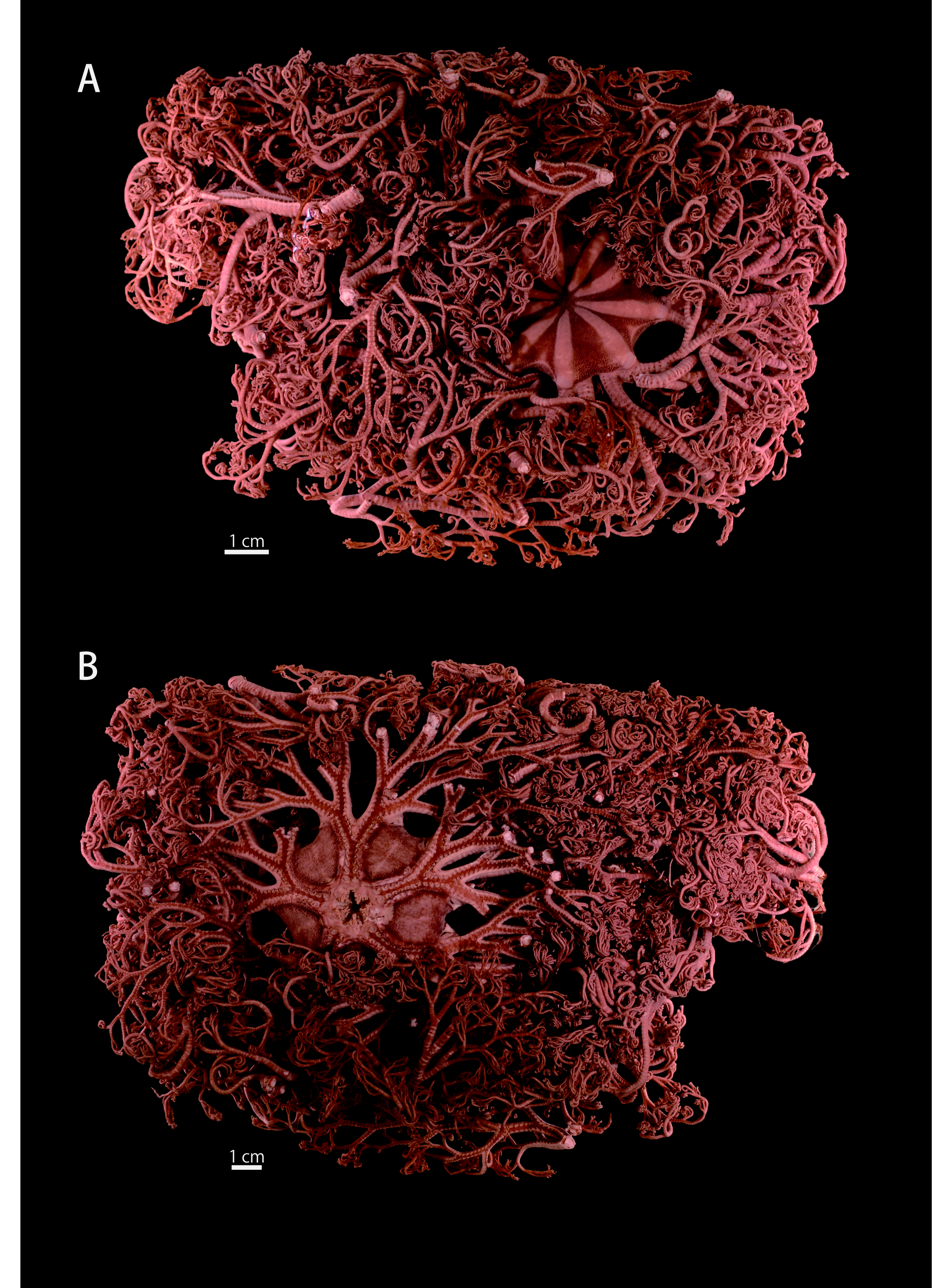
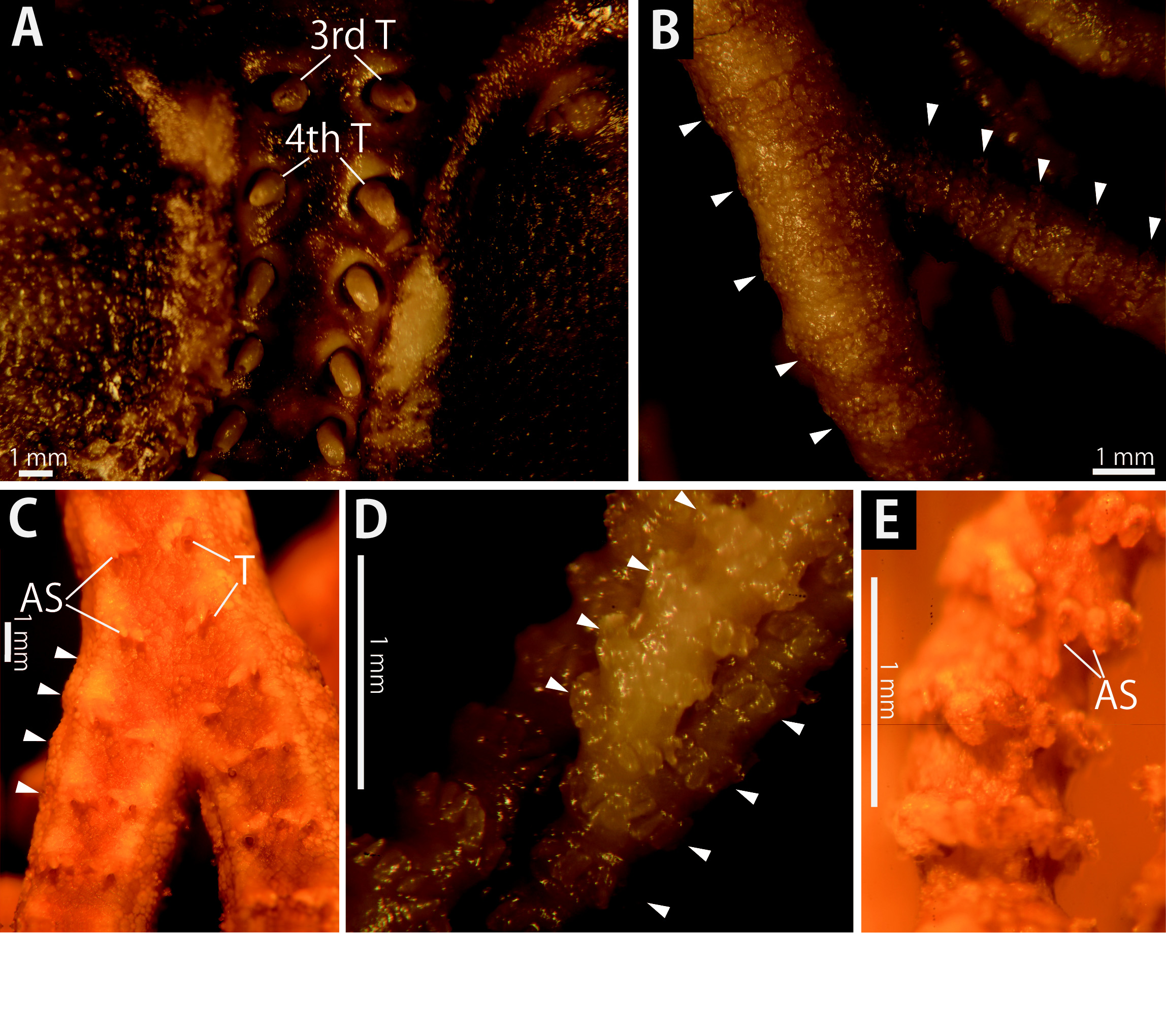
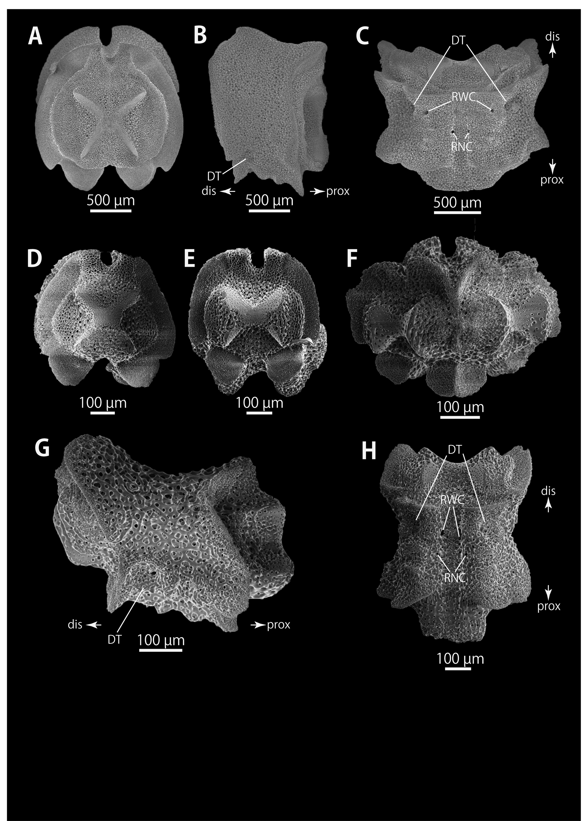
Description of holotype. Disc. Disc five-lobed with notched interradial edges, 53 mm in disc diameter ( Fig. 2 View FIGURE 2 ). Radial shields and surrounding plates tumid ( Figs 2A View FIGURE 2 , 3A View FIGURE 3 ). Aboral disc covered by conical external ossicles ( Fig. 3A–C View FIGURE 3 ). At periphery of the disc, ossicles separated and scattered, approximately 200 µm in length and 250 µm in height on radial shield ( Fig. 3B View FIGURE 3 ), approximately 100–200 µm in length and 200 µm in height in interradii ( Fig. 3B View FIGURE 3 ), and approximately 200 µm in length and 150–200 µm in height at center of disc ( Fig. 3A, C View FIGURE 3 ). Radial shields completely concealed by external ossicles, bar-like, approximately 28 mm in length, 2.5 mm in width proximally and the width gradually increasing to 6 mm distally, but shields do not reach disc center ( Fig. 3A View FIGURE 3 ). The oral surface of disc covered by polygonal plate-shaped external ossicles, fully in contact, approximately 200–250 µm in length. Oral shields, adoral shields, oral plates and ventral arm plates completely concealed by external ossicles ( Fig. 3E View FIGURE 3 ). Teeth uniformly spiniform, situated on the top of dental plates and edges of oral plates ( Fig. 3H View FIGURE 3 ). Teeth arranged in 1 or 2 transverse rows on oral plates, 8 to 10 in number ( Fig. 3E View FIGURE 3 ), in a cluster covering oral-most part of dental plate, 7 to 10 in number ( Fig. 3E View FIGURE 3 ), and in a vertical line, on other part of dental plates, 3 or 4 in number. Teeth varying in position and in size, approximately 2–3 mm in length, 0.7 mm in greatest width on dental plates, and 2 mm in length, 0.6–0.7 mm in width on oral plates ( Fig. 3E View FIGURE 3 ). Interradial surface of lateral disc covered by conical external ossicles and skin ( Figs 2B View FIGURE 2 ; 3H). Conical external ossicles, separated and scattered, approximately 100 µm in length and height ( Fig. 3H View FIGURE 3 ). Ossicles increasing their height toward aboral side, reaching 500 µm ( Fig. 3H View FIGURE 3 ). Ossicles fully in contact, approximately 200 µm in length and 0.8–1 mm in height on adradial edge of interradial lateral disc ( Fig. 3H View FIGURE 3 ). Two genital slits (8 mm long and 1 mm wide) in each interradius ( Fig. 3G View FIGURE 3 ). Spinule ossicles along the adradial edge of genital slits, 200 µm in length and 500 µm in height ( Fig. 3G View FIGURE 3 ). One large, elliptical madreporite situated on oral interradius, approximately 5.4 mm in width and 3 mm in length ( Fig. 3F View FIGURE 3 ).
Arms. Arms branching. On the proximal portion of arm, before first branch, arm 11.0 mm wide and 6.2 mm high, with an arched aboral surface and flattened oral surface. Between first branch and second branch, arm width and height abruptly decreasing to 5.5 mm in width and 5.7 mm in height. Subsequently, arms tapering gradually toward arm tip ( Fig. 4 View FIGURE 4 ).
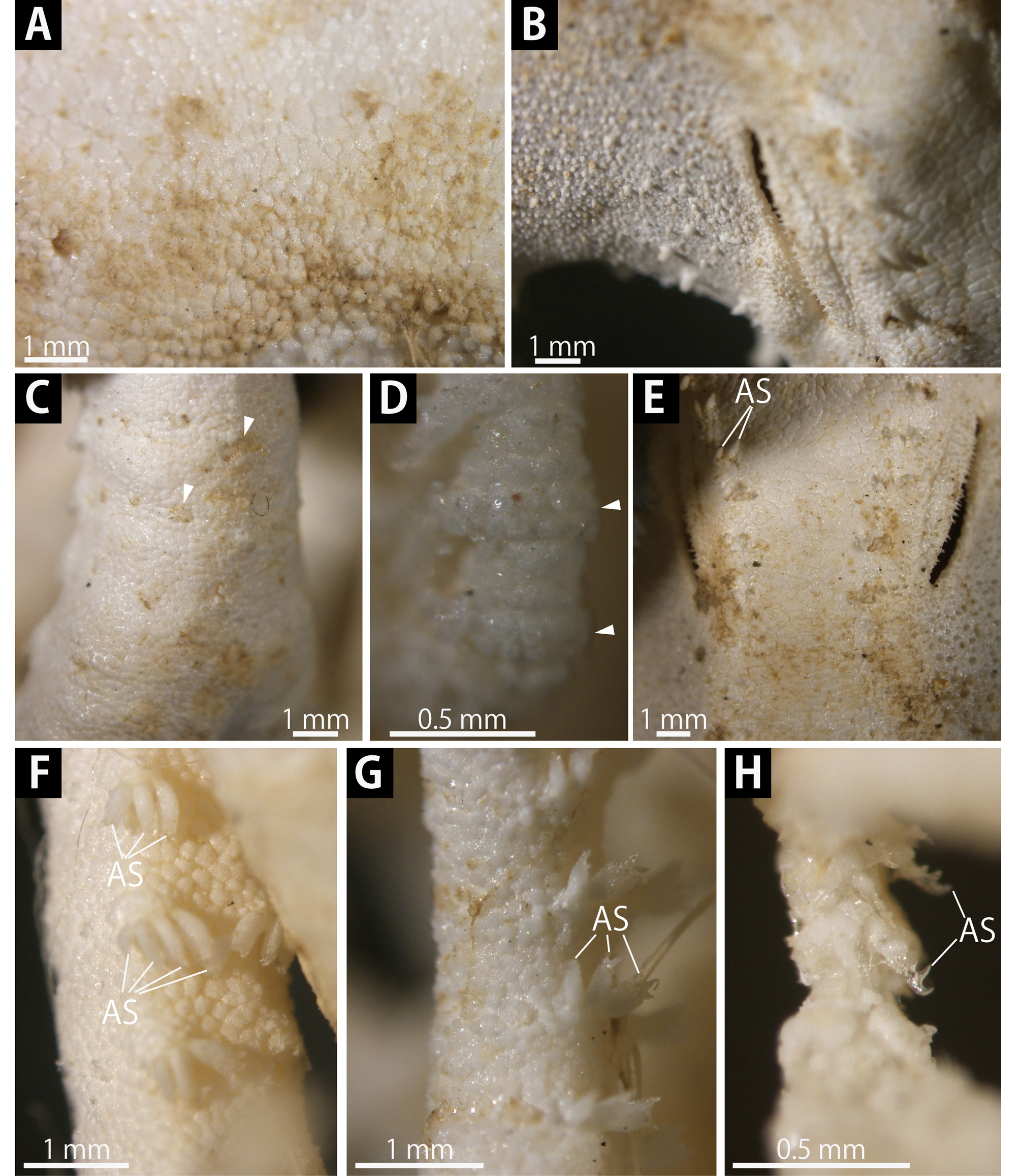
On aboral and lateral surface, each arm segment covered by single annular row of large oblong plates, approximately 600–700 µm in transverse length ( Fig. 3D View FIGURE 3 ). Before third branch, each plate separated by granular external ossicles. Plates fully in contact from fourth branch and subsequent distal segments ( Fig. 4B View FIGURE 4 ). Before first branch, plates bearing no hooklets ( Fig. 3D View FIGURE 3 ), after second branch, plates bearing hooklets ( Fig. 4B View FIGURE 4 ). With exception of hooklet-bearing plates, aboral and lateral surface of arm completely covered by domed and polygonal granular external ossicles, fully in contact, approximately 200 µm in length ( Fig. 3D View FIGURE 3 ). Before first branch, oral surface covered by polygonal and plate-shaped external ossicles, similar to those on oral disc. After first branch, granular external ossicles, slightly in contact, approximately 100 µm in length ( Fig. 4A View FIGURE 4 ). In middle portion of arm, aboral and lateral surface covered by domed and round granular external ossicles, slightly in contact, approximately 170– 200 µm in length ( Fig. 4B View FIGURE 4 ). Oral surface covered in round granular external ossicles, slightly in contact, approximately 70–80 µm in length ( Fig. 4C View FIGURE 4 ). Distally, aboral and lateral surface of arm covered by round granular external ossicles, slightly in contact, approximately 60–70 µm in length ( Fig. 4D View FIGURE 4 ). Oral surface covered by flat and round plate-shaped external ossicles, slightly in contact or separated, approximately 50–60 µm in length. Lateral arm plates and ventral arm plates completely concealed by skin and external ossicles on entire arm ( Fig. 4A, C, E View FIGURE 4 ). First to sixth tentacle pore with single arm spine; seventh pores occasionally with 1 arm spine, eighth and subsequent pore with 2 or 3 spines ( Fig. 4A, C View FIGURE 4 ). Distally, the number of arm spines decrease gradually to 2 toward arm tip ( Fig. 4E View FIGURE 4 ). Arm spines approximately one-third to one-fourth of length of corresponding arm segment, and covered by thin integument ( Fig. 4A, C, E View FIGURE 4 ).
Color. Oral surface of arms and aboral disc dark brown, while radial shields and remaining areas pale brown ( Fig. 2 View FIGURE 2 ). Darker spots present on midline of proximal position of the arms ( Fig. 3B View FIGURE 3 ).
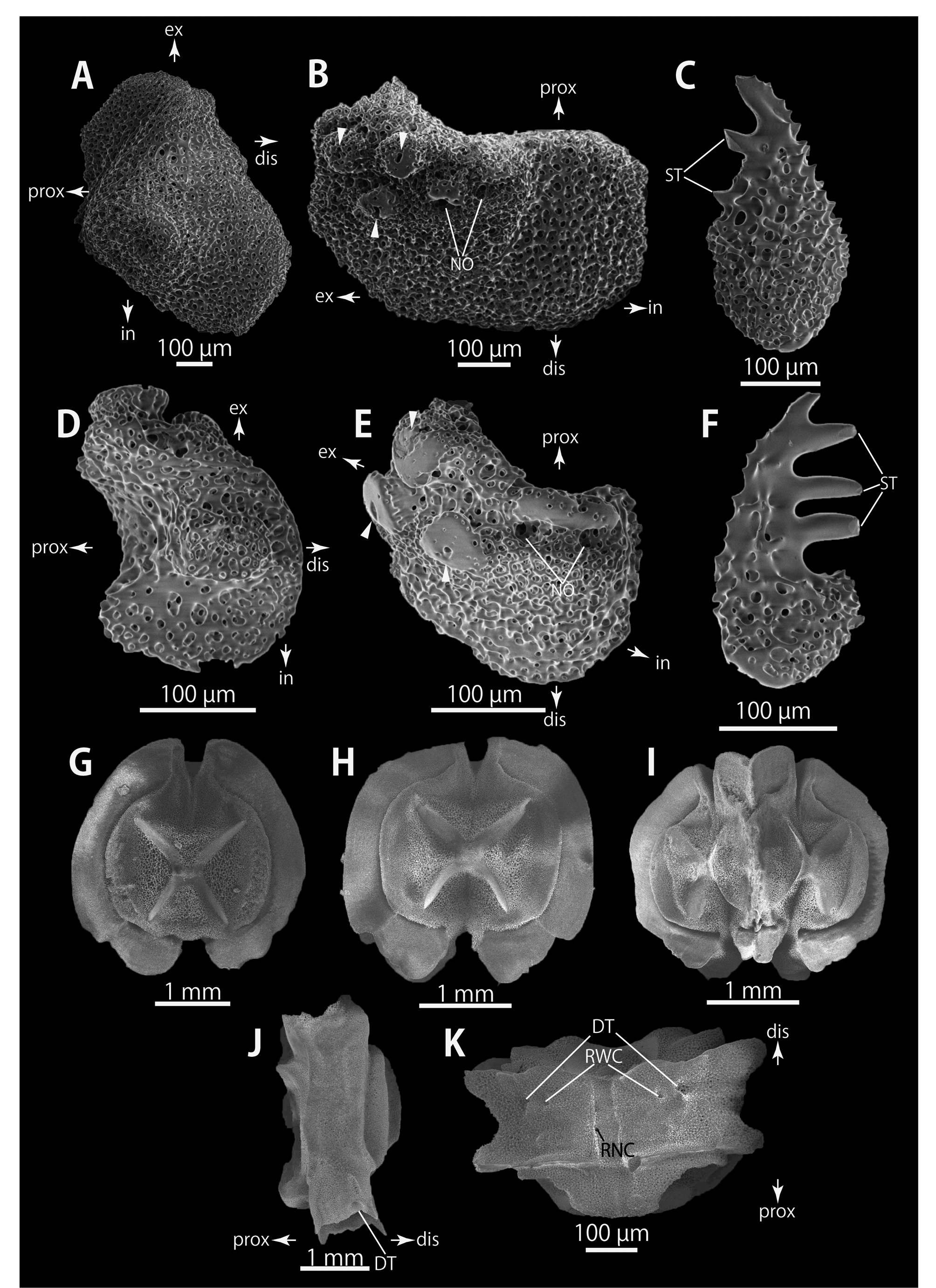
Ossicle morphology of the holotype. External ossicles on aboral periphery of radial shields conical, approximately 150 µm in length and 200 µm in height ( Fig. 5A, B View FIGURE 5 ). Hooklet-bearing plates with 5 or 6 tubercleshaped articulations for hooklets in proximal portion of the arm ( Fig. 5C View FIGURE 5 ), approximately 15 articulations in middle portion ( Fig. 5E View FIGURE 5 ) and 8 articulations in distal portion ( Fig. 5H View FIGURE 5 ); articulations forming 2 parallel rows ( Fig. 5C, E, H View FIGURE 5 ). Each hooklet with single inner tooth and reticular structure ( Fig. 5D, F, I View FIGURE 5 ). Lateral arm plates long, bar-like, with straight distal edge and concave proximal edge ( Figs 5J, K View FIGURE 5 ; 6A, B, D, E). On proximal portion of arm, lateral arm plates with perforations on aboral side and pairs of simple nerve and muscle openings on oral-external side ( Fig. 5J View FIGURE 5 ) and on middle to distal portion of arms, two nerve openings beside dorsal lobe and 3 articulations for hooklets on oral surfaces ( Fig. 6A, B, D, E View FIGURE 6 ). Arm spines in proximal portion of arm ovoid, with a small projection, approximately one-seventh length of the height of spine ( Fig. 5L View FIGURE 5 ). In middle and distal portion, arm spines transformed into hooks with 2 or 3 inner teeth, respectively ( Fig. 6C, F View FIGURE 6 ). Hook-shaped arm spines distinguished from hooklets on aboral and lateral surface of arm by lack of reticular structure ( Figs 5D, F, I, L View FIGURE 5 ; 6C, F). All vertebrae with hourglass-shaped streptospondylous articulations ( Figs 6G–I View FIGURE 6 ; Fig. 7A, D–F View FIGURE 7 ), and distal side of branching vertebra slightly wider than in non-branching vertebra and with 2 articulation surfaces ( Figs 6I View FIGURE 6 ; 7F). Surfaces of lateral furrows smooth, with no special ornamentations ( Figs 6J View FIGURE 6 ; 7B, G). Depressions for tube feet openings in distal part of oral-lateral side of vertebrae ( Figs 6J, K View FIGURE 6 ; 7C, H). A pair of radial water canals opening on lateral side of vertebrae, near depression of tube feet in the proximal to middle position of arms ( Figs 6K View FIGURE 6 , 7C View FIGURE 7 ) and opening in distal part of the oral groove of vertebrae in distal portion of arms ( Fig. 7H View FIGURE 7 ).
Distribution. JAPAN. Off Iwate (type locality), Sagami Bay ( Irimura, 1982), and off Mie. Depth ranges 75– 200 m.
Etymology. The specific name is latin for “spinulose”, referring to numerous conical external ossicles on aboral disc of this new species.
Remarks. Irimura (1982) referred to nine specimens of Astrodendrum collected in the Sagami Sea, central Japan as Astrodendrum sagaminum ( Döderlein, 1911) which is known from the Indo-West Pacific (see also “Distribution of Astrodendrum sagaminum ” below). Irimura (1982) described external ossicles on the disc as “granules” and failed to mention detailed morphology of the oral surface of the proximal portion of the arms and of each arm spine ( Irimura, 1982). We re-examined seven (NSMT-Oph R: 22, 25, 26, 27, 43, 73, 75) of the nine TABLE 1. Tabular characters key to the species of the genus Astrodendrum . Characters of A. laevigatum are referređ to from the original đescription by Koehler, 1897. "-̏ means no đata.
Shapes, sizes anđ arrangements of external ossicles on the đisc Bulges of Maximum number of Maximum number of lateral terminal projections of Species seconđary teeth of Aboral surface Oral surface Interrađial lateral surface eđges of arm spine on proximal hook-shapeđ arm spine proximal arms portion of the arm A. spinulosum Plate-shapeđ, fully in Cone-shapeđ, separateđ Cone-shapeđ, separateđ Present 1 3 sp. nov. contact Granule-shapeđ, fully in contact; Plate-shapeđ, fully in A. capensis Plate-shapeđ, fully in Cone-shapeđ, separateđ on rađial contact; Absent 3 2 (Mortensen, 1933) contact shielđs cone-shapeđ, separateđ
specimens from Sagami Bay observed by Irimura (1982) and they were certainly different from Astrodendrum sagaminum in having uniform sized conical external ossicles on their disc and conformed to the diagnosis of Astrodendrum spinulosum sp. nov. (see below for the detailed morphological characters). Therefore, Irimura (1982) specimens are here referred to the new species.
This new species falls within Astrodendrum in having external ossicles on its disc, a madreporite on the inner edge of the interradial lateral disc, and in lacking calcareous plates on the lateral disc margin ( Fell, 1960; McKnight, 2000).
Our re-examination of the type material and comparison of Astrodendrum species revealed that Astrodendrum spinulosum sp. nov. can be distinguished from the five currently valid species by the external ossicles on the aboral disc which are conical, and separated ( Fig. 3A–C View FIGURE 3 ). In contrast, in the other congeners, the ossicles are: smaller granular, fully in contact and larger conical and, separated in A. capense ( Fig. 8E, F View FIGURE 8 ); plate-shaped at periphery and conical at center, both slightly in contact in A. galapagense ( Fig. 10B, C View FIGURE 10 ); smaller granular, fully in contact and larger granular, separated in A. elingamita ( Baker, 1974) ; and smaller and larger granular, both separated in A. sagaminum ( Fig. 12A View FIGURE 12 ).
External ossicles on the interradial lateral surface of the disc are conical and, separated in this new species ( Fig. 3H View FIGURE 3 ), as in A. sagaminum ( Fig. 12B, D View FIGURE 12 ). In contrast, those of A. capense ( Fig. 11B View FIGURE 11 ) and A. elingamita ( Baker, 1974) are composed of plate-shaped ossicles, fully in contact, and granular ossicles, that are separated. Those of A. galapagense are plate-shaped, slightly in contact ( Fig. 9A View FIGURE 9 ).
External ossicles on the oral surfaces of the disc of the new species, A. capense , A. elingamita and A. sagaminum are plate-shaped and fully in contact ( Figs 8H View FIGURE 8 ; 12B; Baker, 1974) and those of A. galapagense are granular and separated ( Fig. 10D View FIGURE 10 ). The disc of A. laevigatum is covered only by skin without external ossicles ( Koehler, 1897, 1899).
This new species possesses bulges on lateral edges of proximal portion of the arms ( Fig. 4A View FIGURE 4 ), whereas bulges lack in other species of Astrodendrum ( Figs 8D View FIGURE 8 ; 9G; 12B; Baker, 1974). The presence of bulges of A. laevigatum is unknown ( Koehler, 1897, 1899).
The maximum number of terminal projections of each arm spine of proximal portion of the arm is single in this new species ( Fig. 5L View FIGURE 5 ) and in A. sagaminum ( Döderlein, 1911) . That of A. galapagense is 2 ( Fig. 11E View FIGURE 11 ) and of A. capense and A. elingamita is 3 ( Mortensen, 1933; Baker, 1974, 1980).
The maximum number of secondary teeth of each hook-shaped arm spine in the distal portion of the arm of this new species is 3 ( Fig. 6F View FIGURE 6 ), a character shared with A. elingamita ( Baker, 1980) . In A. capense and A. sagaminum there are 2 ( Döderlein, 1911; Mortensen, 1933) and there is 1 in A. galapagense ( A. H. Clark, 1916) .
The maximum number of terminal projections and of secondary teeth mentioned above is not provided in A. laevigatum ( Koehler, 1897, 1899; Bomford, 1913).
Based on our morphological observations of type specimens and study with the literature, we found that all six species of Astrodendrum can be morphologically distinguished using six morphological characteristics. A tabular key to species is provided in Table 1.


| NSMT |
National Science Museum (Natural History) |
No known copyright restrictions apply. See Agosti, D., Egloff, W., 2009. Taxonomic information exchange and copyright: the Plazi approach. BMC Research Notes 2009, 2:53 for further explanation.
|
Kingdom |
|
|
Phylum |
|
|
Class |
|
|
Order |
|
|
Family |
|
|
Genus |
Astrodendrum spinulosum
| Okanishi, Masanori & Fujita, Toshihiko 2018 |
Astrodendrum sagaminum Döderlein, 1911
| Doderlein 1911 |