
Australiatelura tasmanica ( Silvestri, 1949 )
|
publication ID |
https://doi.org/ 10.3853/j.2201-4349.68.2016.1652 |
|
persistent identifier |
https://treatment.plazi.org/id/03A8C40A-FFFD-FFCF-823D-4AE5FCBA5A8D |
|
treatment provided by |
Felipe |
|
scientific name |
Australiatelura tasmanica ( Silvestri, 1949 ) |
| status |
|
Australiatelura tasmanica ( Silvestri, 1949)
Figs 1–41 View Figure 1 View Figures 2–18 View Figures 19–20 View Figures 21–36 View Figures 37–41
Atopatelura tasmanica Silvestri, 1949: 35 .
Australiatelura tasmanica (Silvestri) .— Mendes, 1995: 98.
Type material. Paratypes (all labelled as “cotypi!”, no specimen apparently designated as “typus”; specimen labels only state “ Tasmania ” however Silvestri (1949) lists all material (3.5 mm in length) as collected in Hobart with the ant species Colobopsis gasseri F. or a larger variety (5 mm) collected with Camponotus nigriceps Smith. Being unaware at the time of the presence of a second sympatric Australiatelura species , this latter specimen was not specifically recognised among the material before they were returned to MUSA. Being larger than the typical Australiatelura tasmanica , it is possible that it could belong to another species, such as the second species of Australiatelura described in this paper. Paratypes (4♂♂, 4♀♀, 1 unsexed juvenile): ♂ ( HW 0.71 measured from slide) ( MUSA gbs001868 on single slide); ♂ ( HW 0.89 measured from slide) ( MUSA gbs001869 on single slide), nest of Colobopsis gasseri F.; ♀ ( HW 0.95 measured from slide) ( MUSA gbs001876); ♀ (?) very small, poor condition, very shrivelled, not much distinguishable ( MUSA gbs001882 in alcohol); juvenile, tiny, poor condition, very shrivelled, not much distinguishable ( MUSA gbs001883 in alcohol), same tube as previous specimen; ♂ ( HW 0.54) ( MUSA gbs001884 in alcohol with three ants); ♂ HW 0.57 ( MUSA gbs001885 in alcohol), same tube as previous specimen; ♀ ( HW 0.64) ( MUSA gbs001886 in alcohol with five unidentified ants); ♀ ( HW 0.51) ( MUSA gbs001887 in alcohol) same tube as previous specimen. All collected in Hobart, TAS by A. Lea.
Other topotypic (Hobart) material examined (6♂♂, 4♀♀, 1 unsexed juvenile): ♂ ( HW 0.83) (K260976 K260977 on two slides) TAS: Hobart, Queens Domain , summit loop road, northern most point, 41°51'44.9"S 147°19'08.7"E, 1.vi.2011, S. Bunton, under stones with Iridomyrmex sp. ; ♀ ( HW 0.73) (K 377702 in alcohol), same data as previous GoogleMaps ; ♂ ( HW 0.73) (K261020, K261021 on two slides) same data as previous GoogleMaps ; juvenile ♀ ( HW 0.53) (K 377703 in alcohol), same data as previous GoogleMaps ; juvenile ♂ ( HW 0.58) (K 377705 in alcohol), same data as previous GoogleMaps ; juvenile ♀ ( HW 0.58) (K 377706 in alcohol), same data as previous GoogleMaps ; juvenile ♂ ( HW 0.53) (K 377707 in alcohol), same data as previous GoogleMaps ; ♂ ( HW 0.73) (K261022, K261023 on two slides) TAS: Hobart, Queens Domain , northern end , 42.86246°S 147.31906°E, 141 m asl, 22.xii.2011, G. Smith and S. Bunton, under stone; ♀ ( HW 0.73) (K260974 K260975 on two slides) TAS: Hobart, Mt Stuart lookout GoogleMaps , 42.87452°S 147.29564°E, 253 m asl, 22.xii.2011, G. Smith and S. Bunton, under stone with Camponotus consobrinus (Erichson, 1842) ; 1 juvenile ( HW 0.55) (K 377708 in alcohol), TAS: Hobart, Mt GoogleMaps Nelson, near road, 42.91411°S 147.31437°E, 257 m asl, 22.xii.2011, G. Smith and S. Bunton, under stone with Myrmecia fulviculis Forel, 1913 ; ♂ ( HW 0.78) (K261026, K261027 on two slides) TAS: Hobart, Mt GoogleMaps Nelson, along fence line, 42.91371°S 147.31406°E, 269 m asl, 22.xii.2011, G. Smith and S. Bunton, under stone with “inchman” Myrmecia esuriens F. Smith, 1858 ; ♀ ( HW 0.80) (K 377709 in alcohol) same data as previous GoogleMaps .
Other (non-Hobart) material examined (16♂♂, 6♀♀, 1 unsexed juvenile): juvenile ♀ ( HW 0.50) (K 377680 in alcohol) TAS: Friendly Beaches, at edge of beach, 41.991°S 148.287°E, 24.i.1987, G. Smith and L. Wheeler, with ants Rhytidoponera tasmaniensis Emery, 1911 and Pheidole sp. ; juvenile ( HW 0.50) missing end abdomen (K 377681 in alcohol) same data as previous; ♀ ( HW 0.69) (K 377682 in alcohol) same data as previous; ♂ ( HW 0.70) (K261018, K261019 on two slides) same data as previous; ♂ ( HW 0.68) ( TMAG F 14807 in alcohol) same data as previous; juvenile ♂ ( HW 0.54) (gbs001095 K 377683 in alcohol) same data as previous; juvenile ♂ ( HW 0.50) (K 377684 in alcohol) same data as previous; juvenile ♂ ( HW 0.50) (K 377685 in alcohol) TAS: Friendly Beaches, 41°59'20.8"S 148°17'14.8"E, 31.v.2011, S. Bunton, with ants on edge of beach; juvenile ♀ ( HW 0.50) (K 377686 in alcohol) same data as previous; ♂ in two pieces, missing head ( AMS K 377710 in alcohol) TAS: Freycinet National Park, 24.i.1987, G. Smith and L. Wheeler, under stones; ♂ ( HW 0.60) ( AMS K 377711 in alcohol), same data as previous; ♂ ( HW 0.55) (K 377687 in alcohol) TAS: Bicheno, 41°52'39.1"S 148°18'21.8"E, 30.v.2011, S. Bunton; juvenile ( HW ca 0.50), (K 377689 in alcohol) TAS: Friendly Beaches, 41°59'20.8"S 148°17'14.8"E, 31.v.2011, S. Bunton, same data as previous; juvenile ♂ ( HW 0.50) (K 377690 in alcohol) TAS: Bicheno, 41°52'39.1"S 148°18'21.8"E, 30.v.2011, S. Bunton; subadult ♂ ( HW 0.56) (K 377691 in alcohol) same data as previous; subadult ♂ ( HW 0.55) (K 377692 in alcohol) same data as previous; ♂ ( HW 0.63) (K 377694 in alcohol) Bicheno lookout, 41.87757°S 148.30612°E, 64 m asl, 19.xii.2011, G. Smith and S. Bunton, under granite stones with ants; ♂ ( HW 0.70) (K261024, K261025 on two slides) same data as previous; ♂ ( HW 0.68) (K 377696 in alcohol) same data as previous; ♀ ( HW 0.68) (K 377697 in alcohol) same data as previous; ♀ ( HW 0.50) (K 377699 in alcohol) TAS: Friendly Beaches 41.98912°S 148.28746°E, near sea level, 20.xii.2011, G. Smith and S. Bunton, under stones on sandy soil with Rhytidoponera victoriae (André, 1896) ; juvenile ♂ ( HW 0.50) (K 377700 in alcohol) same data as previous; juvenile ♀ ( HW 0.50) (K 377701 in alcohol) same data as previous; ♂ ( HW 0.60) (K260978 K260979 on two slides) TAS: Friendly Beaches 41.98912°S 148.28746°E, 20.xii.2011, G. Smith and S. Bunton, under stones close to beach with Amblyopone australis Erichson, 1842 , Rhytidoponera victoriae (André, 1896) and Pheidole sp ..
Diagnosis. This species is distinguished from Australiatelura hartmeyeri (Silvestri) , the only other described species with fairly narrow subcylindrical paramera, by the relatively short abiesiform macrochaetae on the thoracic and abdominal terga (much longer in Au. hartmeyeri ).
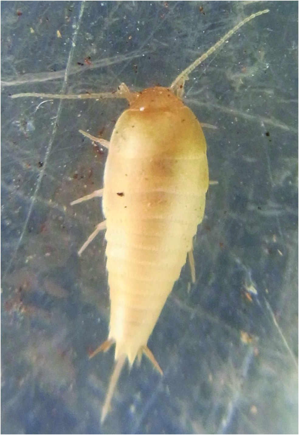
Redescription. Appearance. Pale gold/ochre colour when live ( Fig. 1 View Figure 1 ), becoming off-white in alcohol; lacking pigment. Ateluroid (tear-drop shape) tapering uniformly posteriorly, about 2½ times longer than wide ( Fig. 2 View Figures 2–18 ), head hypognathous ( Fig. 3 View Figures 2–18 ).
Body length. Small species, H+B 4.0 mm in largest specimen available, range of HW 0.53–0.83 mm; antennae up to just over one half H+B; cerci 0.14–0.21 H+B; median dorsal appendage only complete in one specimen (K261027) one third H+B.
Scales. Mostly rounded or pointed apically, multiradiate with about 11–23 rays, the rays on dorsal scales not or only slightly extending beyond the margins but on the urosternites distally free for about one tenth their length ( Figs 4 and 5 View Figures 2–18 respectively). Scales cover surface of all tergites and sternites (including the subgenital plate) and a few scales are present on the coxae of all legs; scales not present on the head and its appendages and the legs (except for a few on the coxae), the paramera, terminal filaments and ovipositor.
Macrochaetae. Macrochaetae simple or apically bifurcated, those along posterior margin of tergites abiesiform ( Fig. 6 View Figures 2–18 ), about 55–90 microns in length in larger specimens and, on average, 1.20 times the length of adjacent scales (range 0.86–1.53).
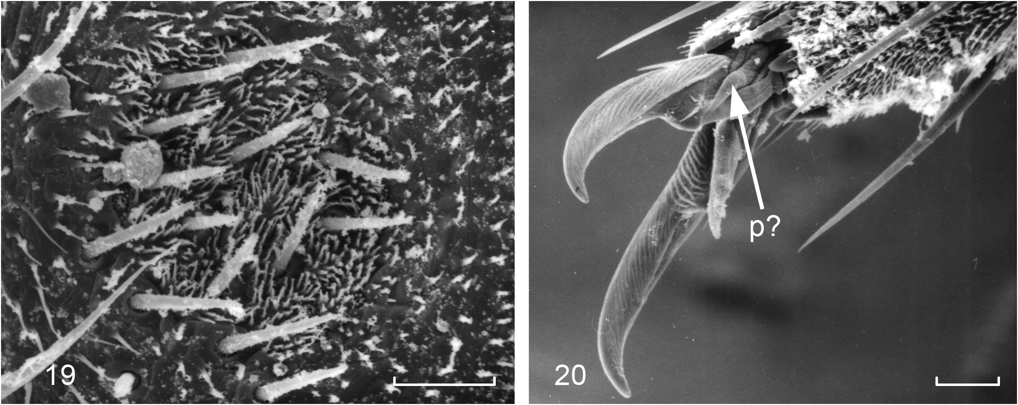
Head. More or less free, only slightly covered by prothorax at hind margin (possibly a preservation effect), vertex with scattered small, fine setae as well as long, fairly strong, minutely apically bifurcated setae arranged in four or five transverse, slightly irregular rows that become less distinct anteriorly where the setae become smaller and more numerous ( Fig. 7 View Figures 2–18 ). Antennae ( Figs 8, 9 View Figures 2–18 ) with 12 to 17 intervals in the flagellum; intervals/annuli from around the 6th to 9th begin to divide into two annuli, with the annuli becoming more distinct distally, and from the 10th to 13th interval the annuli further subdivide; scape with rosette of small setae apically, pedicel ( Fig. 10 View Figures 2–18 ) with an apical macrochaeta longer and stronger than other subapical setae and several shorter macrochaetae; pedicel of adult male with a shallow fovea with about 15 to 25 short setulae ( Figs 10 View Figures 2–18 , 19 View Figures 19–20 ) within or on the margins of the depression; first annulus/interval of flagellum with eight to ten trichobothria in a consistent pattern (see remarks regarding variability of antennae), subsequent intervals with two trichobothria until 8th to 13th and beyond which only a single subapical trichobothrium is present on the most distal annulus of each interval, except on the ultimate annulus which has an terminal papilla ( Fig. 11 View Figures 2–18 ); the ultimate interval may have either two or four annuli. —Labrum with scattered setae, those proximal stronger than those distal. —Mandibles ( Fig. 12 View Figures 2–18 ) with well-developed incisor and molar regions. —Maxillae ( Fig. 13 View Figures 2–18 ) with lacinia about the same length as the galea (although the galea seems to be shrunken in the type material); lacinia with simple pointed apex, pectinate prostheca not or only slightly extending beyond apex of lacinia; galea with single prominent apical conule ( Fig. 14 View Figures 2–18 ). Maxillary palp stout, with two or three feathered papilla distally ( Fig. 15 View Figures 2–18 ); apical article four times longer than wide with sub-parallel sides (range 2.9–5.8) and 1.45 times longer (range 1.14–1.70) than penultimate article and only slightly thinner. Labium with widely rounded posterolateral margins ( Fig. 16 View Figures 2–18 ); ultimate article of palp ( Fig. 17 View Figures 2–18 ) truncated ovate about 1.63 times as long as wide (range 1.22–2.14) with six sensory papillae distally; remaining articles including base with several stronger, apically bifurcate setae.
Thorax. Large ( Fig. 18 View Figures 2–18 ), about 0.43 H+B (range 0.37–0.48), nota not completely enclosing the legs; submarginal posterior row of 19–24 subequally spaced, abiesiform macrochaetae with about ¼–⅓ their length overlapping the posterior margins of the tergites; lateral margins of nota with curved setae, the macrochaeta on each of the posterior angles of the nota about twice as long as the abiesiform setae; disc of nota with some scattered short, delicate setulae among the scales. Pronotum longer than each of the meso- and metanota but not as long as both together.
Legs typical for tribe ( Fig. 21 View Figures 21–36 ), tibia L/W ratio of legs PI 3.7 (range 2.9–4.6), PII 3.5 (3.2–3.7), PIII 3.7 (2.9–4.2); tarsi L/W ratio PI 6.4 (4.8–7.6), PII 7.1 (5.6–8.2), PIII 8.8 (7.5–10.8). Legs becoming progressively longer posteriorly with the tarsus increasing in length more than tibia. Presternum of prothorax with several setae, sternum between coxae with 2+2 long setae; sterna of meso- and metathoracic segments between the coxa with just 1+1 medial setae. Coxa large and flat with some scales; femur with one strong, narrow deeply bifurcated sub-lyriform macrochaeta sub-distally on anterior edge and two thicker longer macrochaetae on angle of ventral/posterior margin as well as many setae over the posterior half of the ventral surface; tibia covered with numerous setae, three distal lyriform macrochaetae ( Fig. 22 View Figures 21–36 ), two stronger spines near ventral apex and some stronger spines distally as well as two strong spines about one third the distance along the tibia, one almost on the posterior/ventral margin, the other more anteriorly on the face of the tibia; tarsus of four articles; pretarsus with two simple, curved, smooth lateral claws and a sharp erect medial empodial claw, pulvillae indistinct, possibly present as small fold externally at the base of the lateral claws ( Fig. 21 View Figures 21–36 ).
Abdomen. Tergites ( Figs 23, 24 View Figures 21–36 ) with submarginal abiesiform macrochaetae similar to thorax, diminishing in number as the segments become narrower, long marginal macrochaetae in each posterolateral corner and four or five setae along the paratergites which fold under the sides of the abdomen ( Figs 23, 25, 26 View Figures 21–36 ); posterolateral corners of urotergite IX produced into subtriangular posterolateral lobes ( Fig. 27 View Figures 21–36 ) with a long macrochaeta in each posterior corner ( Fig. 28 View Figures 21–36 ). Urotergite X with strong 1+1 macrochaetae on the acute apices separated by deep incision, which in both sexes, generally has a distinctly curved medial “corner”, and one or two smaller apically bifurcated setae along inner and a few setae along the outer margins ( Fig. 29 View Figures 21–36 ), underside of urotergite X of males with 1+1 elongated fields of about 20 (range 14–24) modified sclerotised setae (sensory pegs) ( Figs 30–32 View Figures 21–36 ). Urosternites ( Fig. 33 View Figures 21–36 ) I glabrous, II with two very small setae medially on slightly produced margin as well as 2–3 setae on posterolateral margins, III with small medial styli, IV and V with small lateral styli and 1+1 submedial erect macrochaetae plus a few smaller setae as well as four or five setae on posterolateral corners; VI with 1+1 erect submedial macrochaetae and smaller setae, large eversible vesicles with about nine setae on vesicle ( Figs 34, 35 View Figures 21–36 ), lateral styli and lateral macrochaetae; VII similar except the vesicles are pseudovesicles, VIII in male narrower with more protruding posterior margin armed with eight or nine setae between the styli, also with two or three lateral setae ( Fig. 36 View Figures 21–36 ); IX divided into separate coxites ( Fig. 37 View Figures 37–41 ), larger styli (almost twice as long as those on other segments) and long subcylindrical parameres (about 2.6–3.5 times longer than wide) reaching to about half way along the styli; 1+1 setae laterad to the base of each paramere (near base of paramere between paramere and the stylus insertion). Penis with wide longitudinal opening surrounded by small setae and what appears to be large glands.
Cerci ( Fig. 30 View Figures 21–36 ) with eight or nine divisions all longer than wide, basal division not significantly longer than rest, proximal divisions in mature males, medially with stout, pigmented, apically rounded, modified spines (sensory pegs) similar to and in close proximity to those on underside of urotergite X ( Figs 30, 31 View Figures 21–36 , 38. 39 View Figures 37–41 ), the first division with one medium sized peg subapically and rarely an additional smaller peg, second division usually with two larger pegs, but occasionally with an additional smaller peg or in one case a third large peg and the third division with no or just one smaller peg in the proximal third, often only on one cercus and not the other; divisions from fourth divided into two annuli; all divisions with macrochaetae larger and more numerous on laterad surface, trichobothria as shown in Figs 30, 31 View Figures 21–36 , 38 and 39 View Figures 37–41 . —Median dorsal appendage also slender, about twice length of cerci with nine divisions, divided into annuli from the fourth or fifth division ( Fig. 40 View Figures 37–41 ), without pegs.
Female. Same as male except—pedicel lacking fovea, urosternite VIII divided into separate coxites and subtriangular subgenital plate ( Fig. 33 View Figures 21–36 ); ovipositor ( Figs 33 View Figures 21–36 , 41 View Figures 37–41 ) moderately bulbous with about eight to ten indistinct divisions, extending just beyond the end of stylus IX, ultimate division of anterior gonapophyses with two long setae, penultimate division with field of small hooked setae, ultimate division of posterior gonapophyses with distinct subtriangular pointed process.
Juvenile stages. One ♂ (HW 0.56) (K377691) had two very weak pegs developing on the cerci and three pegs beneath urotergite X, while another ♂ (HW 0.55) (K377692) only had one peg on the underside of urotergite X. A juvenile male (HW 0.54) (K377683) had one thickened spine on a cercus but lacked any sensory pegs on the underside of urotergite X while three even smaller males (HW 0.50) (K377684, K377685 and K377690) had no obvious secondary sexual characters.
The valves of the ovipositor were just beginning to develop in one juvenile ♀ (HW 0.53) (K377703) and another (HW 0.58) (K377706), while another juvenile ♀ (HW 0.50) (K377686) showed no development of the ovipositor.
Biology. Panmyrmecophile, collected under stones usually with ants. They are often found walking upside down on the underside of the stone. Ant species include Amblyopone australis Erichson , Camponotus consobrinus (Erichson, 1842) , Camponotus nigriceps Smith, 1842 , Colobopsis gasseri F., Iridomyrmex sp. , Myrmecia esuriens F. Smith, 1858 , Myrmecia fulviculis Forel, 1913 , Rhytidoponera tasmaniensis Emery, 1911 , Rhytidoponera victoriae André, 1896 and Pheidole sp.
Remarks. Silvestri (1908a) erected the genus Atopatelura for the species furcifera collected in the nest of the ant Myrmicaria natalensis eumenoides (Gerstäcker, 1858) in the Democratic Republic of the Congo. It was distinguished from other genera by the presence of medial styli on urosternite III and by the armature (lyriform spines) on the tibia. In the same year, Silvestri (1908b) described Atopatelura hartmeyeri , At. michaelseni and At. kraepelini from south-west Western Australia. Stach (1935) described At. spinifera from Egypt. In 1949, shortly before his death, Silvestri described At. tasmanica from Hobart, Tasmania and At. perarmata from Eritrea. Paclt (1963) suggested that At. tasmanica may be a subspecies of At. kraepelini . Mendes (1995), in his review of the Israeli fauna, redefined the genus, splitting it into three genera, Atopatelura sensu stricto for At. furcifera , Arabiatelura for spinifera , perarmata and Ar. palaestinensis Mendes, 1995 from Israel and without having the opportunity to examine specimens, erected Australiatelura for the four Australian species based on Silvestri’s descriptions. He distinguished Australiatelura , at least partly, on the apparent absence of modified setae or pegs on the cerci and underside of urotergite X.
Silvestri made no reference to modified setae or sensory pegs on any of the Atelurinae he described from Australia, however male specimens collected by the author from eastern New South Wales and Tasmania all showed these pegs. The type specimens of Atopatelura tasmanica were kindly loaned by MUSA allowing confirmation that pegs are indeed present at the base of the cerci as well as beneath urotergite X of all mature males. A redefinition of Australiatelura Mendes would be in order however this should be made with reference to material of the type species of the genus, Atopatelura kraepelini . Apart from the modified spines on the cerci and urotergite X, the species of this genus differ from both Atopatelura and Arabiatelura by the lack of abundant lyriform spines on the legs. While lyriform spines are found on the femur and tibia of the Australian species, these are restricted to just one and three at the dorsal apices of the respective leg segments.
The type material of Atopatelura tasmanica , while mostly in fair condition, has shown some deterioration over the years. The resin used for the slide material has dried out and cracked on several slides allowing air under the coverslip and causing distortion to some parts and there is some level of fungal growth. The material in alcohol is variable. Two specimens are completely shrivelled and unusable, the remainder are intact but the surface of the insects has taken on a granular powdery appearance making it difficult to observe clearly.
Details of the antennae were often used by Silvestri and others when describing species of the subfamily Atelurinae . This is the only subfamily in the Zygentoma where the antennae are relatively short, where distinct intervals and annuli can be counted and the completeness of the antenna confirmed by the presence of a unique terminal trifurcate papilla. Various authors use the chaetotaxy of the scape and pedicel, the presence of secondary sexual modifications on the pedicel in the male (e.g., fovea or apophyses), the number of antennal intervals, as well as the number and arrangement of trichobothria on all annuli, especially the first annulus of the flagellum, and the subdivision of more distal intervals. There are however limitations to the interpretation of these characters as follows:
Trichobothria on first annulus of flagellum. Counting the number of trichobothria on the first annulus can be complicated by the orientation of the antenna on the slide because trichobothria on the lateral margins can be difficult to distinguish especially if the long hair has been lost. More importantly, the delineation between the first and second annuli is often very difficult to discern and in the majority of cases no suture was obvious between the presumed annuli giving the impression that there are ten trichobothria on the annulus. The two most apical trichobothria do however spirally align with the pattern of trichobothria on the following annuli and their alignment with a second partial rosette of setae on the face away from the trichobothria suggests that the most distal trichobothria, when more than eight are counted, belong in fact to the second annulus of the flagellum. Otherwise the arrangement of the trichobothria seems to be consistent.
Number of antennal intervals. There was quite some variation in the number of intervals in “complete” flagellum with as few as 12.5 and up to 17 intervals counted. The apical annulus with its distinct sensilla was counted as a half interval if there was a trichobothrium at the apex of the adjacent annulus or partially divided annulus. There was also a difference between the left and right flagella of some specimens (for example, 16 vs 17). It is possible that such anomalies are the result of damage during previous instars, but does make it difficult to use the number of flagella intervals as a taxonomic character.
Subdivision of intervals and number of trichobothria. While there was an overall pattern there were some differences in the detail. A division into two annuli was generally visible by the sixth to the ninth interval of the flagellum; but there is a degree of subjectivity based on the interpretation of the limits of the first and second annuli and the criteria used for deciding when a subdivision was obvious. A further subdivision of the now distinct intervals into partially divided annuli was visible by the 10th to 13th interval, but the secondary subdivisions remained less distinct than the primary subdivsions. Differences were also noted between the left and right flagella on some individuals.
Secondary sexual characters of the male. There are 1+1 fields of sensory pegs on the underside of urotergite X in mature males. The number can vary between left and right sides of an individual and between individuals but had a roughly similar pattern. Numbers of pegs varied between about 14 and 27 and there was no reliable correlation between specimen size and the number of pegs. There are also pegs on the inner ventral aspect of basal divisions of the cerci. In nearly all cases there was just a large single peg on the first division but in one case there was a much smaller ancillary peg at the same level more dorsally, but only on one cercus. On the second division there were generally two large pegs and often one or two smaller ancillary pegs; in one case there were three large pegs and no ancillary pegs on one of the cerci. On about half the specimens examined, the third division had a small peg on just one cercus. While there seems to be real differences between the species, the variation within a species needs to be taken into account.
Australiatelura tasmanica seems to be most closely related to Australiatelura hartmeyeri (Silvestri) from WA. It is the only other described species with fairly narrow subcylindrical paramera in the male but it has quite long abiesiform macrochaetae on the thoracic and abdominal terga. There is also another “species” commonly found under stones or in soil with ants around Sydney, NSW which also has narrow subcylindrical paramera. While it was initially thought these were the same species, more detailed measurement data suggest the Sydney “species” is generally larger with shorter abiesiform macrochaetae, with some small differences in the arrangement of the secondary sexual characters on the cerci and possibly more trichobothria on the first annulus of the antennae. These differences need to be investigated in more detail.


No known copyright restrictions apply. See Agosti, D., Egloff, W., 2009. Taxonomic information exchange and copyright: the Plazi approach. BMC Research Notes 2009, 2:53 for further explanation.
|
Kingdom |
|
|
Phylum |
|
|
Class |
|
|
Order |
|
|
Family |
|
|
Genus |
Australiatelura tasmanica ( Silvestri, 1949 )
| Smith, Graeme B. 2016 |
Australiatelura tasmanica (Silvestri)
| Mendes, L 1995: 98 |
Atopatelura tasmanica
| Silvestri, F 1949: 35 |