
Balacra (Daphaenisca) daphaena ( Hampson, 1898 )
|
publication ID |
https://doi.org/ 10.11646/zootaxa.4117.2.1 |
|
publication LSID |
lsid:zoobank.org:pub:957C0385-71CE-439A-B541-4453D148EDD1 |
|
DOI |
https://doi.org/10.5281/zenodo.5621327 |
|
persistent identifier |
https://treatment.plazi.org/id/5D21CB45-FF82-4857-FF12-4B65FCCCFD53 |
|
treatment provided by |
Plazi |
|
scientific name |
Balacra (Daphaenisca) daphaena ( Hampson, 1898 ) |
| status |
|
Balacra (Daphaenisca) daphaena ( Hampson, 1898)
( Figs. 5, 6 View FIGURES 5 – 7 , 8–15 View FIGURES 8 – 15 , 20 View FIGURE 20 , 21 View FIGURE 21 )
Pseudapiconoma daphaena Hampson, 1898 , Catalogue of the Lepidoptera Phalaenae View in CoL , vol. 1: 150. Balacra (Daphaenisca) daphaena ( Hampson, 1898) , Kiriakoff 1953, Annales du Musée Royal du Congo Belge, vol. 26: 69– 70.
Balacra (Daphaenisca) daphaena ( Hampson, 1898) , Przybyłowicz 2009, Thyretini of Africa: 13.
Type locality. Nigeria.
Distribution. Cameroon, Nigeria ( Przybyłowicz 2009).
Examined material. Holotype ♂, R. Niger, Asaba [ Nigeria], in coll. BMNH, examined (genitalia not dissected).
Nigeria: 4♂, genitalia prep. n. 740 MAD, in coll. MSNS; 1 ♂ Arct. g.sl. n. 5852 BMNH, in coll. BMNH; 1 ♀, genitalia prep. n. 749 MAD, in coll. MSNS.
Diagnosis. Very similar to affinis and inexpectata sp. n., from which it is distinguishable with certainty with reference to the genital apparatus, the uncus shape being enough to separate affinis and daphaena (trifurcate and bifurcate respectively); it is also adequately diagnostic with respect to inexpectata sp. n., because the bifurcation has a clearly different shape. Moreover they can easily be separated with reference to the general shape of the valva and the saccus, and above all the big cornutus of the aedeagus in daphaena versus the cluster of spines in inexpectata sp. n.
In the female genitalia, the corpus bursae presents two long signa instead of the single one in inexpectata sp. n.; female genitalia of affinis have been not examined, but they are clearly different in the longer ductus missing the stiff knot-like swelling.
It should also be remarked that daphaena presents a much less marked reddish colouration.
Two yellow marks on the third and fourth abdominal tergites, as well as the serrate antennae and the yellow patagia, are clearly distinctive with respect to magnoloi sp. n; the shape and colour of the hindwings (grey-brown) are diagnostic with respect to flava sp. n. (yellow) and magnoloi sp. n. (roughly circular with ground colour darker and red dots or small bands in the middle).
Habitus. ♂ and ♀. Forewing length: male from 15 to 16 mm (n=5), female 17.5 mm (n=1); upperside ground colour dark grey-brown with reduced or absent red scales. Variable presence of thin, incomplete subcostal red line (sometimes obsolescent), with red dot at wing base on inner margin. Forewing underside same colour as upperside, fading towards base.
Hindwings slightly elongated (clearly elongated in the female) with upperside ground colour as in forewings, fading towards base. Underside as upperside with red suffusion along costa, in the female two small stripes on posterior half of wing base.
Fringes same colour as wings.
Frontoclypeus grey-brown with red lateral margin; vertex yellow, bordered by red. Labial palpi dorsally greybrown, ventrally red, with yellow tip. Eyes bordered by red and small number of yellow hairy scales; antennae yellow with red scales around scapus, smoothly serrate, thinner in the female with terminal third greyish.
Thorax same ground colour as wings, with four red marks on mesoscutum, one central yellow mark on mesoscutellum, two more red lateral marks on metathorax; patagia yellow; tegulae grey-brown with small red dot anteriorly at junction with costal margin of forewings. Thorax underside grey-brown.
Legs with red femurs, yellow tibiae and tarsi, brown praetarsi. Spurs formula 0-2-2.
Abdomen same ground colour as wings (densely coloured with blue scales in the female), with two falcate red marks on tergum A1 and small red dot between them (sometimes obsolescent). One yellow mark on both A2 and A3, sometimes with sparse red scales on anterior edge (clearly bordered by red in the female); starting from second urite, only lateral sides of tergites covered by red scales under typically grey-brown layer, red scales emerging only at caudal edge of tergites, surrounding them. The state of this latter character differs from its equivalent in affinis (see description), having been observed only in the three non-dissected males. If confirmed by more material, it could acquire a definitive diagnostic value with respect to affinis .
A8 tergite completely red (in one male, with many yellow scales).
Presence of pleural tufts on every segment.
Underside of abdomen reddish with sparse grey-brown groups of scales, mainly at caudal edge of segments; in the female, presence of large longitudinal median grey-brown fascia.
Anal tuft reddish or grey-brown (even the colour of the anal tuft could be of diagnostic relevance); no tuft in the female.
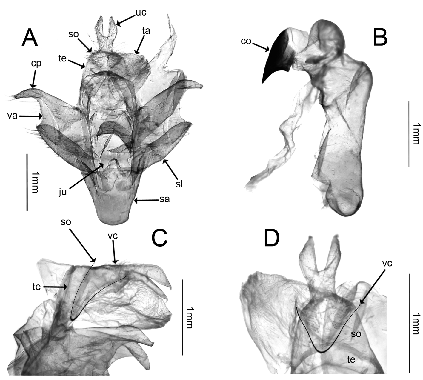
Genitalia. ♂. A8 tergum roughly triangular in dorsal view, almost twice as long as A7. Uncus Y-shaped, in dorsal view extending inside tegumen with V-shaped concavity ( Fig. 20 View FIGURE 20 D), whose arms are considered socii (probably derived from the tenth tergite as the most caudal part of the tegumen, roughly triangular in lateral view, Fig. 20 View FIGURE 20 C). Tegumen-vinculum complex quite strong; saccus large, not very deep, U-shaped. Juxta roughly Xshaped with a central dome, its dorsal arms connected with band-like transtilla, with dorsal corners extending to proximal corners of valvar costa. Tuba analis membranous, quite stiff, large, frustoconical.
Valvae roughly square with clear distal finger-like costal process and pseudosaccus; large membranous area in middle. Sacculus with inner margin folded on itself.
Aedeagus tubular, quite stout with short coecum; vesica with three main lobes: one proximal lobe prolonged in ductus ejaculatorius, two distal subspherical lobes, caudal one bearing small lobe, anterior one bearing large thornlike cornutus.
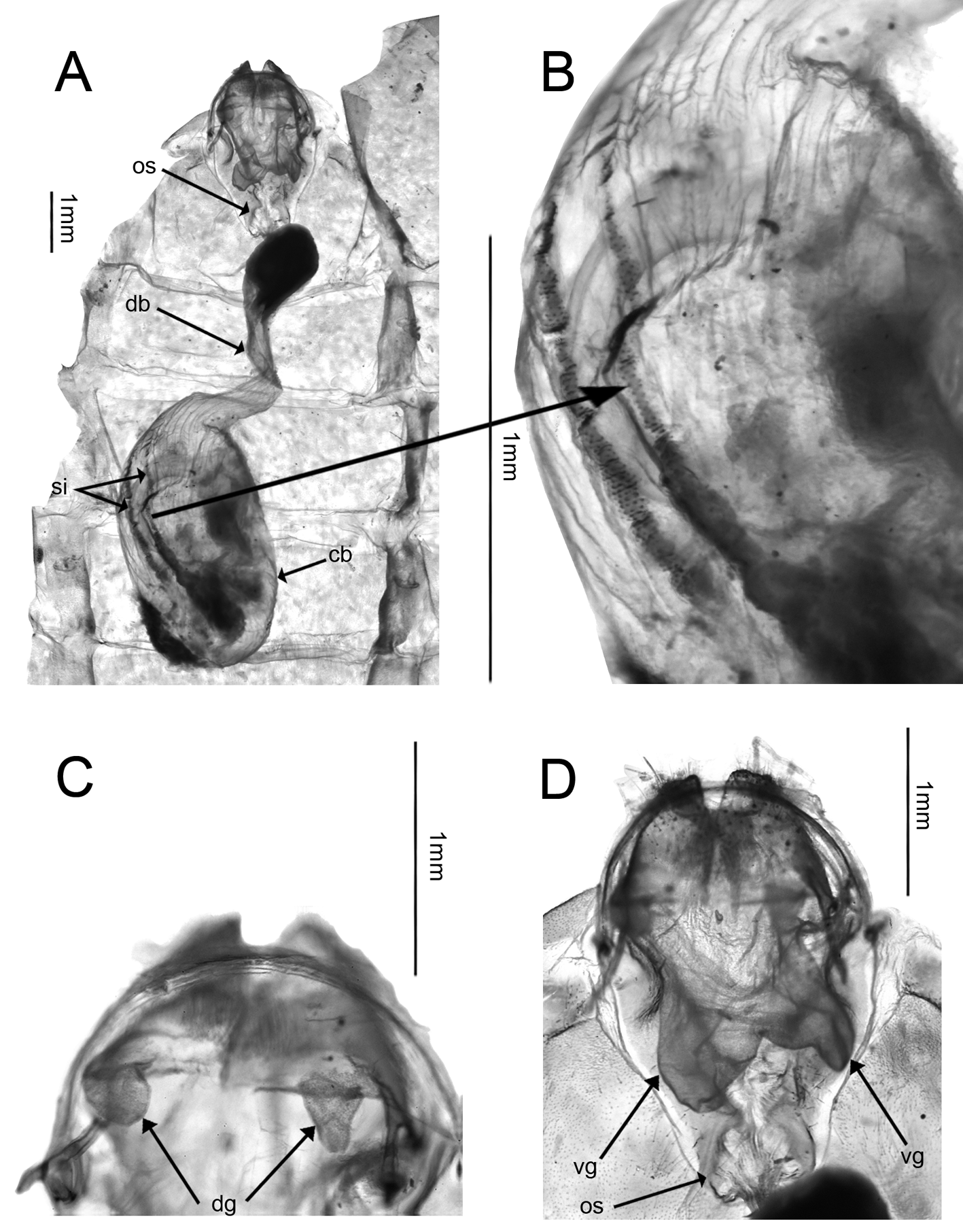
♀. Seventh tergum about twice as long as sixth segment; seventh sternum slightly shorter than seventh tergum. A8 very short, tergum strip-like, sternum about one third shorter than sixth segment. Anterior and posterior apophyses short, anterior ones are half length of posterior ones, which are almost as long as papillae anales. Dorsal pheromone glands ( Fig. 21 View FIGURE 21 C) paired and pouch-like, as short as anterior apophyses. Ventral pheromone glands ( Fig. 21 View FIGURE 21 D) unpaired and double-sack shaped, as long as papillae anales.
Ostium bursae roughly circular, surrounded by sclerotised edges except for caudal membranous duct ( Fig. 21 View FIGURE 21 D). No lamellae. Antrum very short and narrow, membranous; ductus bursae membranous, S-shaped, with stiff knot-like swelling at caudal end. Corpus bursae ovoidal, membranous, with two long parallel signa ( Fig. 21 View FIGURE 21 B).
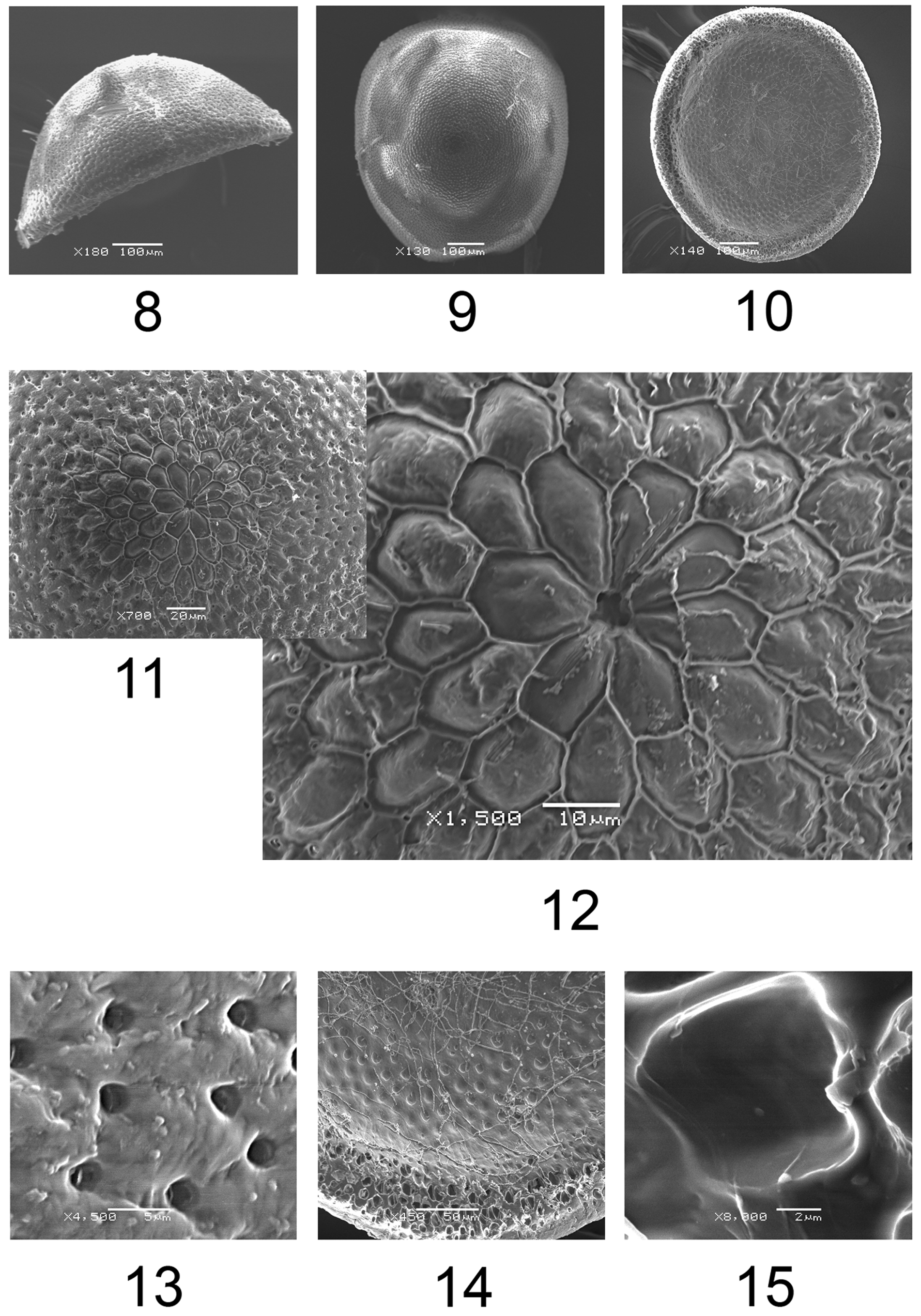
Eggs. Upright type, dome-shaped with base slightly oval, very pale yellow, height about 0.243 mm, diameters at base 0.651 and 0.593 mm ( Figs. 8, 9, 10 View FIGURES 8 – 15 ). Cellular-type exochorion sculpture only on micropylar and transitional areas ( Fig. 11 View FIGURES 8 – 15 ); remaining lateral egg surface orange peel-like with densely disposed aeropyles. Micropylar area represented by three rows of well-marked polygonal cells. Transitional area with one or two rows of polygonal cells or only their fragments, with few roughly circular aeropyles extended more densely on rest of egg surface ( Fig. 13 View FIGURES 8 – 15 ). Micropylar rosette ( Fig. 12 View FIGURES 8 – 15 ) with seven polygonal cells, 1.5–2 times longer than wide, joined along about 2/3 of their length. Central portion of daisy-like rosette about 0.05 mm in diameter and with 5 micropylar openings. Base ( Figs. 10, 14 View FIGURES 8 – 15 ) with raised marginal edge and slightly depressed internal surface; edge with cup-shaped structures with probable adhesive function ( Fig. 15 View FIGURES 8 – 15 ); internal surface with large distal fascia (about 0.08 mm) with small shallow roughly circular pits, and smooth central area.
| MAD |
Madras Museum |
No known copyright restrictions apply. See Agosti, D., Egloff, W., 2009. Taxonomic information exchange and copyright: the Plazi approach. BMC Research Notes 2009, 2:53 for further explanation.
|
Kingdom |
|
|
Phylum |
|
|
Class |
|
|
Order |
|
|
Family |
|
|
Genus |
Balacra (Daphaenisca) daphaena ( Hampson, 1898 )
| Durante, Antonio & Zangrilli, Maria Paola 2016 |
Pseudapiconoma daphaena
| Hampson 1898 |
Balacra (Daphaenisca) daphaena (
| Hampson 1898 |
Balacra (Daphaenisca) daphaena (
| Hampson 1898 |