
Brachyrhynchus triplostylis Artois & Schockaert
|
publication ID |
https://doi.org/ 10.11646/zootaxa.3635.2.3 |
|
publication LSID |
lsid:zoobank.org:pub:7D2BD85D-4E5E-4E70-9995-06845DC14108 |
|
DOI |
https://doi.org/10.5281/zenodo.5683953 |
|
persistent identifier |
https://treatment.plazi.org/id/03A6F45B-FFB8-FFBA-2482-C9592DCEB08E |
|
treatment provided by |
Plazi |
|
scientific name |
Brachyrhynchus triplostylis Artois & Schockaert |
| status |
sp. nov. |
Brachyrhynchus triplostylis Artois & Schockaert n. sp.
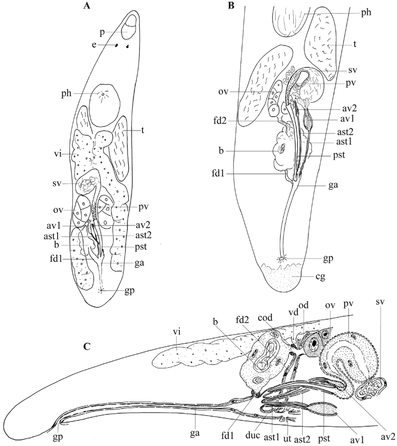
( Figs 1 View FIGURE 1 B–C, 2, 3A, 4B)
Localities. Italy, Sardinia, Stintino, Punta Negra, 40°57’11.6”N, 8°13’43.2”E, on algae from rocks, partially shaded, 0.5 m deep, 19 August 1994 (TYPE LOCALITY); Alghero, Calabona beach, 40°32’38.9”N, 8°19’13.1”E, on algae from rocks in a small bay, 28 March 2010.
Studied material. Several animals studied alive, seven whole mounts, one chosen as holotype (SMNH, no. 7834), the others voucher specimens (HU, nos VI.1.34–VI.1.39). Two serially-sectioned specimens designated paratypes (HU, nos 539–540).
Etymology. The species epithet refers to the presence of three stylets.
Description. Long, slender and rather slow animals. They are about 0.8 mm long (measured on whole mounts), unpigmented and have two eyes. The epidermis is syncytial, containing numerous optically-empty vacuoles and flattened, lobate nuclei. The epithelium is about 3 µm high with cilia about 4 µm long. In the apical part of the epithelium there are many globular rhabdites, which are only about 1/10–1/15 of the epithelium height in diameter.
The proboscis closely resembles that of Phonorhynchoides somaliensis Schockaert, 1971 (for a detailed description, see Schockaert 1971). It is extremely small, only about 1/20 to 1/12 of the body length long, with an indistinct apex. The proboscis sheath is rather short and covered with a high epithelium containing two nuclei. It is surrounded by longitudinal muscles only. There are no nuclei at the junction of the sheath and cone epithelia. The number of protractor muscles could not be determined. The internal circular muscle layer of the bulb is very thin.
Two pairs of integument retractors are present: one dorsal and one ventral pair, the former only weakly developed. There are three pairs of proboscis retractors: a latero-dorsal, a lateral and a ventrolateral pair. At each side of the body and caudally from the brain, the lateral and dorsolateral retractors fuse.
The pharynx is situated in the first body-half and is slightly inclined forwards. The prepharyngeal cavity is lined with a very low anucleated epithelium. The pharynx bulb is of the normal polycystidid construction with four sclerotized teeth around the proximal pharyngeal opening (for a detailed description, see Meixner 1925). The pharynx has 24 internal longitudinal muscles.
The gonads are paired. Testes ( Fig. 1 View FIGURE 1 B: t) lie dorsally at each side of the body and extend from the caudal end of the pharynx to the level of the seminal vesicle and the ovaries. In sectioned material they are situated completely behind the pharynx. The ovoid ovaries ( Figs 1 View FIGURE 1 B–C; 2: ov) are relatively large, and are situated on each side of the prostate vesicle. Both vitellaria ( Fig. 1 View FIGURE 1 C: vi) are situated dorsally and extend on each side of the body from the caudal end of the pharynx to the level of the common genital atrium. Just behind the pharynx they lie very near to each other.
The common genital pore ( Fig. 1 View FIGURE 1 B–C: gp) is situated ventrally, subterminally, at about 95%. A long and narrow common genital atrium ( Figs 1 View FIGURE 1 B–C; 2: ga) runs straight rostrally from this pore, broadening proximally, where it receives the uterus and the male and female atrial systems. It is lined with a very low, nucleated epithelium, which is degenerated at some places, leaving isolated spots of bare basement membrane (pseudocuticula), and it is surrounded by a thin longitudinal muscle layer.
The copulatory organ is of the conjuncta-simplex type (terminology of Karling 1956). The large unpaired seminal vesicle ( Fig. 1 View FIGURE 1 B–C: sv) is more or less globular and is lined with a flat, nucleated epithelium. The seminal duct enters the interposed prostate vesicle (of type IV, see Artois & Schockaert 2003) ( Fig. 1 View FIGURE 1 B–C: pv) through a muscular pore and continues axially through this vesicle. The prostate vesicle is surrounded by relatively thick, spirally-running, almost circular muscles. Distally the prostate vesicle narrows to a long ejaculatory duct that enters the single-walled prostate stylet (of type IV, see Artois & Schockaert 2003) ( Figs 1 View FIGURE 1 B–C; 2; 3A; 4B: pst). This stylet is 139–192 µm long (x = 157 µm; n = 6), single-walled and has a blunt end. It lies in the narrow male atrium, which has no visible epithelium and is surrounded by weak longitudinal muscles only. The male atrium enters the common atrium dorso-anteriorly.
Apart from the sperm-conducting stylet described above, there are two accessory stylets in the male system, each connected to its own glandular vesicle. The first accessory stylet is 63–79 µm long (x = 73 µm, n = 6) ( Figs 1 View FIGURE 1 B–C; 2; 3A; 4B: ast1). It lies in a narrow duct without a visible epithelium and is surrounded by longitudinal muscles only. This stylet is connected to an ovoid glandular vesicle, which is surrounded by spirally-running muscles ( Fig. 1 View FIGURE 1 B–C: av1). It contains two kinds of eosinophilic secretion, the darker of which lies peripherally. The nucleated parts of these glands are extracapsular. The second accessory stylet is 75–130 µm long (x = 100 µm; n = 6) ( Figs 1 View FIGURE 1 B–C; 2; 3A; 4B: ast2), also situated in a duct without a visible epithelium and surrounded by weak longitudinal muscles. This stylet is connected to a small pyriform glandular vesicle ( Figs 1 View FIGURE 1 B–C; 3A; 4B: av2), which is surrounded by a more or less circular muscle sheath, which forms a sphincter at the proximal end of the stylet. The vesicle contains a pale basophilic secretion. The nucleated parts of the secretory glands are (probably) extracapsular. Three different stylet-to-stylet ratios can be calculated: 1) (length of the first accessory stylet / length of the prostate stylet) x 100 = α = 40–52% (x = 47%; n = 6), 2) (length of the second accessory stylet / length of the prostate stylet) x 100 = β = 54–68% (x = 63%; n = 6) and 3) (length of second accessory stylet / length of first accessory stylet) x 100 = γ = 119–168 % (x = 135%; n = 6).
All three stylets lie very close to each other, ventral to the female genital organs. They enter through the anterior wall of the common genital atrium, latero-dorsally at the right hand side. At the place where the stylets enter, the epithelium of the common genital atrium is reduced to a pseudocuticula. The first accessory stylet lies most ventrally; the prostate stylet lies at the left from the second accessory stylet.
The oviducts are short and broad ( Figs 1 View FIGURE 1 C; 2: od) and surrounded by thin longitudinal muscles. Distally they join to form the female duct of type II (terminology of Artois & Schockaert 2005) ( Figs 1 View FIGURE 1 B–C; 2: fd2), which runs straight in the ventro-caudal direction towards the atrium. The epithelium of this female duct is high and nucleated and surrounded by weak longitudinal muscles. Just before the female duct opens into the antero-dorsal wall of the genital atrium, it narrows and receives the uterus ( Figs 1 View FIGURE 1 C; 2: ut), which is of the normal polycystidid type (for a detailed description see Meixner 1923, 1925), and continues towards the common genital atrium as a ductus uterocommunis ( Figs 1 View FIGURE 1 B–C; 2: duc). Eosinophilic uterine glands open into the uterus just at its junction with the female duct. The bursa ( Figs 1 View FIGURE 1 C; 2: b) is situated caudally and is connected with the common genital atrium through a short and broad bursal stalk, which actually is a female duct of type I (terminology of Artois & Schockaert 2005) ( Figs 1 View FIGURE 1 B–C; 2: fd1). This stalk departs from the ventro-caudal wall of the bursa and enters the genital atrium dorsally. It is surrounded by a very thick, circular muscle sheath and lined with a pseudociliation. Two very short muscular ducts depart from the anterior wall of the bursa towards each of the oviducts, representing a double common oviduct (terminology of Artois & Schockaert 2005) ( Figs 1 View FIGURE 1 C; 2: cod). The epithelium of these ducts is high and nucleated, with a thick basement membrane. A circular muscle sheath surrounds both ducts and extends over the bursal wall, on which it thins out.
Diagnosis. Species of Brachyrhynchus with a bluntly-ending prostate stylet type IV, 139–192 mm long. Accessory stylets 63–79 µm and 75–130 µm long. Stylet ratios (symbols: see description): α = 40–52%, β = 54–68% and γ = 119–168%.
No known copyright restrictions apply. See Agosti, D., Egloff, W., 2009. Taxonomic information exchange and copyright: the Plazi approach. BMC Research Notes 2009, 2:53 for further explanation.