
Caloptilia aesculi, Liao & Ohshima & Huang, 2019
|
publication ID |
https://doi.org/ 10.11646/zootaxa.4586.3.13 |
|
publication LSID |
lsid:zoobank.org:pub:2E6B0959-E391-40BC-96C7-A1A53316E531 |
|
persistent identifier |
https://treatment.plazi.org/id/9CF8C4FB-280F-4B72-847B-9D0A3A28A00F |
|
taxon LSID |
lsid:zoobank.org:act:9CF8C4FB-280F-4B72-847B-9D0A3A28A00F |
|
treatment provided by |
Plazi |
|
scientific name |
Caloptilia aesculi |
| status |
sp. nov. |
Caloptilia aesculi View in CoL sp. nov.
http://zoobank.org/ 9CF8C4FB-280F-4B72-847B-9D0A3A28A00F
Diagnosis. This species can be distinguished by its forewing pattern with conspicuous yellow fascia on the dark brown to black background ( Fig. 1A, B View FIGURE 1 ). Male genitalia of this species is somewhat similar to C. fidella (Reutti) and C. celtidis Kumata , as having a row of cornuti in phallus but differs in the number of cornuti (15 to 20) from the two species (11 or 12 in C. fidella and 5 or 6 C. celtidis ).
Description. Adult ( Figs. 1A, B View FIGURE 1 ). Wingspan 8.7–10.8 mm.
Head: Vertex covered with appressed, dark brown scales; face with bright yellow scales (but specimens of Badagongshan with yellowish scales as Fig. 1B View FIGURE 1 ). Antenna about 6 mm length, slightly longer than forewing (about 5.3 mm); scape slightly stout; pedicel short, wider than length; flagellum slender, more than 70 segments, dark brown and yellow scales to alternate with each segment, first flagellomere distinct extended, about twice as long as other flagellomeres. Maxillary palpus dark brown, mixed some yellow scales; four-segmented, segmental ratio about 1:1.5:3:3. Galeae very long, well developed, about 3 times as long as labial palpus. Labial palpus yellow dorsally and black ventrally, with 3 segments, segmental ratio about 1:4:4; second segment distinct concave; third segment slightly curved, sharp-pointed apically.
Thorax: Tegula and thorax covered with dark brownish or black scales. Foreleg and midleg covered with black scales mixed brownish scales at base of each tarsus; hindleg covered with yellow scales mixed dark brown scales at apex of each tarsus. Forewing lanceolate ( Figs. 1A, B View FIGURE 1 ), maximum width about 0.15× as long as length; ground color dark brown to black, with the following bright yellow and brownish markings: an oblique brownish spot near wing base reaching posterior margin; a parallelogram-shaped transverse yellow spot arising from basal 1/4 of anterior margin outward-inclined laterally, and connecting with a brownish spot reaching posterior margin; a small brownish spot placed at the middle of posterior margin; a triangular yellow spot placed at near midpoint of anterior margin, the inner angle reaching half width position; a small brownish spot placed at apical 1/3 of posterior margin; a small yellow spot placed at apical 1/4 of anterior margin; distal part with two very small yellow spot. Fringe fuscous, longest fringe placed at apical 1/3 of posterior margin, about 1.5× as long as width of forewing. Venation with Rs weak on basal part just beyond the point where the vein R 1 branch off, M 2 and M 3 short-stalked, CuP wholly weak as a fold, R 2 branches from the cell more basal than the CuA 2, 1A+2A simple, fused for entire length ( Fig. 1C View FIGURE 1 ). Hindwing lanceolate, basal 1/4 approximately equal in width, then gradually narrowing, maximum width about 0.1× as long as length; ground color fuscous but at apex black, with fuscous fringe except apex of hindwing bearing black fringe. Sc+R 1 parallel to costal margin, R 2+3 short and parallel to Sc+R 1, R 4+5 nearly reaching to apex of hindwing, M 1 and M 2 stalked with almost absent base, cell between M 2 and M 3 open, M 3 fused with CuA 1 basally, A present ( Fig. 1C View FIGURE 1 ).
Abdomen: Dorsally covered with dark brown and black scales, ventrally with yellow to bright yellow scales, except apex with brown slender scales.
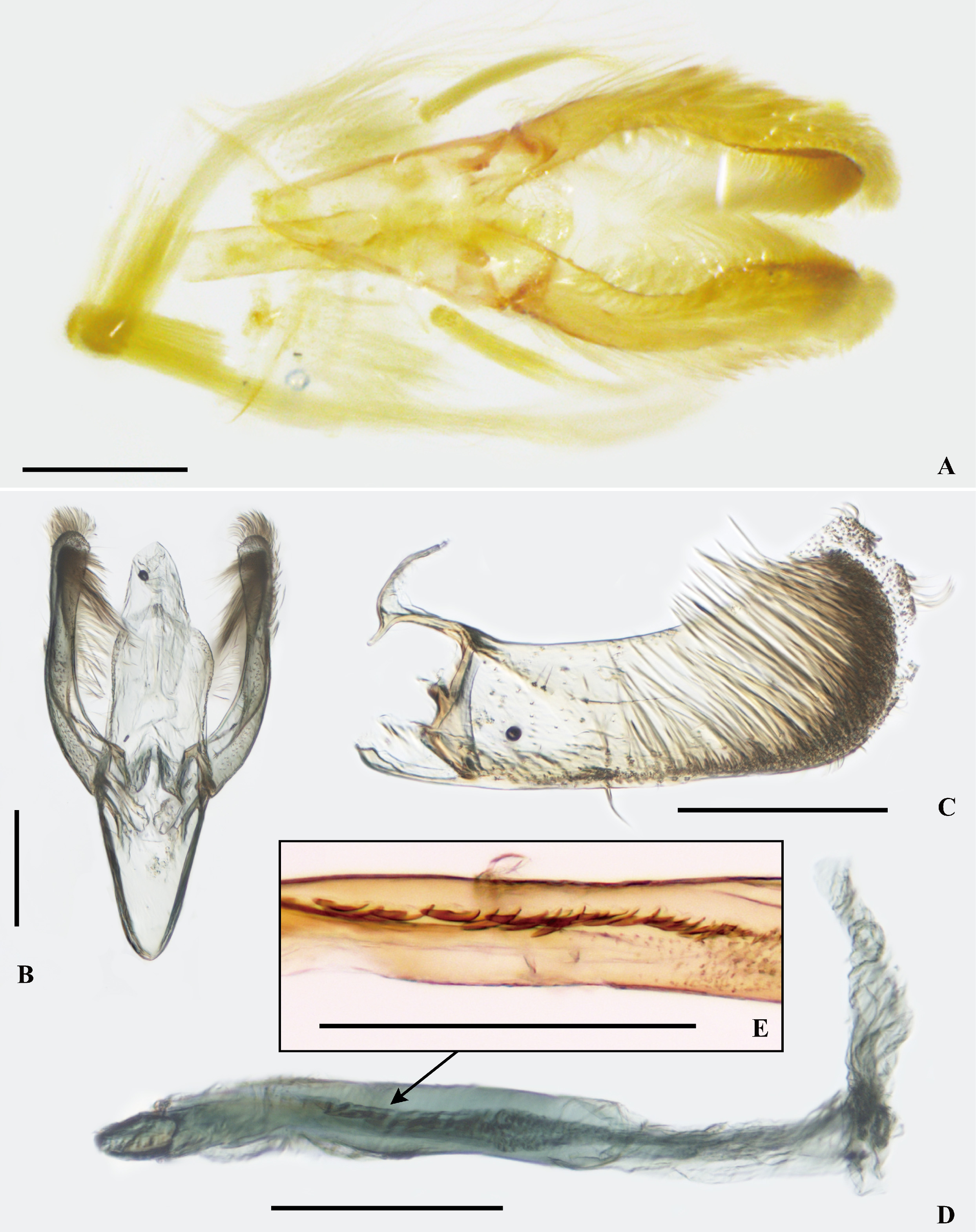
Male genitalia ( Fig. 2 View FIGURE 2 ): Tegumen quadrangular with a slightly bluntly acute apex, almost wholly membranous except lateral margins more or less weakly sclerotized; vinculum almost as wide as tegumen ( Figs. 2A, B View FIGURE 2 ). Saccus slightly shorter than tegumen, long triangular, slightly rounded apically. Uncus and gnathos fully membranous. Valva broad lobate ( Fig. 2C View FIGURE 2 ), almost equally broad in basal 2/3, cucullus slightly broader, with oblique terminal margin; inner surface with numerous very slender setae and short, slightly stout setae along lateral margin arising from basal 1/3 of dorsal margin, with two short and pointed setae placed in basal 1/5; basal processes slender with an additional branch at apex, and forming sickle shape; slightly slenderer upper (dorsal) arms forming transtilla which connected to each other by membranous structure. Phallus long, tubular, straight except apical 1/4 slightly curved, membranous basally ( Fig. 2D View FIGURE 2 ), with 15 to 20 small cornuti arranged as one column in the middle 1/4 ( Fig. 2E View FIGURE 2 ). Abdominal segments VII and VIII membranous, each bearing a pair of coremata of long piliform scales, about 1.5× and 0.6× as long as valva respectively; interior process of 7 th sternite absent ( Fig. 2A View FIGURE 2 ).
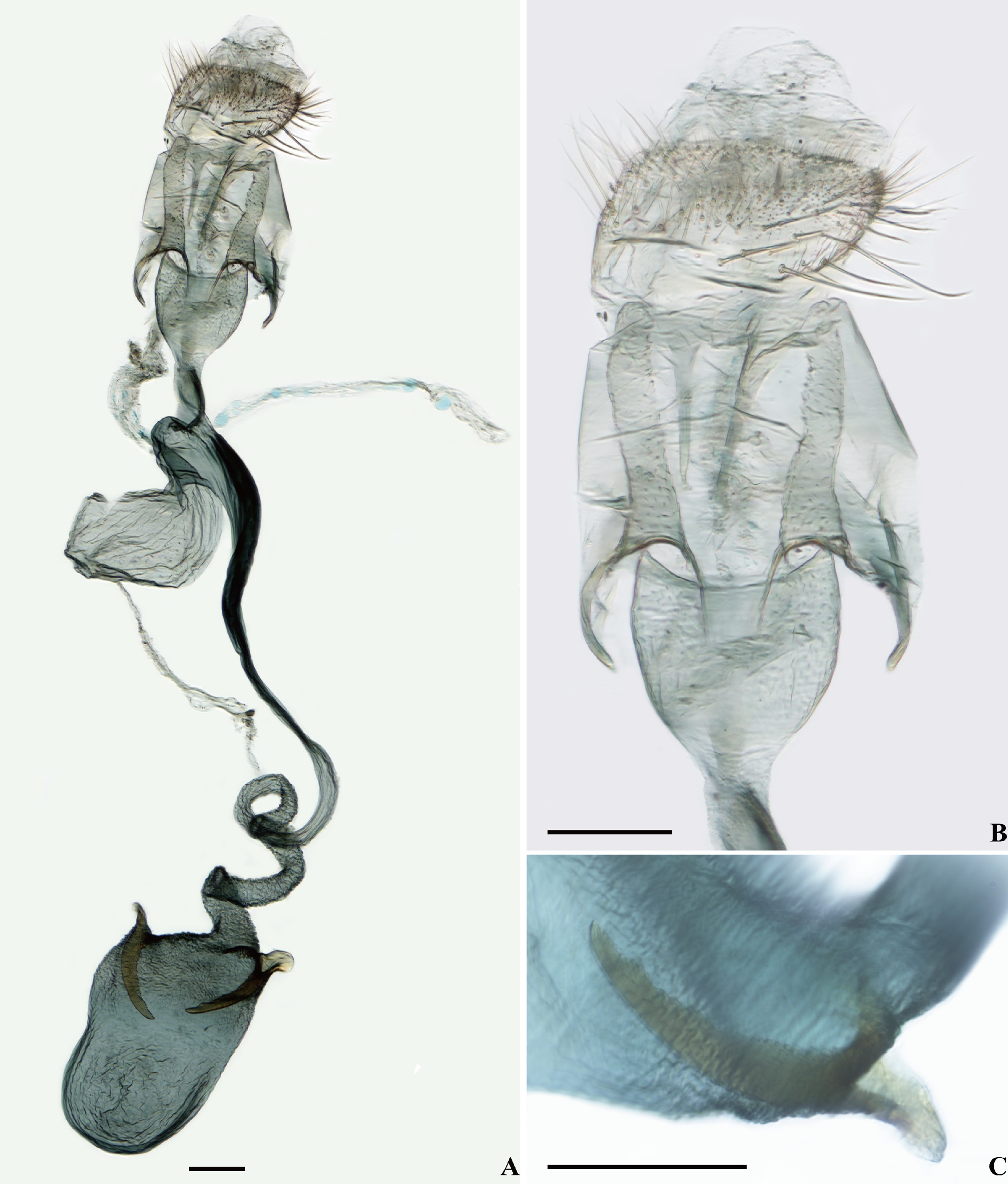
Female genitalia ( Fig. 3 View FIGURE 3 ): Papillae anales membranous with short and pointed setae medially and anteriorly, longer laterally and posteriorly ( Figs. 3A, B View FIGURE 3 ). Apophyses posteriores moderately long, slightly widened on basal half, and then gradually narrowing. Eighth abdominal segment rather long, about 1.6× as long as papillae anales. Apophyses anteriores about 0.7× as long as apophyses posteriores, short stumps, widened on basal half, distinctly bent apically. Ostium bursae large; antrum ellipsoidal, dilated, weakly sclerotized, cephalic half narrowed rapidly, the caudal margin being very concave; ductus bursae slender, long, weakly sclerotized on caudal 2/3, then remaining cephalic 1/3 membranous and coiled; corpus bursae almost quadrangular and flattened with more or less rounded cephalic margin, with a pair of signa placed in base of corpus bursae laterally, equal in size, scimitarshaped, well sclerotized, bearing numerous small teeth on inner margin ( Figs. 3A, C View FIGURE 3 ). Ductus seminalis arising from a rather short distance from the cephalic margin of antrum, weakly sclerotized and sinuate; bulla seminalis ellipsoidal, membranous and swollen, about 0.6 times as large as corpus bursae.
Holotype: ♂, China: Huameiguan, Liuba County, Hanzhong City , Shaanxi Province, ex Aesculus chinensis , 10.VIII.2018 (larva), 20.VIII.2018 em. HAUHL017 642, Rearing number IsO-1026, I. Ohshima, C.Q. Liao leg. ( HUNAU).
Paratypes: 4♂ 7♀, same data as holotype, except 22.VIII.2018 em. (a single male specimen was deposited in HUNAU and three legs of the specimen were used for COI sequencing, and the remaining ten specimens were in KSU) ; 2♂, same data as holotype, except 23.VIII.2018 em. ( KSU) ; 1♂, same data as holotype, except 28.VIII.2018 em. ( HUNAU) ; 2♂ 1♀, China: Tianpingshan, Badagongshan National Nature Reserve , Sangzhi County, Zhangjiajie City , Hunan Province, ex Aesculus chinensis , 23.VIII.2018 (larva), 11.IX.2018 em. C.Q. Liao leg. ( HUNAU) .
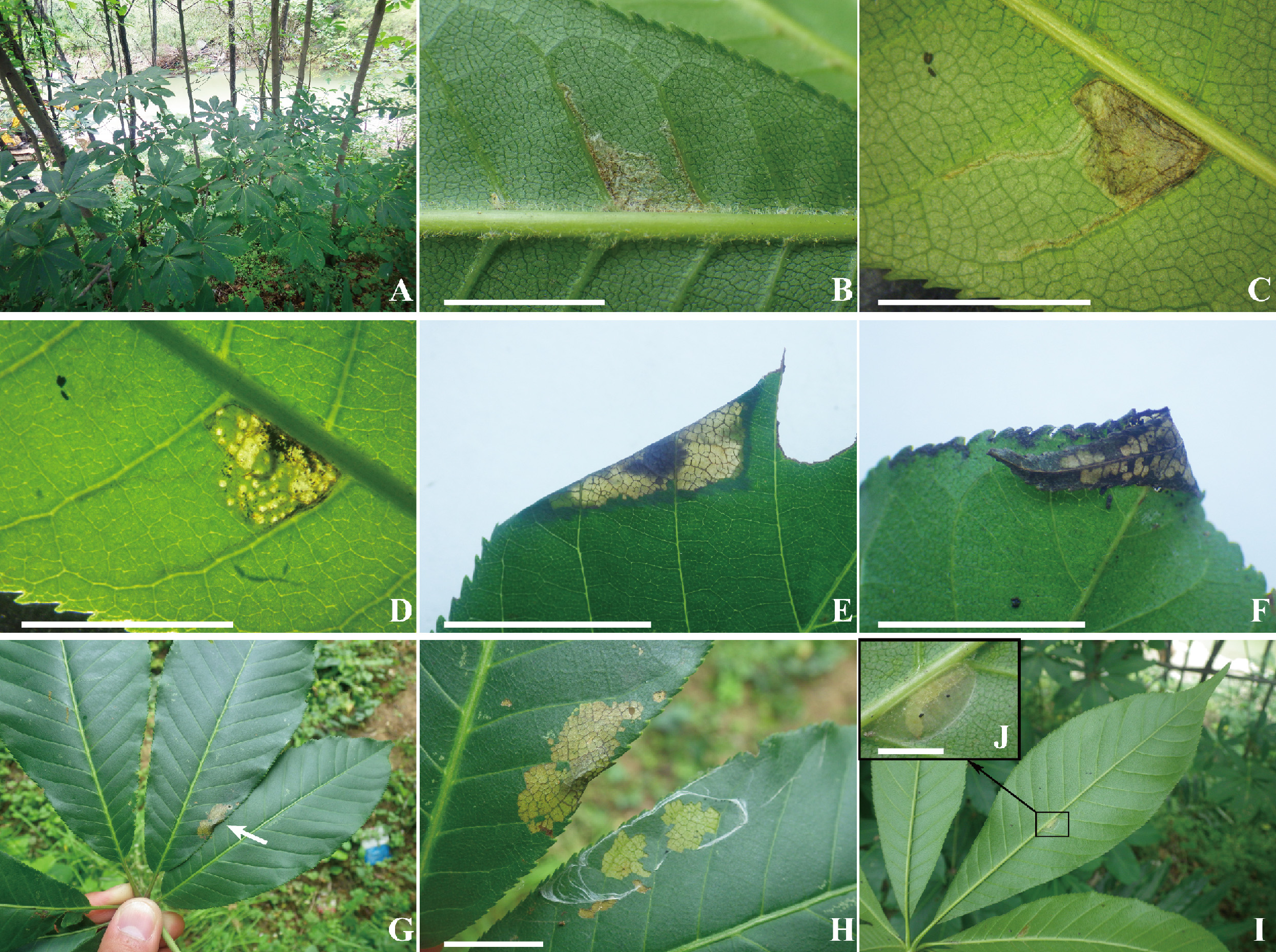
Host: Aesculus chinensis Bunge (Hippocastanaceae) growing in roadsides near houses of localities ( Fig. 4A View FIGURE 4 ).
Leaf mine, leaf shelter and pupal cocoon: Eggs are laid on the underside of leaflets, generally middle part near midvein, and first instar larvae make epidermial linear mines along leaf veins ( Fig. 4B View FIGURE 4 ). Second instar larvae live in blotch mines but still feed on epidermis. Third instars begin feed on parenchyma touching both palisade and spongy layers ( Fig. 4C View FIGURE 4 ), thus mines become transparent ( Fig. 4D View FIGURE 4 ). After molt into a forth instar, larvae vacate the mines and move to the side margin ( Fig. 4E View FIGURE 4 ) or the apical part of mined leaflets ( Fig. 4F View FIGURE 4 ) for making leaf folds, and then continue feeding on it as Parornichini ( Gracillariinae ) does ( De Prins et al., 2019). Sometimes larvae pull together neighbor leaflets by spinning that results in nest-like structure as Tortricidae ( Figs. 4G, H View FIGURE 4 ) instead of leaf folds. Before pupation, larvae leave their shelters and move on the underside of a leaflet to make an oval transparent cocoon along the main vein, where pupation takes place ( Figs. 4I, J View FIGURE 4 ).
Distribution. China (Shaanxi, Hunan).
Etymology. The specific epithet is derived for the host plant genus, Aesculus .
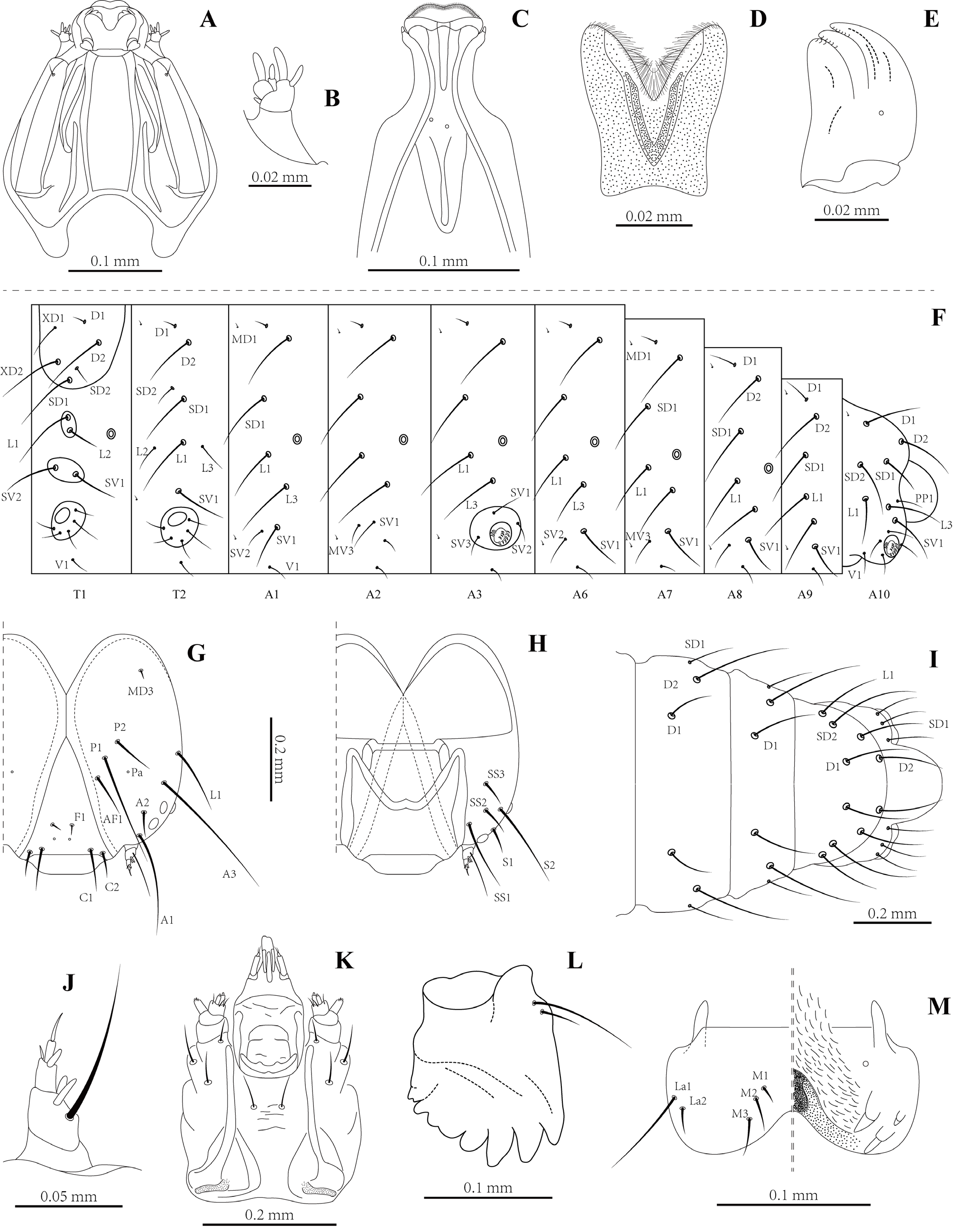
Immature stages. Larva: Sap-feeding (first and second) instar ( Figs. 5 View FIGURE 5 A–F). Leaf miner. Head: Distinctly flattened dorsoventrally, prognathous, partially retracted into pronotum ( Fig. 5A View FIGURE 5 ), most setae lost or reduced; stemma absent; antenna short, 3-segmented, with 4 moderately large and one minute basiconic sensillae ( Fig. 5B View FIGURE 5 ); labrum slightly longer than width, deeply bilobed in the half part, anterior margin with dense labral setae especially at middle part ( Fig. 5D View FIGURE 5 ); mandibles a pair of flat, respectively blunt lobes each
with 3 teeth ( Fig. 5E View FIGURE 5 ); maxillae and labium connate, labium rounded apically, anterior margin slightly emarginate ( Fig. 5C View FIGURE 5 ), labial palpi absent, maxillary palp two-segmented. Setae generally absent or reduced. Legs invisible.
Tissue-feeding last (fifth) instar ( Figs. 5 View FIGURE 5 F–M). Head: well sclerotized, approximately round with full complement of mouthparts; four pairs of stemmata present, three dorsal-laterally and one ventrally, approximately equal in size ( Figs. 5G, H View FIGURE 5 ); antenna four-segmented, with five slender basiconic sensillae and one long pointed seta totally ( Fig. 5J View FIGURE 5 ); frons elongate, nearly extending to epicranial suture. Ecdysial cleavage line terminating near epicranial suture. AF2 absent; P2 and Pa present; Setae P1, A1, A2, S2 and SS1 the most elongate, AF1, P2 and L1 moderately elongate, F1 and MD3 almost reduced ( Figs. 5G,H View FIGURE 5 ); labrum with five pairs of setae, with La3 absent, inner surface with numerous small spines medially ( Fig. 5M View FIGURE 5 ); mandibles heavily sclerotized, with multiple teeth and two setae ( Fig. 5L View FIGURE 5 ); maxillae and labium connate, each maxillae with three slender setae, postmentum with a pair of setae medially ( Fig. 5K View FIGURE 5 ). Thorax: prothorax with six pairs of setae including D1, D2, XD1, XD2, SD1 and SD2; two lateral setae present anterior to spiracle; SV1 and SV2 close to each other; three lateral setae present on meso- and metathorax. Bases of thoracic legs well separated; tarsal claw with broad base terminating in slender, slightly curved claw (Fig. F). Abdomen: D1 anterodorsal to D2 on segments A1–9; SD2 absent on A1–A9; lateral setae L1 and L3 present; SV3 absent on the A1 and A6–9 ( Fig. 5F, I View FIGURE 5 ). Prolegs present on A3–5 and A10; with an outer penellipse row of 28–33 crochets and 7–8 crochets internally on abdominal prolegs 3–5; anal prolegs with 15 crochets arranged forward in a semicircle ( Figs. 5F, I View FIGURE 5 ).
Pupa ( Figs. 6 View FIGURE 6 A–D). Length 4.9–5.1 mm (n = 2); maximum width about 1.1 mm. Vertex with frontal process (cocoon cutter) short, acute, with one pair of setae placed close to scape of antenna. Forewing extending to middle of A6; antenna slightly longer to middle of A7; hindleg extending to posterior margin of A8. Thoracic tergite smooth, meso- and metanotum each with a pair of setae laterally; abdomen mostly covered dorsally with dense, minute spines; A1–A9 with 3 pairs of setae. Cremaster of A10 greatly reduced, consisting of 5 pairs of minute tubercle. The main difference between male and female pupae is that the genital pore on A8 and the oviporus on A9 of female pupae connect into a longer suture, while only one shorter suture formed by genital pore on A9 of male pupae, as show in Figs. 6 View FIGURE 6 B–C.
Note: The sign “S” indicate different specimens.


| KSU |
King Saud University |
No known copyright restrictions apply. See Agosti, D., Egloff, W., 2009. Taxonomic information exchange and copyright: the Plazi approach. BMC Research Notes 2009, 2:53 for further explanation.