
Chimarra ventrospina, Johanson, Kjell Arne & Espeland, Marianne, 2010
|
publication ID |
https://doi.org/ 10.5281/zenodo.198496 |
|
DOI |
https://doi.org/10.5281/zenodo.6196429 |
|
persistent identifier |
https://treatment.plazi.org/id/03F23313-FF8A-9561-C4E8-0E28FDAAFDC3 |
|
treatment provided by |
Plazi |
|
scientific name |
Chimarra ventrospina |
| status |
sp. nov. |
Chimarra ventrospina , new species
Figs 8 View FIGURES 2 – 10 , 41–45 View FIGURES 41 – 45
Diagnosis. Chimarra ventrospina is distinguished from other Chimarra species by the presence of a long, strong, well-sclerotized posteroventrad-oriented tooth distally on the phallotheca. In addition, the posterior margins of segment IX each forms a large triangle; the mesal lobe of tergum X has a nearly identical shape as the lateral lobes in lateral view; and the lateral lobes of tergum X originate at mid-height of the genitalia. The inferior appendages resemble those of C. aureofusca Kimmins, 1957 , but C. ventrospina is easily separated from C. aureofusca by the much shorter segment IX.
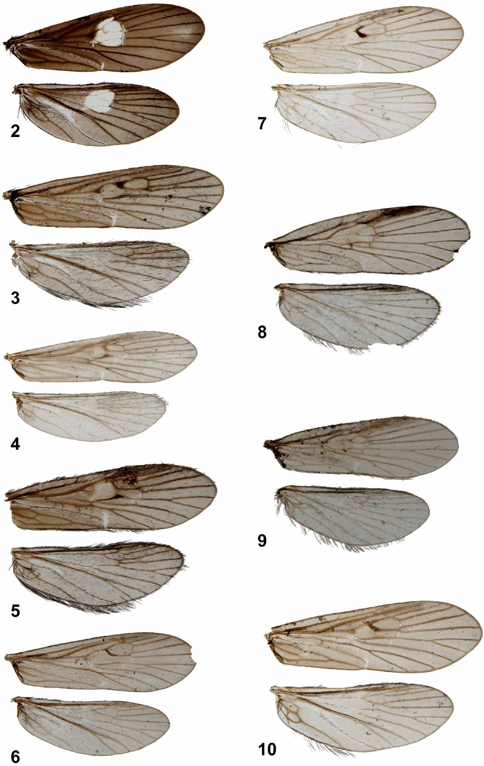
Description: Wings ( Fig. 8 View FIGURES 2 – 10 ): Forewings each 5.0 mm long, membrane uniformly grey; veins brown. Hind wings each 4.2 mm long, membrane uniformly pale grey; veins brown.
Male Genitalia ( Figs 36–40 View FIGURES 36 – 40 ): Segment IX rather short, each side with anteroventral part slightly produced anterodorsad in lateral view ( Fig. 41 View FIGURES 41 – 45 ); anterodorsal margins concave, except with 2 pairs of triangular projections subdorsally; ventral margin strongly convex and rounded in lateral view, without ventral process; below each preanal appendage posterior margin forming large, right-angled triangular plate pointing posterad; setae restricted to marginal area of posterior triangular plate; in ventral view ( Fig. 43 View FIGURES 41 – 45 ) segment IX about uniformly wide along its length, with undulated lateral margins and widely and shallowly concave posterior margin; anterior margin concave; in dorsal view ( Fig. 42 View FIGURES 41 – 45 ) tergum IX very short, forming narrow, transverse band above tergum X; lateral margins nearly parallel, each lateral margin tapering at anterior apex. Anterior apodemes absent. Preanal appendages originating from basodorsal margin of lateral lobes of tergum X, about 0.05 mm long, directed dorsad; irregularly rounded in lateral view ( Fig. 41 View FIGURES 41 – 45 ), narrower in dorsal view ( Fig. 42 View FIGURES 41 – 45 ). Tergum X divided into thick, membranous mesal lobe and pair of sclerotized lateral lobes ( Figs 41, 42 View FIGURES 41 – 45 ). Mesal lobe triangular in lateral view, about half as long as and located well above lateral lobes ( Fig. 41 View FIGURES 41 – 45 ), pointing posterad, without setae; nearly rectangular in dorsal view ( Fig. 42 View FIGURES 41 – 45 ), with posteromesal incision. Lateral lobes without setae, originating from mid-height of segment IX; in lateral view ( Fig. 41 View FIGURES 41 – 45 ), each lateral lobe about as high as mesal lobe, weakly curving posterodorsad, uniformly narrowing apically, apex rounded; in dorsal view ( Fig. 42 View FIGURES 41 – 45 ), lateral lobes straight, slightly converging, each forming narrow lateral plate outside mesal lobe, and broader mesal plate below mesal lobe; lateral and mesal margins nearly straight. Lateral lobes well separated basally; sensillae on lateral lobes not detected. Inferior appendages with long central plate ( Fig. 41 View FIGURES 41 – 45 ); anteriorly pointed in lateral view; inferior appendages forming rounded plates, slightly longer than high, each appendage with 3 mesad-oriented dark, triangular teeth along posterior margin, the ventral one forming a posteroventral tooth in lateral view ( Fig. 41 View FIGURES 41 – 45 ); dorsal and ventral margin above posteroventral tooth hyperboloid; in ventral view ( Fig. 43 View FIGURES 41 – 45 ) inferior appendages oriented posterad, almost parallel, with smoothly convex lateral margins; mesobasal plate long, broad, with irregular mesal margin; posteroventral tooth located at posterior end of meso-basal plate. Sclerotized portion of phallic apparatus long, thick; phallobase occupying almost 2/3rds of phallotheca, almost 3 times thicker than cylindrical posterior part of phallotheca ( Figs 44, 45 View FIGURES 41 – 45 ); posteroventral margin of phallotheca with long, strong, well-sclerotized posteroventradoriented tooth ( Figs 44, 45 View FIGURES 41 – 45 ). Phallotremal sclerite complex small, 3-branched, vertical in lateral view; in ventral view nearly H-shaped with small additional posterior spine on transverse bar; numerous endothecal spines present distally in retracted phallotheca; about 7, anterad-pointing spines in group immediately anterior of ventral marginal tooth; 3 posterad-pointing spines in group at transverse zone between phallotheca and endotheca, immediately posterior of ventral marginal tooth.
Holotype male: Solomon Islands: Western Province, Kolombangara Island, stream crossing main road, 200 m N road L 2, 158 m, loc 0 6, 8 °04.520'S 157°08.845'E, Malaise trap, 11–15.i.2008 [M Espeland].
Etymology. Ve n t ro s p i n a, from venter, “underside” in Latin; and spina, thorn or spine in Latin; referring to the ventral spine of the phallobase.


No known copyright restrictions apply. See Agosti, D., Egloff, W., 2009. Taxonomic information exchange and copyright: the Plazi approach. BMC Research Notes 2009, 2:53 for further explanation.