
Chimarra vitapinensis, Johanson, Kjell Arne & Espeland, Marianne, 2010
|
publication ID |
https://doi.org/ 10.5281/zenodo.198496 |
|
DOI |
https://doi.org/10.5281/zenodo.6196425 |
|
persistent identifier |
https://treatment.plazi.org/id/03F23313-FF8D-9563-C4E8-09A7FB44FEAD |
|
treatment provided by |
Plazi |
|
scientific name |
Chimarra vitapinensis |
| status |
sp. nov. |
Chimarra vitapinensis , new species
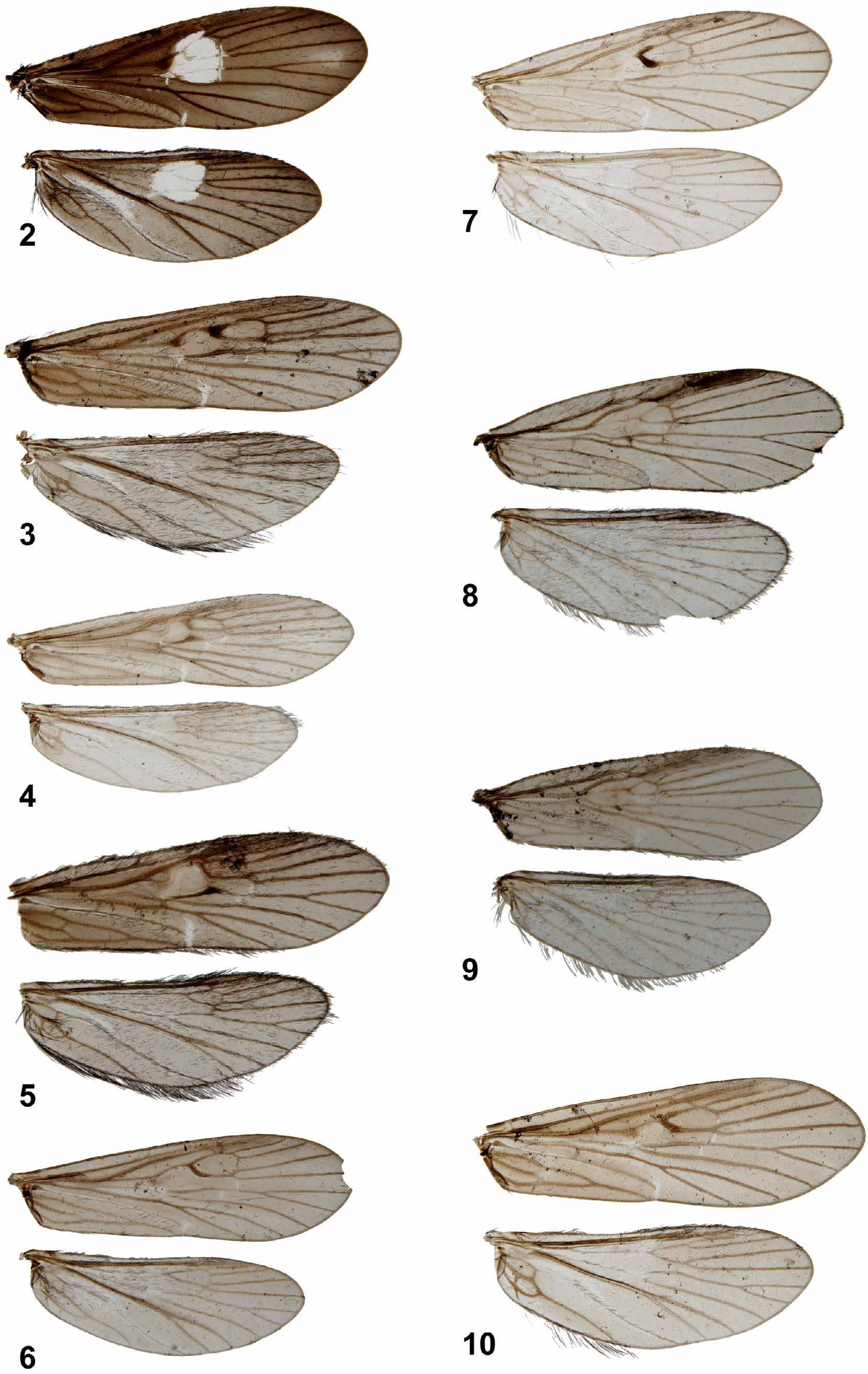
Figs 7 View FIGURES 2 – 10 , 36–40 View FIGURES 36 – 40
Diagnosis. The genitalia of C. vitapinensis are unique among Solomon Island Chimarra species, in particular segment IX is nearly drop-shaped in lateral view, the inferior appendages are sigmoid shaped in lateral view, and the membranous mesal lobe of tergum X is triangular in dorsal view. Some of the New Guinean Chimarra species have similar inferior appendages, like C. papuana, Kimmins, 1962 , and C. sabrona Kimmins, 1962 , but all these species have an anteriorly produced segment IX, and a large ventral process.
Description: Wings ( Fig. 7 View FIGURES 2 – 10 ): Forewings each 4.4 mm long, membrane uniformly pale grey; veins brown to dark brown; veins and neighbourhood membrane of basal part of Dc nearly black. Hind wings each 3.7 mm long, membrane uniformly pale grey; veins brown to dark brown.
Male Genitalia ( Figs 36–40 View FIGURES 36 – 40 ): Segment IX rather short, nearly drop-shaped, with anteroventral part ellipsoid in lateral view ( Fig. 36 View FIGURES 36 – 40 ); each anterodorsal margin undulated; ventral margin slightly convex and angled in lateral view, without ventral process; each posterior margin undulated, with rounded outgrowth immediately above inferior appendage; setae restricted to mid-height of posterior margin; in ventral view ( Fig. 38 View FIGURES 36 – 40 ) segment IX slightly narrowing posteriorly, with undulated lateral margins and widely and shallowly concave posterior margin; anterior margin convex, without mesal incision; in dorsal view ( Fig. 37 View FIGURES 36 – 40 ) with nearly parallel lateral margins, each lateral plate tapering at anterior apex; tergite membranous. Tergite IX short, without process near base of tergum X. Anterior apodemes absent. Preanal appendages located on posterior margin of segment IX well below dorsal margin ( Fig. 36 View FIGURES 36 – 40 ), about 0.05 mm long, directed dorsolaterad; smoothly rounded in lateral view ( Fig. 36 View FIGURES 36 – 40 ), narrower in dorsal view ( Fig. 37 View FIGURES 36 – 40 ). Tergum X divided into long, membranous mesal lobe and pair of sclerotized lateral lobes ( Figs 39, 37 View FIGURES 36 – 40 ). Mesal lobe sharply triangular in lateral view, about half as long as and located well above lateral lobes ( Fig. 36 View FIGURES 36 – 40 ), triangular in dorsal view ( Fig. 37 View FIGURES 36 – 40 ); about half as long as inferior appendages, without setae. Lateral lobes without setae, originating from near bases of preanal appendages; in lateral view ( Fig. 36 View FIGURES 36 – 40 ), each lateral lobe straight, with basal half parallel-sided, distal half tapering into up-curving apex; in dorsal view, lateral lobes straight, slightly converging; mesal margins slightly convex, lateral margins irregular along distal half, apex rounded ( Fig. 37 View FIGURES 36 – 40 ). Lateral lobes widely separated basally by mesal lobe; distal half of each lateral lobe with 2 sensillae along dorsal margin. Inferior appendages with compact, short central plate ( Fig. 36 View FIGURES 36 – 40 ); basal half of each inferior appendage nearly straight, oriented posteroventrad; lateral part bending dorsad to form vertical, narrowing plate, and long, cylindrical, posteromesad-oriented apex ( Figs 36, 38 View FIGURES 36 – 40 ); in ventral view ( Fig. 38 View FIGURES 36 – 40 ) each inferior appendage with long mesobasal plate with irregular mesal margin. Sclerotized portion of phallic apparatus long, slender; phallotheca nearly cylindrical from before half its length in lateral and ventral views ( Figs 39, 40 View FIGURES 36 – 40 ); phallobase about as thick as rest of phallotheca in lateral view, and twice as thick as rest of phallotheca in ventral view; posteroventral margin of phallotheca apparently not strongly produced posterad in lateral view ( Fig. 39 View FIGURES 36 – 40 ). Phallotremal sclerite complex not observed; pair of equally long, slightly curved endothecal spines present distally in retracted phallotheca.
Holotype male: Solomon Islands: Guadalcanal Province, Guadalcanal, Weather Coast, Kusumba Region, small stream 400 m S Vutapinau Village, loc 22, 9 °36.713'S 159°40.598'E, light trap, 26.i.2008 [M Espeland].
Paratypes: Solomon Islands: 11 males, same data as holotype; 4 males, Western Province, Kolombangara Island, stream crossing main road, 200 m N road L 2, 158 m, loc 0 6, 8 °04.520'S 157°08.845'E, Malaise trap, 11–15.i.2008 [M Espeland].
Etymology. Vitapinensis , derived from Vitapinau Village, the type locality of the species.

No known copyright restrictions apply. See Agosti, D., Egloff, W., 2009. Taxonomic information exchange and copyright: the Plazi approach. BMC Research Notes 2009, 2:53 for further explanation.