
Conostigmus minimus Trietsch & Mikó, 2020
|
publication ID |
https://doi.org/ 10.11646/zootaxa.4792.1.1 |
|
publication LSID |
lsid:zoobank.org:pub:326F6A15-216E-439A-AD59-3CDF7551D3F6 |
|
persistent identifier |
https://treatment.plazi.org/id/039687D1-FFFF-658E-9FA4-FD3540A0C364 |
|
treatment provided by |
Plazi |
|
scientific name |
Conostigmus minimus Trietsch & Mikó |
| status |
sp. nov. |
Conostigmus minimus Trietsch & Mikó sp. nov.
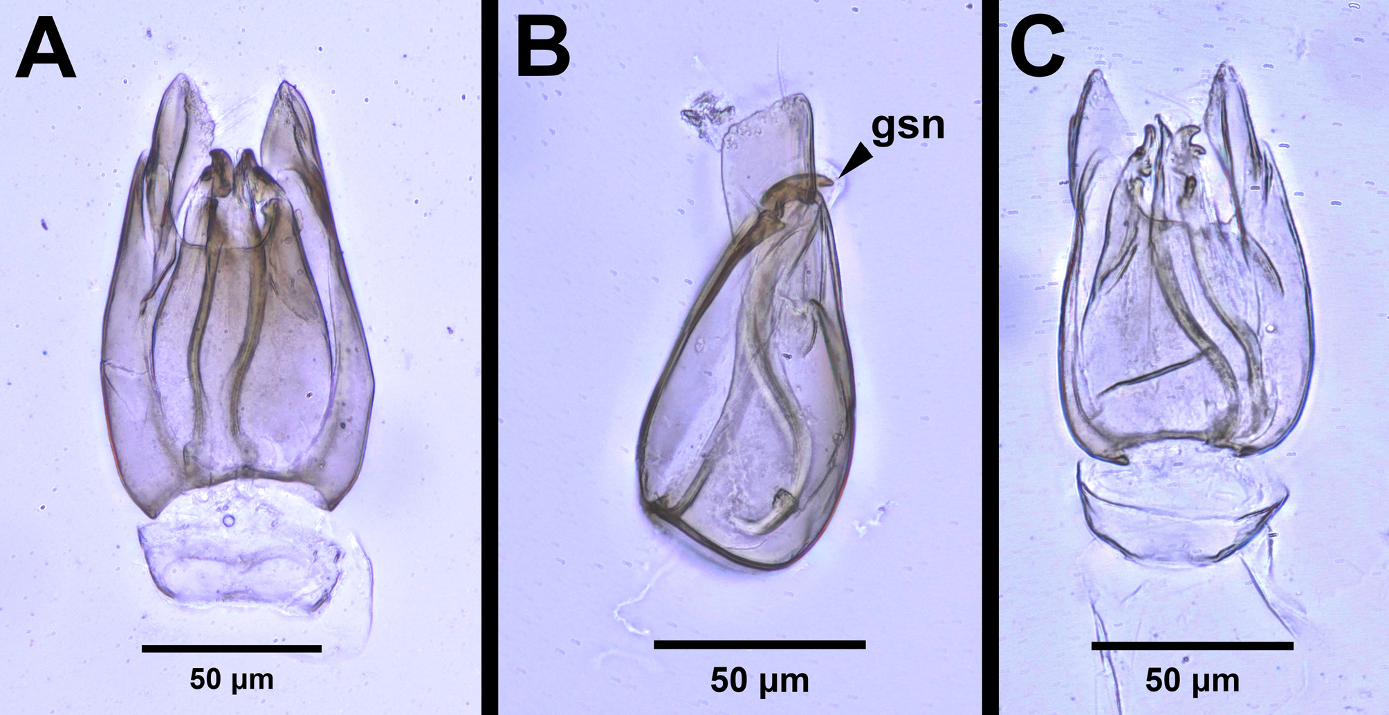
Figs. 70 View FIGURE 70 , 71 View FIGURE 71
Diagnosis. This species shares several characters in common with C. laeviceps , C. musettiae , C. franzinii , and C. bipunctatus , including the following: facial pit present; preoccipital furrow present; median process on the intertorular carina present and acute; sternaulus present and elongate, exceeding 3/4 of the mesopleuron length at the level of the sternaulus; ventral projection of the metapleural carina present; medioventral conjunctiva absent and parossiculi fused; and gonocondyle present and acute. Conostigmus minimus can be differentiated from C. laeviceps , C. musettiae , C. franzinii , and C. bipunctatus by the male genitalia characters, specifically by the absence of the medioventral ridge of the gonostyle–volsella complex and the gonossicular spines of similar lengths (one spine not more than 2× as long as the others).
The female of this species is unknown.
It is worth noting that this species is known from only four specimens, and that these specimens are much smaller than most C. franzinii . It is possible that the medioventral ridge and gonossicular spines are simply not as developed in specimens this small, and that these specimens may be the same species as C. franzinii . However, if this were the case, we would also expect other characters, such as the sternaulus, the median process of the intertorular carina, and the ventral projection of the metapleural carina to be less developed or absent in C. minimus , but these features are as fully developed in C. minimus as in C. franzinii . In fact, the ventral projection of the metapleural carina is more than or equal to 2× as long as wide in C. minimus and less than 2× as long as wide in C. franzinii . In addition, the genitalia differences can still be seen in smaller C. franzinii specimens of the same size as C. minimus specimens (UCRC_ENT 00457072, INHS Insect Collection 287574, INHS Insect Collection 287580), so these differences in male genitalia do not appear to be a function of body size. Based on these differences, it is our hypothesis that C. minimus and C. franzinii are separate species.
Variability. Other than slight differences in size and coloration, no variations were observed between the four specimens.
Description. Body length: 1.425 –1.475 mm. Color hue pattern in male: cranium, mesosoma, posterior part of metasoma brown; scape, F1–F9, pedicel, forelegs, midlegs, hindlegs, neck of petiole and anterior portion of metasoma yellow. Color intensity pattern in male: cranium darker than mesosoma, flagellomeres darker than legs. Color intensity dorsal and ventral to the site of the sternaulus: concolorous. Color intensity pattern of syntergite: petiole neck and anterior region of syntergite lighter in coloration than the posterior region of the syntergite. Foveolate sculpture on body count: absent. Rugose sculpturing count: absent. Rugose region on upper face count: absent.
Antennae: Male scape length vs. pedicel length: 3.6–5.0. Male scape length vs. F1 length: 1.9–2.1. Male F1 length vs. pedicel length: 1.8–2.6. Male F1 length vs. male F2 length: 1.0–1.3. Longest male flagellomere: F1. Length of setae on male flagellomere vs. male flagellomere width: setae shorter than width of flagellomeres; setae as long as width of flagellomeres. Sensillar patch of the male flagellomere pattern: F6–F9.
Head: Head width, dorsal view: equal to or only slightly wider than mesosoma (less than 1.3× wider than mesosoma). Head height (HH, lateral view) vs. eye height (EHf, anterior view): HH:EHf=1.72–1.86. Head height (HH) vs. head length (HL): HH:HL=0.9–1.1. Head width (HW) vs. interorbital space (IOS): HW:IOS=1.5–1.62. Head width (HW) vs. head height (HH): HW:HH=1.3–1.43. Cephalic size (csb): Mean: 225–310 μm. Maximum eye di- ameter vs. minimum eye diameter: 1.1–1.3. POL:OOL: POL equal to or shorter than OOL and ocellar triangle with short base. Male ocular ocellar line (OOL) vs. lateral ocellar line (LOL): OOL:LOL=2.3–3.0. Male ocular ocellar line (OOL) vs. posterior ocellar line (POL): OOL:POL=1.7–3.0. Male ocular ocellar line (OOL): posterior ocellar line (POL): lateral ocellar line (LOL): 2.3–3.0:1.0–1.4:1.0. Head shape (anterior view): circular or triangular. Preoccipital lunula count: present. Preoccipital carina count: absent. Occipital carina structure: occipital carina complete. Occipital carina sculpture: crenulate. Preoccipital furrow count: present. Preoccipital furrow anterior end: preoccipital furrow ends inside ocellar triangle, but ends posterior to the anterior ocellus. Preoccipital furrow sculpture: crenulate. Postocellar carina count: absent. Dorsal margin of occipital carina vs. dorsal margin of lateral ocellus in lateral view: occipital carina ventral to lateral ocellus in lateral view. Transverse scutes on upper face count: absent. Transverse frontal carina count: absent. Randomly sized areolae around setal pits on upper face count: absent. Setal pit on vertex size: smaller than diameter of scutes. Ventromedian setiferous patch and ventrolateral setiferous patch count: absent. White, thick setae on upper face count: absent. Antennal scrobe count: absent. Facial structure count: facial pit present. Facial pit count: present. Facial sulcus count: absent. Median facial keel count: absent. Supraclypeal depression count: absent. Intertorular area count: present. Intertorular carina count: present. Median process on intertorular carina count: present. Median process on intertorular carina shape: acute. Median process of intertorular carina structure: process extends across intertorular area towards dorsal margin of clypeus. Median region of intertorular area shape: convex. Ventral margin of antennal rim vs. dorsal margin of clypeus: not adjacent. Torulo–clypeal carina count: present. Subtorular carina count: absent. Mandibular tooth count: 2. Mandibular lancea count: absent.
Mesosoma: Weber length: WL=310–460 μm. Anterior mesoscutal width (AscW) vs. posterior mesoscutal width (PscW): AscW/PscW=0.64–0.71. Mesoscutal length (MscL) vs. anterior mesoscutal width (AscW): MscL/ AscW=1.6–2.0. Mesoscutal length (MscL) vs. mesoscutellar length (MscIL): MscL:MscIL= 1.0–1.2. Wing count: present. Fore wing size: wings present and macropterous with apex extending past petiole. Pronotum median length: less than longest median anatomical line of the mesoscutum. Notaulus count: present. Crenulae of notaulus width: width of the crenulae does not increase more than 2× anteriorly. Notaulus posterior end location: adjacent to transscutal articulation. Posterior region of notaulus orientation: posterior end of notaulus does not curve and is not adjacent to median mesoscutal sulcus. Median mesoscutal sulcus count: present. Median mesoscutal sulcus posterior end: adjacent to transscutal articulation. Scutoscutellar sulcus vs. transscutal articulation location: adjacent. Axillular carinae count: present. Axillular carinae shape: the left and right carinae are separated posteromedially. Speculum ventral limit: not extending ventrally of pleural pit line. Metapleural sulcus shape: straight. Mesometapleural sulcus count: present. Ventrolateral invagination of the pronotum count: present. Sternaulus count: present. Sternaulus length: elongate and exceeding 3/4 of mesopleuron length at level of sternaulus. Sternaulus sculpture: smooth. Epicnemial carina count: complete. Epicnemium posterior margin shape: anterior discrimenal pit present; epicnemial carina curved. Transverse striations on the ventral metapleural area count: absent. Scutes on posterior region of mesoscutum and dorsal region of mesoscutellum convexity: flat. Ventral projection of the metapleural carina count: present. Ventral projection of the metapleural carina length: more than or equal to 2× as long as wide. Lateral propodeal carina count: present. Lateral propodeal carina shape: inverted “Y” (left and right lateral propodeal are adjacent medially posterior to antecostal sulcus of the first abdominal tergum, and connected to the antecostal sulcus by a median carina representing the median branch of the inverted “Y”). Mesopostscutellum count: absent (scutellum flat). Anteromedian projection of the metanoto–propodeo–metapecto–mesopectal complex count: absent. Posterior margin of nucha in dorsal view shape: straight.
Metasoma: Transverse carina on petiole shape: concave. Paired blue iridescent ovoid patches on the syntergite count: absent. Shortest width of petiole neck vs. syntergal translucent patch maximum width: 1.6–2.2. Shortest width of petiole neck vs. synsternal translucent patch maximum width: 1.8–2.0. Syntergal translucent patch maximum width vs. minimum width: 1.2–1.7. Synsternal translucent patch maximum width vs. minimum width: 1.3–1.8. Syntergal translucent patch maximum width orientation: anterolaterally. Synsternal translucent patch maximum width orientation: anterior–posteriorly. Synsternal setiferous patch shape: linear. Synsternal setiferous patch structure: comprised of a single row of setae posterior to the synsternal translucent patch and widening to a double row of setae anterior to the synsternal translucent patch. Synsternal setiferous patch anterior end: synsternal setiferous patch begins anterior to the synsternal translucent patch anterior margin. Synsternal setiferous patch posterior end: synsternal setiferous patch ends lateral to the synsternal translucent patch posterior margin. Synsternal setiferous patch length vs. synsternal translucent patch maximum width: synsternal setiferous patch at least 2× as long as the maximum width of the synsternal translucent patch. S1 length vs. shortest width: S1 wider than long.
Male Genitalia: Distal margin of male S9 shape: convex. Proximolateral corner of male S9 shape: acute. Male S9 distal setal line/setal patch count: distal setae composing transverse setiferous line or lines. Male S9 distal setal line / setal patch structure: patch of setae occurring medially. Distomedian hairless area interrupting transverse row of setae or patch on male S9 count: absent with distal setiferous patch/line continuous medially. Submedial projections on proximal margin of S9 count: absent. Cupula length vs. gonostyle–volsella complex length: cupula less than 1/2 the length of gonostyle–volsella complex in lateral view. Proximodorsal notch of cupula count: absent. Proximolateral projection of the cupula shape: blunt. Gonocondyle count: present. Gonocondyle shape: acute. Distodorsal margin of cupula shape: straight. Dorsomedian projection of the gonostyle–volsella complex count: absent. Dorsomedian conjunctiva of the gonostyle–volsella complex count: present. Dorsomedian conjunctiva of the gonostyle–volsella complex length relative to length of gonostyle–volsella complex: dorsomedian conjunctiva extending equal to or less than 1/3 of length of gonostyle–volsella complex in dorsal view. Dorsomedial margin of gonostyle–volsella complex shape: straight without a median projection. Proximal end of dorsomedian conjunctiva of the gonostyle–volsella complex shape: blunt or straight. Parossiculus count or parossiculus and gonostipes fusion: present and parossiculi not fused with the gonostipes. Medioventral conjunctiva of the gonostyle–volsella complex count or fusion of parossiculi: medioventral conjunctiva absent and parossiculi fused. Medioventral ridge of the gonostyle–volsella complex count (only applicable if medioventral conjunctiva of the gonostyle–volsella complex absent): absent. Apical parossicular setae count: two. Distal projection of the parossiculus count: present. Distal projection of the penisvalva count: absent. Gonossiculus spine count: 2. Gonossiculus spine length: one spine not more than 2× as long as the other(s) (spines of similar lengths). Harpe length: harpe shorter than gonostipes in lateral view. Harpe shape: simple and not bilobed. Harpe orientation: medial. Lateral margin of harpe shape: widest point of harpe is in its proximal 1/3rd. Distal margin of harpe in lateral view: blunt or straight. Lateral setae of harpe count: absent. Lateral setae on harpe density: setae sparse. Dense patch of setae on the distoventral edge of the harpe count: present. Distal setae on harpe length: setae not of equal length, longer setae present on distoventral edge of harpe. Distodorsal setae of sensillar ring of harpe length vs. harpe width in lateral view: setae as long as or shorter than harpe width. Distodorsal setae of sensillar ring of harpe orientation: distally. Sensillar ring area of harpe orientation: distoventrally. Sensillar ring shape: elongate. Distoventral margin of harpe in lateral view: straight; convex.
Distribution. Nearctic.
Etymology. The name for this species is derived from the Latin minimus meaning “smallest”. This species is the smallest macropterous species of Conostigmus found in the Nearctic. Other species of similar size, such as C. muesebecki and C. dimidiatus , lack fully-formed (macropterous) wings.
Material Examined. Holotype male: USA: Tennessee: CMNHENT0022741 ( CLEV) . Paratypes (3 males): USA: Tennessee : 3 males. CMNHENT0022732, 0022743 ( CLEV); CMNHENT0022733 View Materials ( PSUC) .

No known copyright restrictions apply. See Agosti, D., Egloff, W., 2009. Taxonomic information exchange and copyright: the Plazi approach. BMC Research Notes 2009, 2:53 for further explanation.