
SCOLECOMORPHIDAE, Taylor, 1969
|
publication ID |
https://doi.org/ 10.1111/j.1096-3642.2012.00838.x |
|
persistent identifier |
https://treatment.plazi.org/id/03DB87B7-FFE0-FFA8-FF1F-96E2FD236032 |
|
treatment provided by |
Marcus |
|
scientific name |
SCOLECOMORPHIDAE |
| status |
|
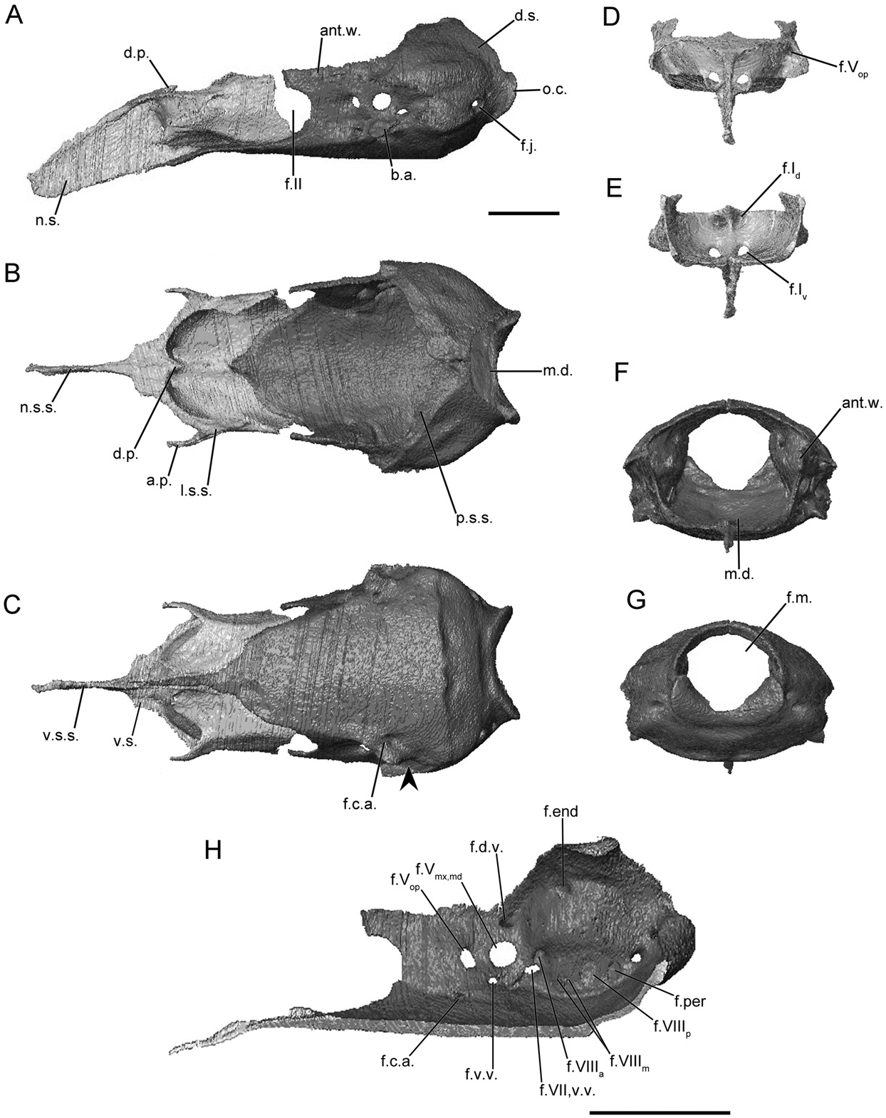
SCOLECOMORPHIDAE View in CoL ( FIGS 8 View Figure 8 , S16-S17)
The main body of the sphenethmoid is long and accounts for roughly half the total length of the sphenethmoid in scolecomorphids ( Fig. 8A View Figure 8 ). The wall is capped by a narrow sutural surface that receives the parietal posteriorly and frontal anteriorly ( Fig. 8B View Figure 8 ). A long, ossified anterolateral process extends toward the medial surface of the prefrontal ( Fig. 8B View Figure 8 ). A very small dorsomedial process is present in Cr. lamottei , but a slightly longer one is present in the species of Scolecomorphus . In all scolecomorphids the process is completely covered dorsally. The posterior margin of the long lateral wall is only slightly incised by the anterior margin of the small optic foramen ( Fig. 8A View Figure 8 ). The floor of the sphenethmoid is deeply incised anteriorly, and is further incised at the midline by a thin slit ( Fig. 8B View Figure 8 ).
The nasal region is divided by a tall, blade-like nasal septum ( Fig. 8A View Figure 8 ) that extends anterior to the posterior limit of the external naris. The dorsal sutural surface is broad at its base, but tapers distally, producing a thin ridge-like dorsal surface along most of its length ( Fig. 8B View Figure 8 ). Ventrally the nasal septum is also very thin. Long sheet-like sola nasi are present in the species of Scolecomorphus (Fig. S16B). These are weakly developed in Cr. lamottei ( Fig. 8B View Figure 8 ).
Paired anterior foramina are present. The ventral pair is more laterally located from the midline in Sc. vittatus (Fig. S17E) than those of Scolecomorphus kirkii (Fig. S16E) and Cr. lamottei ( Fig. 8E View Figure 8 ). A single anterolateral foramen pierces the base of each anterolateral process ( Fig. 8D View Figure 8 ).
The antotic wall of the os basale is angled towards the midline anteriorly ( Fig. 8B View Figure 8 ), except for Sc. vittatus in which it is roughly parallel to the contralateral wall (Fig. S17B). When viewed anteriorly the wall is dorsolaterally orientated, and is smoothly continuous with the floor of the os basale ( Fig. 8F View Figure 8 ). The dorsal sutural surface is narrow to moderately broad, as in Sc. vittatus . It expands slightly posteriorly and is continuous with the sutural surface along the anterior margin of the otic capsule ( Fig. 8B View Figure 8 ). The anterior margin of the antotic wall is deeply incised in Cr. lamottei ( Fig. 8A View Figure 8 ), but only very weakly incised in the species of Scolecomorphus (Fig. S16A), by the posterior margin of the optic foramen. The antotic foramina of Cr. lamottei are of the pattern referred to here as Pattern 5, with an additional dorsal foramen for a vein present ( Fig. 8H View Figure 8 ). Two, and variably a third, foramina are present in the antotic wall of Scolecomorphus (Fig. S16H). This is referred to here as Pattern 7.
The dorsal surface of the otic-occipital complex of the os basale is only slightly tilted posteroventrally. Only a very thin region is posteriorly exposed in dorsal view near the midline; the remainder forms the sutural surface that receives the parietal ( Fig. 8B View Figure 8 ). A pointed ridge is present on the anterolateral surface of the otic capsule in Cr. lamottei ( Fig. 8A View Figure 8 ), but not Scolecomorphus . In lateral view the occipital condyle only slightly projects beyond the posterior limit of the otic capsule ( Fig. 8A View Figure 8 ), and the jugular foramen is partially visible in lateral view. An additional, smaller foramen is present just posterior to the jugular foramen in Cr. lamottei ( Fig. 8C View Figure 8 ).
The medial wall contains the four foramina ( Fig. 8H View Figure 8 ) in their common locations (endo- and perilymphatic foramina, and the anterior and posterior vestibulocochlear nerve foramina), found in all species examined here. Variably within the family two or three additional foramina transmit components of the medial branch of the vestibulocochlear nerve ( Maddin, 2011).
The anterior portion of the floor of the os basale is triangular in outline (Fig. S16C), except for Cr. lamottei in which it is very thin and rod-like ( Fig. 8C View Figure 8 ). In all scolecomorphids the floor reaches to contact the nasal septum. The floor is concave rather than flat. The posterior median depression is weakly defined. In lateral view the floor projects far ventral to the ventral limit of the otic capsules ( Fig. 8A View Figure 8 ). The basicranial articulation is a knob-like projection capped with cartilage. In ventral view there is no constriction of the lateral margin of the floor posterior to the basicranial articulation (arrowhead; Fig. 8C View Figure 8 ). A ventral wing-like projection is absent from the ventral surface of the otic capsule. The posterior margin of the floor is represented by a weakly expressed, transverse boundary that does not approach the ventral margin of the foramen magnum ( Fig. 8C View Figure 8 ).
The foramen that leads to a canal that conducts the carotid artery is located more anteriorly, relative to the otic capsule, than is the case for most other species ( Fig. 8C View Figure 8 ). The canal terminates at lateral and medial foramina located anterior to the antotic foramina ( Fig. 8H View Figure 8 ).
The stapes is absent from scolecomorphids.
No known copyright restrictions apply. See Agosti, D., Egloff, W., 2009. Taxonomic information exchange and copyright: the Plazi approach. BMC Research Notes 2009, 2:53 for further explanation.
|
Kingdom |
|
|
Phylum |
|
|
Class |
|
|
Order |
|
|
Family |