
Ctenophilothis altus, (Lewis, 1885)
|
publication ID |
https://doi.org/ 10.11646/zootaxa.3691.2.6 |
|
publication LSID |
lsid:zoobank.org:pub:BC20CC30-59B7-428C-840C-0DA252954BA6 |
|
DOI |
https://doi.org/10.5281/zenodo.6148026 |
|
persistent identifier |
https://treatment.plazi.org/id/03889A47-8A48-A429-FF7A-FE66FD2CFEF9 |
|
treatment provided by |
Plazi |
|
scientific name |
Ctenophilothis altus |
| status |
|
Ctenophilotis altus (Lewis, 1885) View in CoL
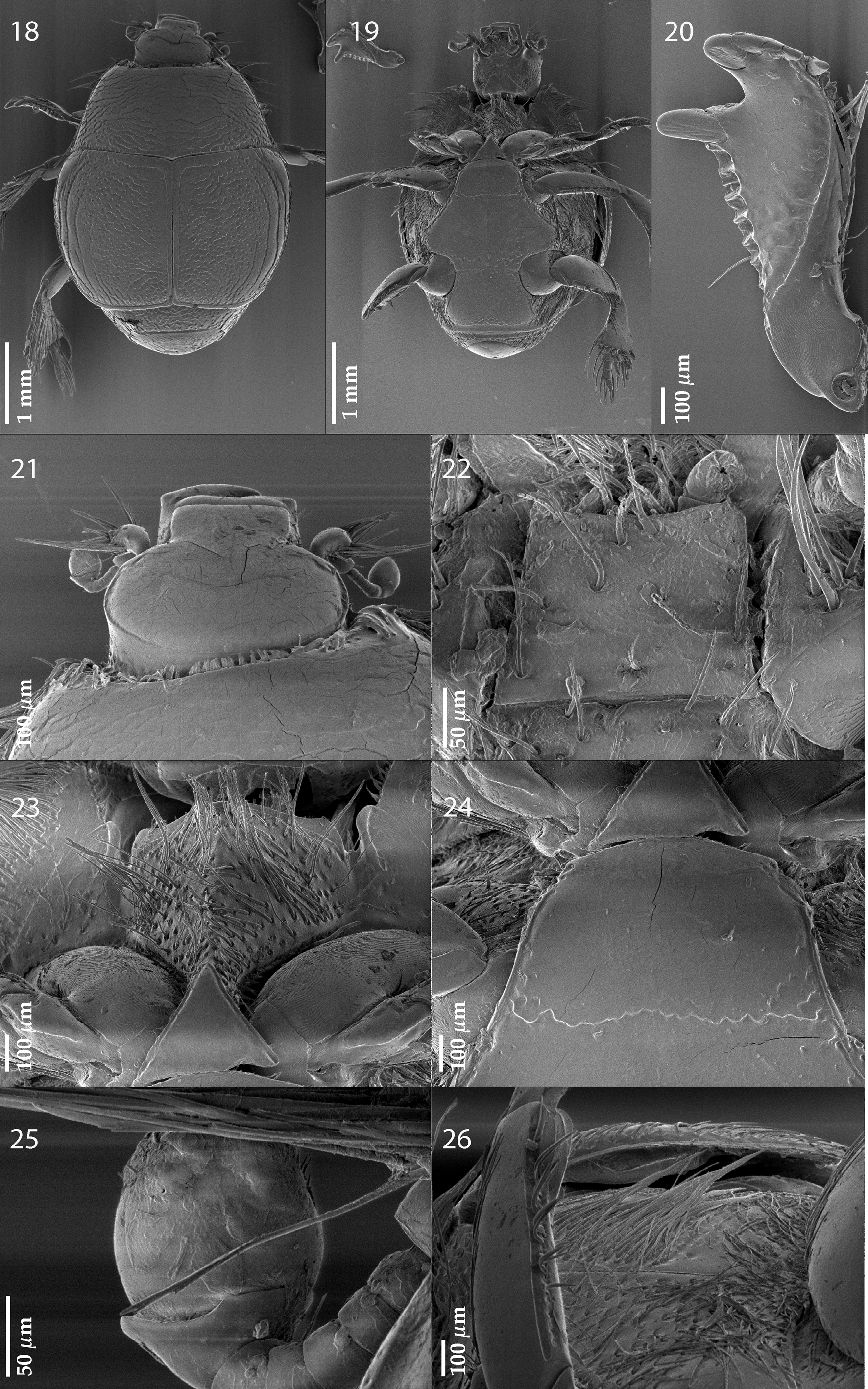
( Figs. 18–26 View FIGURES 18 – 26 )
Xenonychus altus Lewis, 1885: 468 .
Styphrus altus Bickhardt, 1910: 107 .
Philotis altus Reichardt, 1930 ; Peyerimhoff, 1936: 225 (keyed), fig. 2; Mazur, 1984: 109.
Ctenophilotis altus Kryzhanovskij, 1987: 25 , fig. 1; Olexa, 1990: 146, 153 (keyed), figs. 6, 9, 10, 22, 27, 52, 53; Mazur, 1997: 268; Mazur, 2004: 91; Mazur, 2011: 213.
Type material examined: Holotype, female, right protibia, right metafemur and right mesotarsus broken off, glued onto the same carton as specimen, left protarsus and last segment of left mesotarus missing, glued on a triangular point, followed by: “ Egypt ” (round hand-written blue label), followed by “ Xenonychus / altus / Type Lewis” (written); followed by “ Type ” (round red-margined printed label); with another label: “G. Lewis Coll. / B.M. 1926- 369” (printed), followed by “D07-074, female sign” (pink label, pencil written), and another label “ LECTOTYPE / Xenonychus altus / Lewis, 1885 / Designated by / T. Lackner, 2008” (red, hand-written label), added by the author (MNH).
Additional material examined: Egypt. 1 ♀, Central Asfar / 11.4.1933; Coll. Alfieri. (NHMB).
Body length: PEL: 2.50–2.65 mm; APW: 1.00 mm; PPW: 2.00– 2.125 mm; EL: 1.50–1.75 mm; EW: 2.10–2.40 mm.
Redescription. Body ( Fig. 18–19 View FIGURES 18 – 26 ) roundly oval, moderately convex from above, underside very convex, pronotum conspicuously narrower than elytra; cuticle rufocastaneous, feebly shining; legs, mouthparts castaneous, antennal club yellow. Antennal scape thickened, rounded, with numerous long setae ( Fig. 21 View FIGURES 18 – 26 ); penultimate segment (before club) cupuliform; club ( Fig. 25 View FIGURES 18 – 26 ) moderately-sized, sub-conical, without visible articulation, almost completely glabrous apart from two vaguely visible latero-apical sensory areas that are covered with irregular short setae; sensory structures of the antenna not examined. Mouthparts: mandibles stout, outer-margin straight, abruptly curved inwardly; apical part elongated and pointed; labrum deeply incised under clypeus, more than three times wider than long, smooth; pits and setae arising from them absent; mentum ( Fig. 22 View FIGURES 18 – 26 ) sub-trapezoid, with a very shallow antero-median emargination; anterior and lateral margins with sparse short setae; several setae present also on disk of mentum; cardo of maxilla on lateral margin with few short setae; stipes triangular, with numerous much longer setae; ultimate maxillary palpomere somewhat thickened, about twice as long as penultimate, its width shorter than half its length. Clypeus ( Fig. 21 View FIGURES 18 – 26 ) rectangular, about 1.5 times as wide as long, slightly elevated laterally, smooth; frontal stria absent; supraorbital stria finely impressed; frontal disk ( Fig. 21 View FIGURES 18 – 26 ) broad, circular, impunctate, with a single well-impressed transverse chevron; eyes very flattened, invisible from above.
Pronotal sides ( Fig. 18 View FIGURES 18 – 26 ) moderately narrowing anteriorly, apical angles conspicuous; anterior incision for head deep, semicircular, marginal pronotal stria laterally carinate, broadly interrupted behind head; surface along lateral margins fringed with short setae; disk moderately convex, covered with undulate or angular transverse wrinkles, area along anterior and lateral margins smooth; pronotal hypomeron with long amber setae; scutellum very small, almost invisible.
Elytral humeri ( Fig. 18 View FIGURES 18 – 26 ) prominent, epipleura ciliate ( Fig. 26 View FIGURES 18 – 26 ); marginal epipleural stria complete, thin, slightly continued along elytral apex; marginal elytral stria well impressed, carinate, shortly prolonged along elytral base, apically continued as vaguely impressed apical stria. Humeral stria vaguely impressed, connected to rather long internal subhumeral stria that is present medially; only first and second dorsal striae well impressed, in punctures; first almost reaching elytral apex, apically bent inwardly, basally continued along elytral base as straight undulate line, somewhat distanced from elytral base and linked to sutural elytral stria; second elytral stria abbreviated on basal fourth and apical tenth, apically bent inwardly, interrupted on apical third; sutural elytral stria well impressed, apically faintly connected with apical elytral stria; elytral disk with deep coarse punctures that are becoming elongate wrinkles on apical third, area between sutural stria and elytral suture, and area between elytral base and prolonged first dorsal elytral stria as well as elytral flanks smooth. Propygidium completely exposed, covered with elongate confluent wrinkles; pygidium convex, covered with elongate confluent wrinkles less coarse than those of propygidium, laterally and apically smooth. Anterior margin of prosternum ( Fig. 23 View FIGURES 18 – 26 ) obtuse-angulate; marginal prosternal stria vaguely impressed; keel setose, abruptly and strongly depressed behind striate triangular prosternal apophysis; lateral prosternal striae absent. Anterior margin of mesoventrite ( Fig. 24 View FIGURES 18 – 26 ) outwardly emarginate; marginal mesoventral stria laterally well impressed, carinate, anteriorly erased; disk smooth; meso-metaventral suture well impressed, irregularly undulate; intercoxal disk of metaventrite smooth, posterior margin with an irregular row of punctures; lateral metaventral stria well impressed, curved outwardly, shortened; lateral disk of metaventrite excavate, covered in punctures fringed with long setae; metepisternum setose, punctuation almost unrecognizable beneath setae.
Intercoxal disk of the first visible abdominal sternite laterally with a thin complete stria; disk smooth; dorsal and lateral disks of all visible abdominal sternites setose.
All femora on outer margin with long setae, pro-femur covered with setae on entire surface. Protibia ( Fig. 20 View FIGURES 18 – 26 ) dilated, outer margin with two large widely separated distal teeth topped by denticle, followed by several long movable thorn-like denticles (all of them are broken off on right protibia on Fig. 20 View FIGURES 18 – 26 , but well-visible on left protibia where some of them are preserved; Fig. 18 View FIGURES 18 – 26 , as well as on the drawing by Peyerimhoff 1936, fig. 2); inner row of setae on inner margin of protibia comparatively long; protibia on posterior surface smooth, surface divided approximately medially by a definite complete stria; apical margin ventrally with two short apical denticles; protarsi absent; protibial spur minute. Mesotibia slightly dilated, but not particularly thickened, outer margin with two dense rows of long denticles abutting each other, denticles grow in size apically; posterior surface fringed with a dense brush of long setae; mesotarsus thickened, short, each tarsomere with two strongly sclerotized setae; claws of apical tarsomere broken off. Metatibia triangularly dilated, but not particularly thickened, without separately extended ventro-lateral part; generally similar to mesotibia.
Differential diagnosis. C. altus differs from C. chobauti chiefly by the shape of protibia (in case of C. chobauti the outer margin of protibia is with two low approximate distal teeth, topped by stout denticle, whereas in C. altus the distal teeth are widely separated and denticles that top them are much larger; compare Figs. 10 View FIGURES 2 – 10 and 20 View FIGURES 18 – 26 and see also Peyerimhoff 1936, fig. 2) and the shape of mandibles (rounded in case of C. chobauti and almost straight and abruptly curved inwardly in this species; compare Figs. 5 View FIGURES 2 – 10 and 21 View FIGURES 18 – 26 ). Further differences are found in the length of elytral striae: in case of C. chobauti the internal subhumeral stria is longer and almost parallel to first dorsal elytral whereas that of C. altus is shorter, furthermore C. altus has more prominent elytral humeri (in case of C. chobauti pronotum is hardly narrower than elytra, whereas in C. altus it is much narrower).
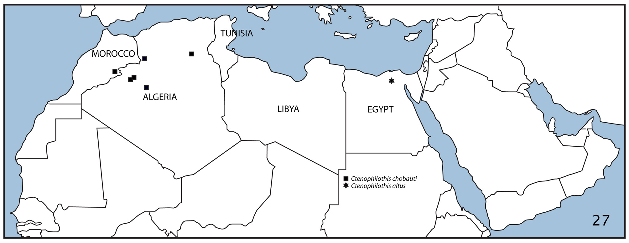
Distribution. Egypt: Central Asfar [=Gabal Asfar, 25 km NE of Cairo, Qalioubeya Province, approximately N 30°13’01.32”, E 31°23’ 15.56, 17 m a.s.l.]. ( Fig. 27 View FIGURE 27 ).
Biology. Unknown, presumably similar to C. chobauti .
Remarks. The only known exact locality of this rare species is Gabal Asfar in northern Egypt, vicinity of Cairo. According to M. Saleh Saleem (Riyadh, Saudi Arabia), this used to be a part of desert at the eastern edge of the Nile Delta, but was turned into an agricultural area in 1950s. Unfortunately, both examined specimens were female, therefore the depiction of male genitalia has not been possible, although it would undoubtedly further clarify the taxonomic status of both species.

No known copyright restrictions apply. See Agosti, D., Egloff, W., 2009. Taxonomic information exchange and copyright: the Plazi approach. BMC Research Notes 2009, 2:53 for further explanation.