
Deltochilum tenuistriatum, González-Alvarado & Vaz-De, 2021
|
publication ID |
https://doi.org/ 10.5852/ejt.2021.775.1551 |
|
publication LSID |
lsid:zoobank.org:pub:976D7020-5904-4951-97CE-B4FE58DA12A8 |
|
DOI |
https://doi.org/10.5281/zenodo.5589719 |
|
persistent identifier |
https://treatment.plazi.org/id/03F48795-DC4D-FFA2-A613-A31BFB53F9ED |
|
treatment provided by |
Felipe |
|
scientific name |
Deltochilum tenuistriatum |
| status |
sp. nov. |
Deltochilum tenuistriatum View in CoL sp. nov.
urn:lsid:zoobank.org:act:9BBED604-6552-43D2-B7AF-44B2DF95DDF7
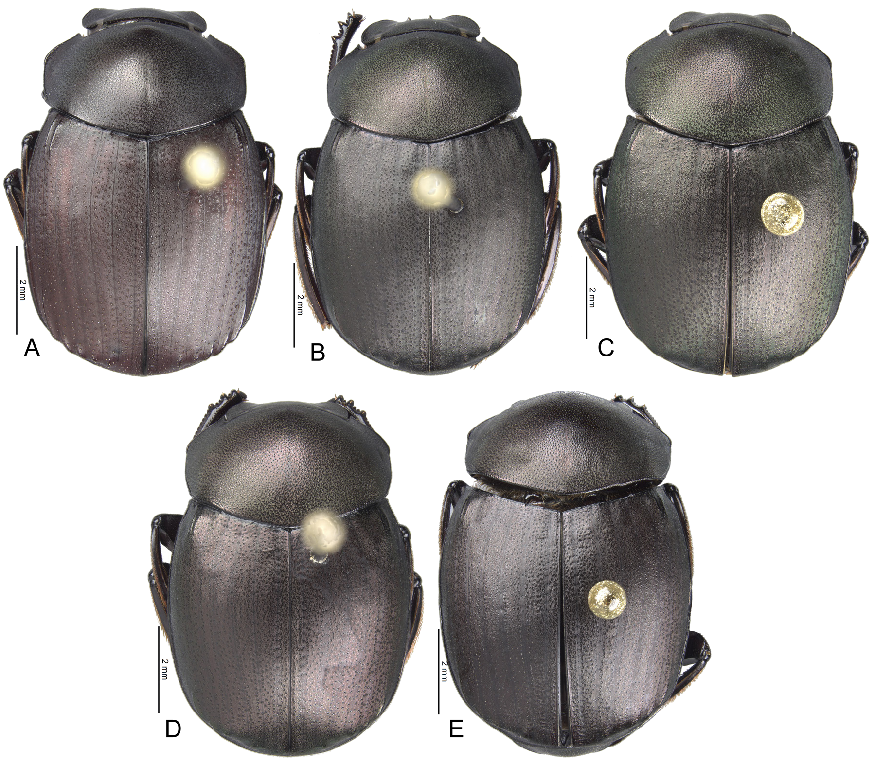
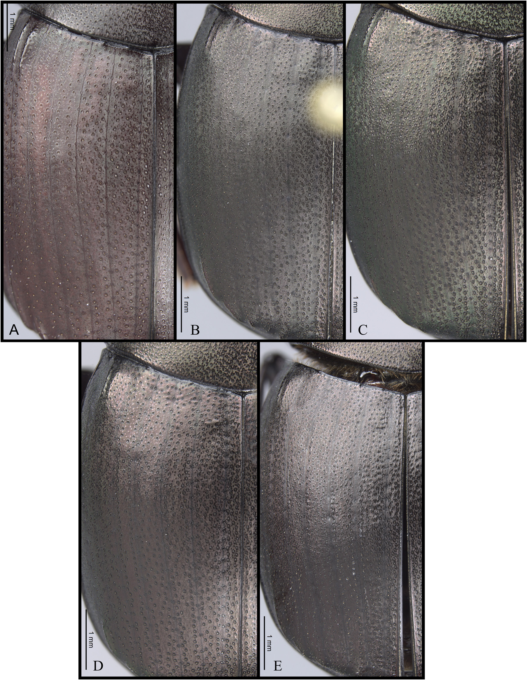
Figs 1E View Fig , 2E View Fig , 3E View Fig , 4E View Fig , 5E View Fig , 6E View Fig , 7E View Fig , 8E View Fig , 10 View Fig
Diagnosis
Close to D. quasistriatum sp. nov. by having the striae I–VII conspicuous ( Figs 1D–E View Fig , 4D–E View Fig ), but III–VII successively thinner and more ill-defined and by the pronotal disc with irregular shiny points contiguous to punctures ( Fig. 3D–E View Fig ). However, it can be distinguished by having the first stria subequal to second stria, smallest interstrial punctures ( Fig. 4E View Fig ) and by pygidial punctures which are smallest and most disperse ( Fig. 5E View Fig ).
Etymology
From Latin tenuis 'weak' + stria in reference to the ill-defined striae.
Type material
Holotype VENEZUELA • 1 ♂; Bolívar, 10 km E of S. [San] F. [Francisco] Yuruani; 5°1′34″ N, 61°2′34″ W; alt. 1300 m; 8–10 Jul. 1987; S. and J. Peck leg.; carrion traps, Gran Sabana, forest; [aedeagus and endophallus extracted]; CMNEN WSD00041745 . GoogleMaps
Description
MEASUREMENTS AND COLOR. Holotype male, length 8 mm, humeral width 5.2 mm. Dark green with red reflections dorsally ( Fig. 1E View Fig ). Black ventrally, with shiny red reflections on metaventrite, metaventral process, meso- and metafemora and ventrite VI ( Fig. 6E View Fig ).
HEAD ( Fig. 2E View Fig ). Dorsal inter-ocular distance approximately eight times width of the eye. Punctures on frons separated by less than one diameter of each puncture. Punctures on head disc separated by less than one diameter of each puncture, almost contiguous.
PRONOTUM ( Fig. 3E View Fig ). Medial angle projected. Punctures on disc separated by less than one diameter, almost contiguous. Shiny points on disc irregular and contiguous to punctures.
ELYTRA ( Figs 1E View Fig , 4E View Fig ). Carina of ninth interstria reaching middle of elytral length. Punctures on first stria subequal in size to second stria, but denser. Striae I–VII conspicuous. Width of first stria subequal to second stria. Striae III–VII narrow and ill-defined, successively narrower and more ill-defined, with VII almost inconspicuous. Width third stria approximately 1/40 th the distance between striae II and III. Stria VIII conspicuous only laterally, discontinuous in some parts and reaching apex of carina of the ninth interstria. Punctures of second interstria on disc separated by less than one diameter, on third by one diameter. Punctures of third interstria on disc occupying about 1/14 th the distance between striae II and III. Apical tubercles on interstriae III, V–VII ( Fig. 5E View Fig ).
ABDOMEN ( Fig. 6E View Fig ). Width of expansion of ventrite I, on ventrite III, subequal to distance between clypeal teeth; expansion reaching distal margin of ventrite IV. Margins of expansion between ventrites II–III forming an acute angle. Expansion on ventrite IV narrower than on ventrite III, and margins almost parallel. Apex of expansion rounded. Basal area of expansion with punctures separated by less than one diameter, almost contiguous.
LEGS. Apex of mesotibia on ventral-internal margin with a small spatulate expansion. Expansion of metafemur 1.7 × wider than the width of metafemur basal to expansion. Internal margin of metatibia with large tubercles, occupying almost all metatibial length.
PYGIDIUM ( Fig. 5E View Fig ). Most of the punctures separated by one diameter; punctures basally denser than punctures on disc. Discal punctures occupying approximately 1/38 th the width on middle of pygidium.
GENITALIA ( Figs 7E View Fig , 8E View Fig ). Aedeagus as described in the gilli species group. Medial endophallite sinuate. Sub-medial area of endophallus with scales.
Remarks
This species is only known from the holotype.
Known distribution
Venezuela. Bolívar: 10 km E San Francisco Yuruani ( Fig. 10 View Fig , white eight-point star).


No known copyright restrictions apply. See Agosti, D., Egloff, W., 2009. Taxonomic information exchange and copyright: the Plazi approach. BMC Research Notes 2009, 2:53 for further explanation.
|
Kingdom |
|
|
Phylum |
|
|
Class |
|
|
Order |
|
|
Family |
|
|
Genus |