
Doryphorybius flavus ( Iharos, 1966 )
|
publication ID |
https://doi.org/ 10.5281/zenodo.277335 |
|
DOI |
https://doi.org/10.5281/zenodo.5630120 |
|
persistent identifier |
https://treatment.plazi.org/id/03D7474B-CF5C-A901-71BA-F95AD0BCE102 |
|
treatment provided by |
Plazi |
|
scientific name |
Doryphorybius flavus ( Iharos, 1966 ) |
| status |
|
Remarks on Doryphorybius flavus ( Iharos, 1966)
Material examined. 12 paratypes (slide codes " HNHM Tard-398", with seven examples, and " HNHM Tard-399", with five examples); according to Iharos (1966) the specimens were extracted from moss on rock in Eplény, Ungary; no other data available.
I examined type material of the species which is, unfortunately, in very poor condition. In all the specimens examined the body is contracted by the mounting media and the reticulate sculpture of the cuticle, which is one of the most important diagnostic characters, has become invisible. Apart from the body length, it was not possible to find suitable specimens for the correct measurement of the other structures. I have, therefore, chosen to omit the metric characters in order to avoid confusion; however, the pt index of the stylet supports insertion point on the buccal tube is about 68–70%.
As reported in the original description ( Iharos, 1966), there is a smooth swelling on the anterior side of the first three pairs of legs and the posterior side of the hind legs [“Die Beine haben über den Krallen einen glatten Buckel” - The legs have a smooth swelling over the claws]. It is not possible to confirm or deny the presumed absence of reticulate sculpture or dots on legs (a character which might not have been noticed), as none of the cuticular sculpture is now visible.
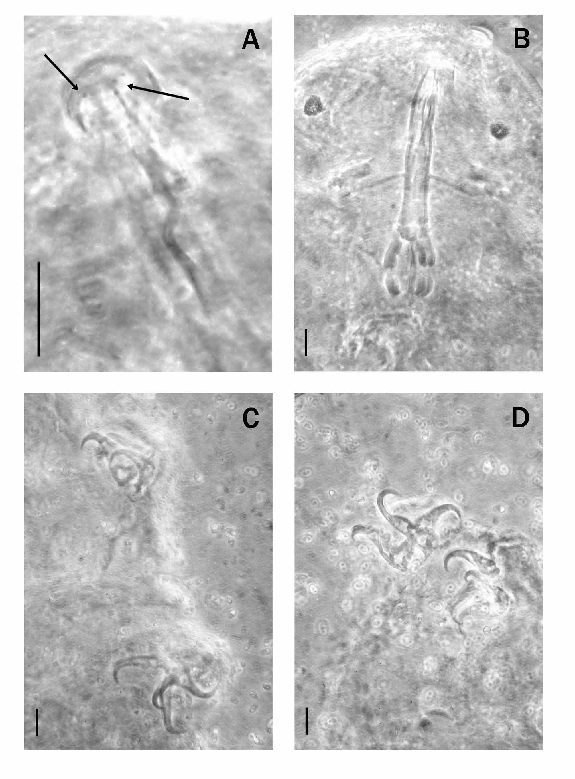
Although Kaczmarek & Michalczyk (2004) examined paratypes of D. flavus they reported that a buccal armature was not visible and assumed that it was absent. However, dorso-lateral teeth are visible in some specimens ( Fig. 1 View FIGURE 1. D A, arrows), but it is not possible to establish whether the number of the teeth is constant. There appears to be at least four of per side, deposited along the anterior margins of the stylet sheaths and therefore could be considered residuals of a subdivided transverse ridge, which is present in many species of eutardigrades. Peribuccal structures were not visible. The first macroplacoid shows a light incision, not previously reported ( Fig. 1 View FIGURE 1. D B).
Claws of the Isohypsibius type ( Fig. 1 View FIGURE 1. D C, D), slightly different in shape and size on each leg; basal portions of the external claws clearly wider than those of the internal ones ( Fig. 1 D View FIGURE 1. D ). Small accessory points on main branches. Smooth lunules present, with those of the inner claws, small and narrow, not always visible and for this reason not previously noticed. No other sclerified structures were visible on the legs.
| HNHM |
Hungarian Natural History Museum (Termeszettudomanyi Muzeum) |
No known copyright restrictions apply. See Agosti, D., Egloff, W., 2009. Taxonomic information exchange and copyright: the Plazi approach. BMC Research Notes 2009, 2:53 for further explanation.