
Dryadosaura nordestina, Rodrigues & Freire & Pellegrino & Sites Jr, 2005
|
publication ID |
https://doi.org/ 10.1111/j.1096-3642.2005.00177.x |
|
DOI |
https://doi.org/10.5281/zenodo.10545377 |
|
persistent identifier |
https://treatment.plazi.org/id/03A81E4C-EB1D-F419-FC77-7668FC5CFC87 |
|
treatment provided by |
Diego |
|
scientific name |
Dryadosaura nordestina |
| status |
sp. nov. |
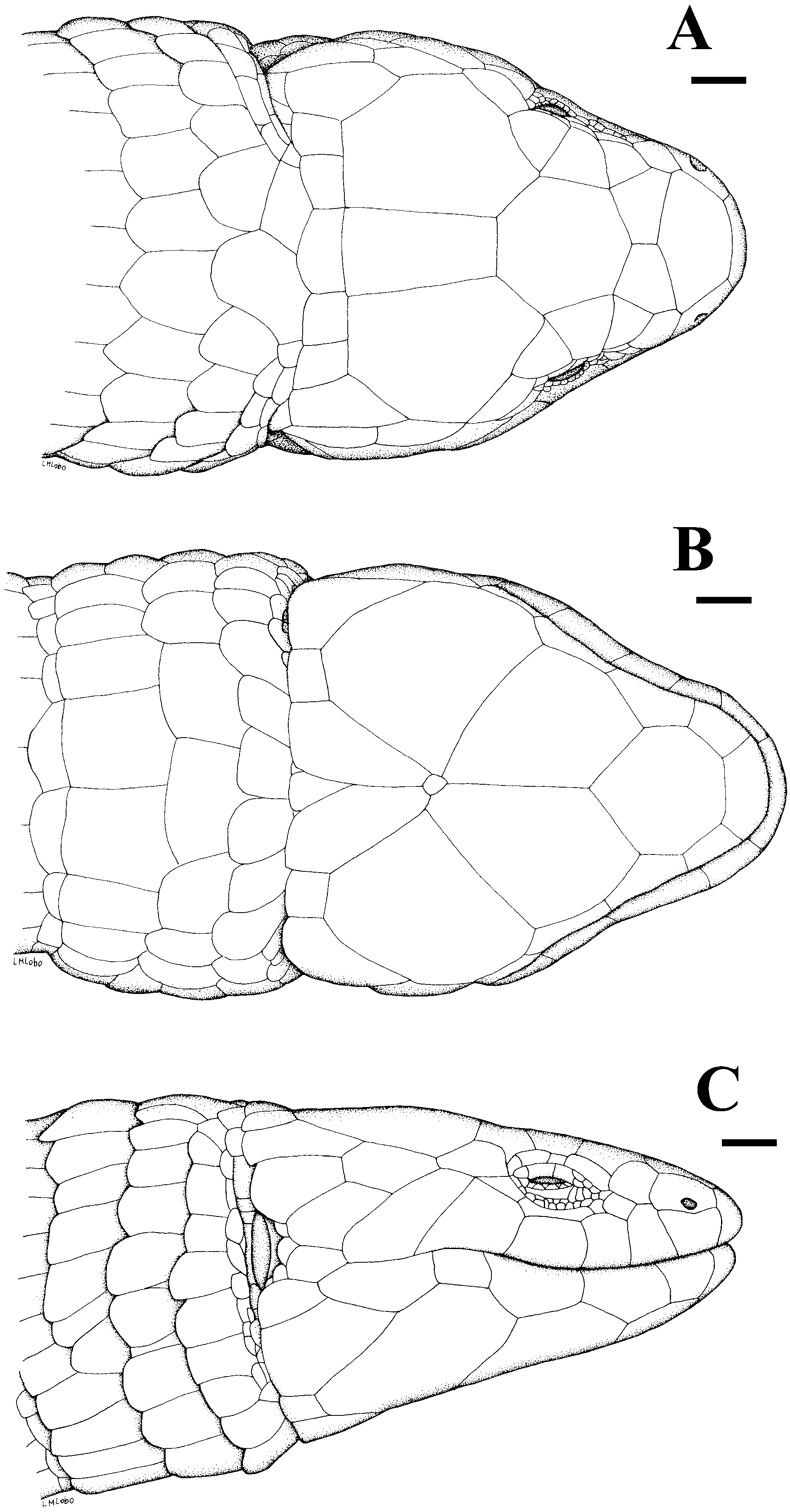
DRYADOSAURA NORDESTINA SP. NOV. ( FIGS 1 - 4 View Figure 1 View Figure 2 View Figure 3 View Figure 4 )
Holotype: MZUSP 60635 View Materials , an adult male from João Pessoa (07 ∞¢ 07S, 34 ∞ 52 ¢ W): state of Paraiba: Brazil, collected by Miguel T. Rodrigues 6 September 1983.
Paratypes: MZUSP 60334 View Materials , 60336–60339 View Materials , 61132 View Materials , 65983 View Materials , 65984 View Materials , 65987 View Materials , 65988 View Materials , from João Pessoa , state of Paraiba ; MZUSP 66352 View Materials , 87709–87719 View Materials , from Serra dos Cavalos, state of Pernambuco ; MNRJ 9931 View Materials , from Recife (Parque Dois Irmãos), state of Pernambuco ; MZUSP 93218–93222 View Materials , and MUFAL 1537 , 1540–1546 , Murici ( Fazenda Bananeiras ) ; MUFAL 293–296 , Pontal do Peba, Piaçabuçú, State of Alagoas ; MUFAL 1547 , from Mata do Catolé , Maceió, state of Alagoas ; MUFAL 1534–1536 , 1538 , 1539 , Mata do Cedro, Rio Largo , state of Alagoas ; MUFAL 055 , from Natal (Parque Estadual das Dunas), state of Rio Grande do Norte ; MUFAL 007 , Praia de Cutuvelo , Parnamirim, state of Rio Grande do Norte ; MUFAL 052 , Tibaú do Sul , State of Rio Grande do Norte .
Etymology: Nordestina refers to north-eastern Brazil, the geographical region where the species occurs.
Diagnosis: Microteiid characterized by an elongate body, five toes and fingers, and short stout limbs. Collar fold, ear opening, and eyelid present. Frontonasal single; prefrontals, parietals and interparietals present; frontoparietals absent. Two pairs of chin shields; three supraoculars. Dorsal scales in 23–25 regular transversal rows; anteriorly smooth, becoming progressively hexagonal and mucronate; keeled at rump level. Lateral scales similar to dorsals, smooth, rectangular, laterally imbricate. Ventral scales smooth, juxtaposed or slightly imbricate, rectangular, in 15–17 rows; 26–33 scales around midbody. Infradigital lamellae mostly divided 6–9 in finger IV, and 13–16 in toe IV. Preanal and femoral pores number four and six, respectively, in males; 2 - 4 preanal pores in females.
Description of holotype ( Fig. 1A - C View Figure 1 ): Rostral broad, wider than high, contacting first supralabial, nasal and frontonasal. Frontonasal pentagonal, wider than long, contacting rostral, nasal, loreal and prefrontals. Prefrontals pentagonal, almost as long as wide, in broad contact at midline; midline suture shorter than suture with frontonasal. Frontal heptagonal, slightly longer than wide, wider posteriorly; anterior margin angulose, indenting prefrontals, lateral margins almost parallel, slightly divergent posteriorly, contacting second and third supraoculars, posterior margins diagonally contacting parietals and in straight contact with interparietal. Frontoparietals absent. Interparietal subrectangular, longer than wide, as long as or slightly longer than, and narrower than frontal, narrower than parietals. Lateral margins of interparietal slightly diverging posteriorly; left suture of the scale incomplete posteriorly. A pair of very large irregularly hexagonal parietals in straight lateral contact with interparietal and also anteriorly contacting the frontal, and laterally contacting third supraocular and postocular; posterior part of parietals almost rounded and contacting two large temporal and two smaller occipital scales.
Three supraoculars, first slightly smaller than third, second the largest, as large as prefrontal; first supraocular contacting prefrontal, second supraocular in broad contact with frontal, third supraocular in broad contact with parietal. Nasal mostly above first supralabial, and also contacting second infralabial, loreal, frontonasal and rostral, large, longer than high, with nostril centrally placed in the lower part of scale, and slightly indenting suture with first labial. Loreal posterior to nasal, narrower and diagonally orientated; contacting frontonasal, prefrontal, first supraocular, a small frenocular, and second supralabial. Frenocular small, longer than high, followed posteriorly by two suboculars, the second the largest. Six supralabials; fourth under the eye, fifth and sixth the highest, fourth and sixth the largest, first and second subequal, smaller. Fourth supralabial separated from the eye by a large subocular posteriorly followed by a scale which contacts fifth supralabial and a postocular inserted between third supraocular and parietal. Sixth supralabial followed posteriorly by an elongate granule which contacts anterior margin of ear. Four superciliaries, second smallest, first the largest, wider anteriorly, longer than first supraocular, contacting loreal, first and second supraoculars, second superciliary and upper eyelid. Central part of eyelid with a semitransparent undivided disc surrounded by small and slightly pigmented and irregularly shaped smooth granules. Lower eyelid with 8-9 strongly pigmented palpebrals. Temporal region with four large, smooth and juxtaposed scales between parietal, sixth supralabial, and the ear, two of them diagonally contacting the sixth labial. Ear opening surrounded by a series of small and juxtaposed granules contacting anteriorly two large temporals, and two much smaller elongate and juxtaposed flat granules; external auditory meatus small, tympanum distinct, subovoid with anterior margin rounded. Lateral surface of neck with smooth scales, irregular in size and shape, varying from juxtaposed to slightly imbricate, and arranged in irregular transverse series between ear and shoulder. All head scales smooth and juxtaposed with scattered sensorial organs.
Mental broad, wider than high. Postmental heptagonal, longer than wide. Two pairs of genials, both contacting infralabials; the first smaller and in broad contact at midline, the second separated by an enlarged pair of flat and elongate pregulars, which contact at midline, preventing contact between second pair of chin shields. Five infralabials, first smallest, all others subequal. Gulars smooth, imbricate, quadrangular, juxtaposed to slightly imbricate, irregular in size, in five transverse rows. Gulars followed by a distinct interbrachial region with eight larger and elongate scales. A distinct collar fold characterized by some granules and reduced scales in the second row of gulars preceding the interbrachial row.
Dorsal scales disposed in regular transversal rows, anteriorly smooth, imbricate, rounded in the occipital region, becoming progressively narrower, more elongate and rectangular towards the arm level, and then progressively hexagonal, with lateral sides almost juxtaposed, keeled at rump level. Twenty-five transverse rows of dorsals between interparietal and the posterior level of hind limbs. Lateral scales about the same size as dorsals but smooth, rectangular, imbricate laterally, not acuminate and more diagonally orientated than dorsals; those closer to ventrals larger. A series of transversally arranged granules in the skin separating transverse series of lateral scales. A distinctive area with granular scales surrounds the area of arm insertion. Scales around midbody 28. Ventral scales smooth, laterally juxtaposed, slightly imbricate anteroposteriorly, rectangular, about twice as long as wide; 15 transverse rows from interbrachials (excluded) to preanals. Four scales in precloacal region, the central ones the largest. Ten pores (four preanal, six femoral).
Tail scales shorter and more imbricate anteroposteriorly than midbody dorsals, otherwise identical to them, keeled, lanceolate; those from ventral part of tail base wider than those of dorsal part, becoming gradually identical in size towards tip of tail. Tail regenerated with rectangular and smooth scales.
Fore limbs extremely robust with large, smooth and imbricate scales, larger and flat dorsally; those from ventral part of brachium smaller. Forearm as long as thick. Anterior and ventral parts of hind limbs with irregularly large, smooth and imbricate scales, largest scales ventrally. Posterior part of hind limbs with granular and juxtaposed scales. Dorsal part of tibia and femur with keeled, imbricate scales. Carpal and tarsal scales large, imbricate; supradigital lamellae smooth, imbricate. Palmar and plantar surfaces with smooth, small granules; infradigital lamellae mostly divided, seven on finger IV and 15 on toe IV. Fingers and toes clawed, respectively, with the following relative sizes: 1 <2 = 5 <4 <3 and, 1 <5 <2 <3 <4.
Dorsal surfaces of body and tail and lateral part of tail dark brown with an irregularly distributed yellowish reticulate or interrupted pattern. Yellow pigment is concentrated in parietal area of head and along dorsolateral part of body where irregular and enlarged spots occupy the central part of scales, delimiting a discontinuous pale line. Flanks predominantly yellowish to cream, as the ventral parts of body and tail, but strongly mottled with an irregular dark brown pattern. Lateral parts of head predominantly dark brown with scattered yellow spots concentrated in the suture region of posterior labials; ventral part of the head creamy yellow, immaculate, except for infralabials and scattered and irregular dark brown pigmentation on the posterior part of last pair of genials. Ventral parts of body and tail creamy yellow, immaculate. Ventral part of tail becomes gradually darker distally. Limbs dark brown, irregularly mottled with yellow dorsally, creamy yellow and immaculate ventrally. Palmar and plantar surfaces greyish.
Measurements of holotype: SVL 54 mm; tail length 31 mm, regenerated.
Variation: Regardless of sex, range variation in scale counts are as follows: 23–25 dorsal, 15–17 ventral, 23– 33 around the body; 6–9 lamellae in finger IV, and 13– 16 lamellae in toe IV. Apart from differences in scale counts there is little variation in scalation in the type series. The shape of the lateral margins of the interparietal varies from straight to slightly divergent, as does the condition of the second pair of genials; these are in contact at midline in most specimens, although the extent of contact varies. Although in most specimens prefrontals are in broad contact, in two specimens ( MUFAL 007 , 293 ) the frontonasal and frontal are in slight contact, preventing contact between prefrontals. Number of supra- and infralabials is fairly constant, but some variation also occurs. In two specimens ( MZUSP 60638 View Materials , 60636 View Materials ) sutures between supralabials are incomplete between 4th and 5th supralabials; in two specimens there are seven ( MZUSP 60637 View Materials ) or four ( MUFAL 1536 ) supralabials in one side, instead of six. As in the holotype, most of specimens have five infralabials, but some have four due to a fusion between an infralabial and the second pair of genials. Some specimens show only two superciliaries due to a fusion between first and second .
All adult males have a total of ten pores, four preanal plus six femoral; these are also observed in juveniles but are not so conspicuous. Most adult females have two rather inconspicuous preanal pores (exceptionally four); femoral pores are absent in females. Males are slightly larger than females (maximum SVL, respectively, 57 mm and 52 mm) and have a massive musculature in the temporal region (less developed in females). Tail length varies between 1.14 and 1.45¥ SVL. There is little variation in colour pattern except for the brown pigmentation on the lateral part of the head, which can be more or less extensive, and the ventral colour of live adult males, which is more reddish compared with the creamy venter of adult females.
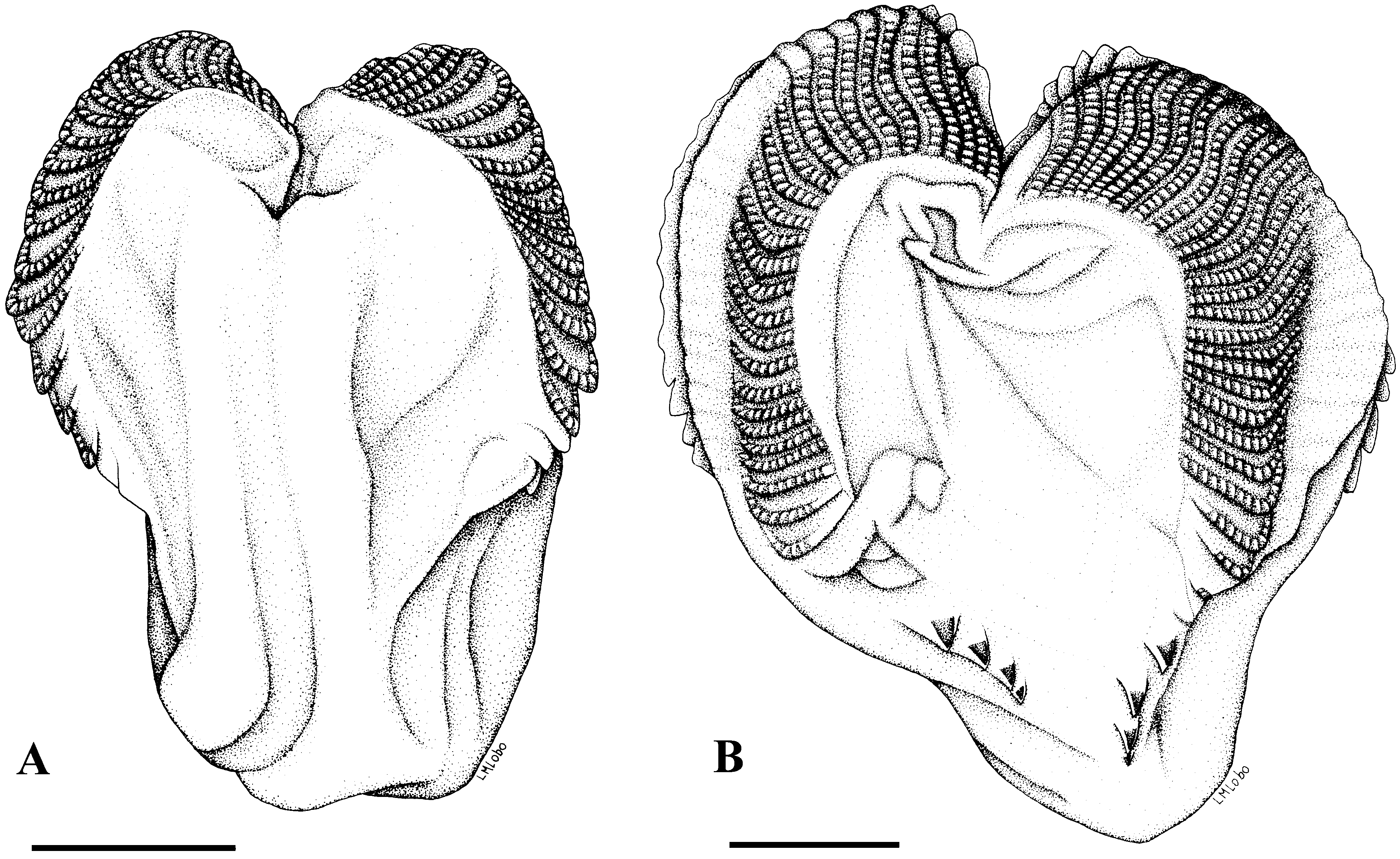
Hemipenis ( Fig. 2 View Figure 2 ): The hemipenis of four specimens ( MZUSP 65983, 87711, 87712, 87718) although without the apex totally everted, shows distinctive bilobation. The sulcate face of the organ lacks ornamentation and is characterized by a conspicuous sulcus spermaticus, which bifurcates centripetally towards the apex of each lobe. The opposite face has two longitudinally aligned series of 4–5 large spines, separated by naked intervals of about the same size as the spines, which converge at the base of the organ. The right and left lobes are symmetrical, with two longitudinal series of about 20 flounces with evident comb-like spines separated by areas without ornamentation.
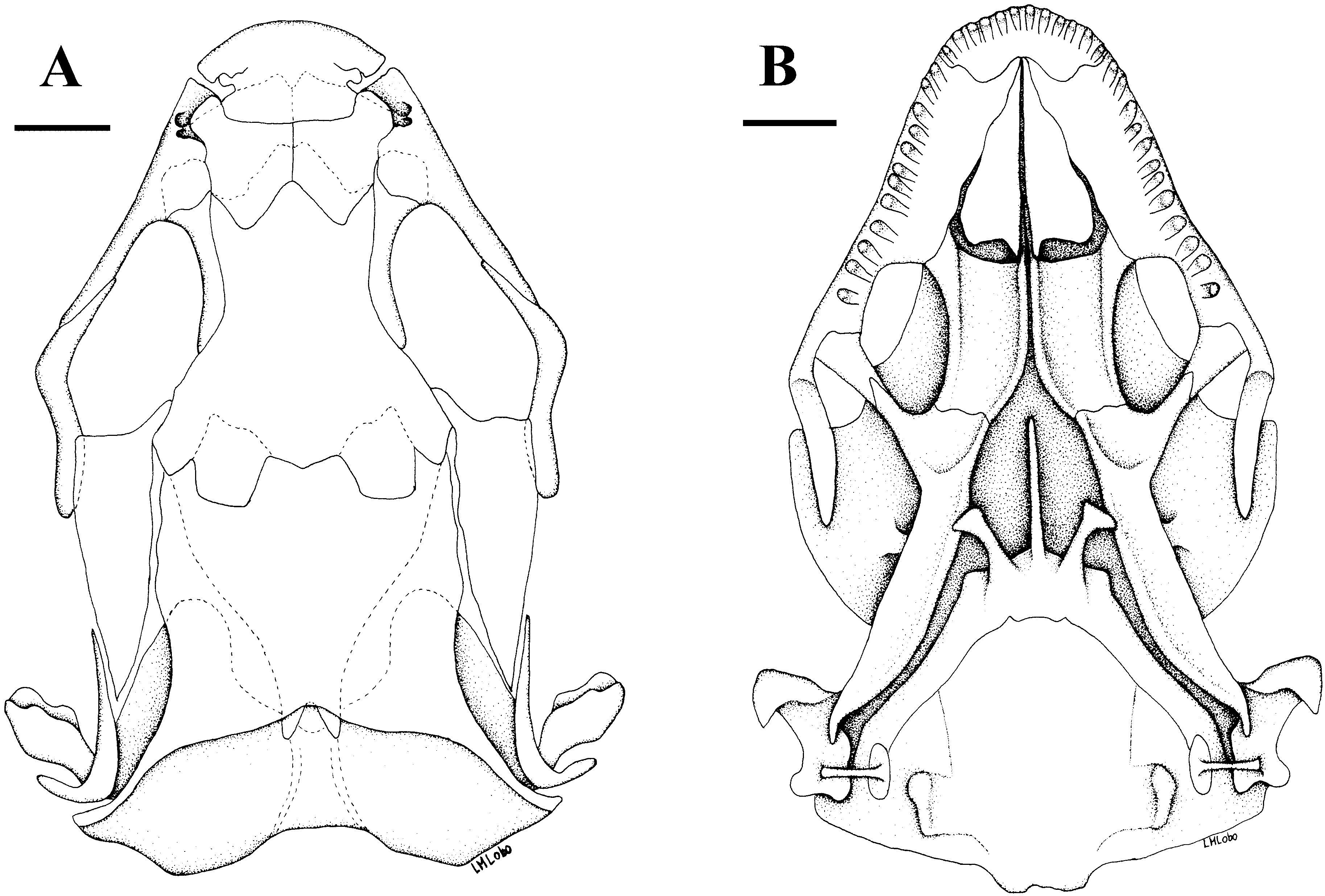
Osteology ( Figs 3A, B View Figure 3 , 4A - D View Figure 4 ): The following description is based on three alizarin prepared skeletons ( MZUSP 66230, 93422, 93423). Premaxillary broader than long, touching but not articulating laterally the maxillary, its posterior border straight, covering the anterior part of nasals; 11 premaxillary conical teeth. Nasals large, longer than wide, wider anteriorly, in broad contact at midline and covering the anterior margin of the frontal. Frontal as long as wide, wider posteriorly, covering parietal by a pair of frontoparietal tabs. Parietal longer than wide, wider posteriorly and covering the lateral margin of the occipital region. Epipterygoid wider above, contacting externally the descending epipterygoid process of the parietal. Maxillary contacting nasal dorsally, almost without overlapping, covering extensively the prefrontal and posteriorly contacting the jugal; 12–13 maxillary teeth.
Prefrontal large, posterior process long, in broad contact with frontal and forming the major part of the dorsal part of the orbital region. Lacrimal absent, apparently fused to the prefrontal and forming a slight median protuberance at the anterior part of the orbita. Postfrontal and postorbital fused in an anteriorly very wide postorbitofrontal, closing posteriorly the orbita and contacting dorsally the frontal and parietal, ventrally the jugal, and externally and posteriorly the squamosal. Squamosal posteriorly curved, fitting at the top of the quadrate. Supratemporal fenestra opened, but constrained anteriorly by the parietal and postorbitofrontal, and posteriorly by the parietal. Supratemporal present, in straight contact with posterior part of parietal. Thirteen scleral ossicles in the eye. Vomer, palatine, pterygoid and ectopterygoid present. Vomer, palatine, premaxillary and maxillary in contact, restricting the fenestra exochoanalis. Infraorbital fenestra large, bordered posteriorly by the ectopterygoid. Stapes rod-like, wider at the base. Sutures between the supraoccipital, exoccipital, basioccipital and otic area of the skull are not as visible as those between the basioccipital and basisphenoid. Processes basipterygoides and parashynoidales are well developed.
In the lower jaw the dentary, articular, splenial, angular, and supra-angular are distinct; there are 14– 15 dentary teeth, conical anteriorly, bicuspid or tricuspid posteriorly.
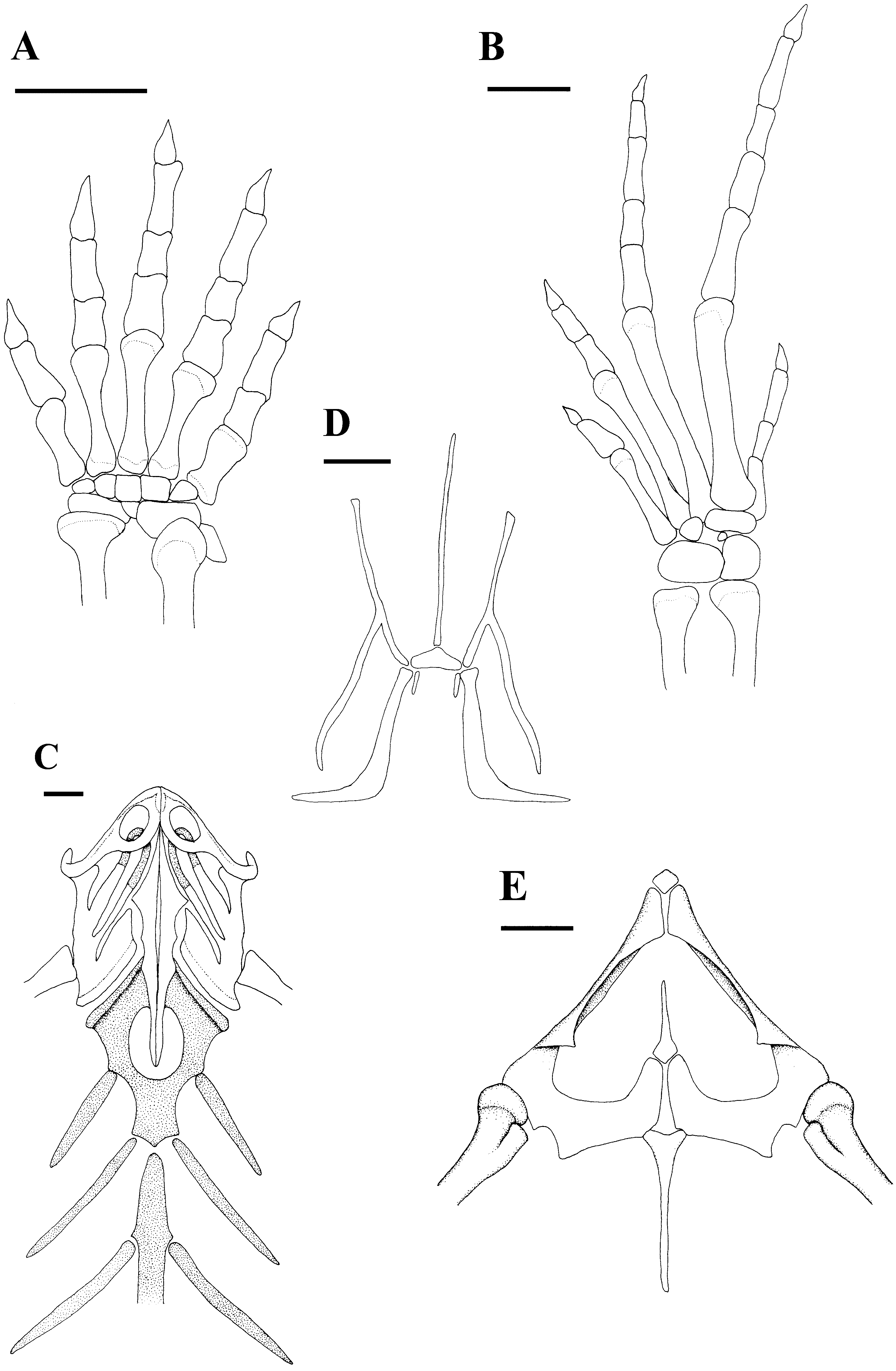
Glossohyal long, separated from basihyal. First ceratobranchial curved and wider posteriorly; hypohyal and ceratohyal present.
Anterior part of clavicle flattened, enlarged, enclosing a fenestra. Interclavicle long, elongate, with lateral processes extremely reduced, reaching the posterior part of sternal fenestra. Coracoid, scapular, and scapulocoracoid fenestrae present in the scapulocoracoid; suprascapula present. Sternum with a large fenestra, two sternal ribs and a xiphisternum receiving two inscriptional ribs. Ilium, ischium and pubis present, the latter with a conspicuous pectinate apophysis. Hypoischium long, almost reaching the preanal border; a small preischium present.
Twenty-five procoelous presacral vertebrae, neural spines low, higher anteriorly; hypopophyses present in the first six vertebrae; zygantrum-zygosphene present. Last presacral vertebra lacking ribs. Two sacral vertebrae.
Humerus and femur slightly larger than radius and ulna and tibia and fibula. Remaining elements of fore and hind limbs as in Figure 4A, B View Figure 4 .
Ecology and distribution: Dryadosaura nordestina occurs in the remnant patches of Atlantic Forest domain ( Ab’Saber, 1977) from the state of Rio Grande do Norte to the northern bank of Rio São Francisco in the state of Alagoas. All specimens were collected on the forest floor, either by hand under logs, or searching leaf litter, or with pitfall trap; we never saw active individuals except for one specimen crossing a trail at Mata da Salva (municipality of Rio Largo, state of Alagoas) at 16.00 hrs. We suggest that their massive head and forelimb musculature might be associated with fossorial and digging habitats. Dryadosaura nordestina is apparently restricted to the Atlantic forests near the coast and does not extend inland to the isolated forest islands of the open semiarid Caatingas of north-eastern Brazil. These forest remnants are relicts from times when the Atlantic forest covered a larger area in the semiarid Caatingas ( Vanzolini, 1981; Rodrigues, 2003). The westernmost record for D. nordestina is Serra dos Cavalos, a forested mountain near the Atlantic forest/ Caatinga transition in the state of Pernambuco.
Phylogenetic analyses
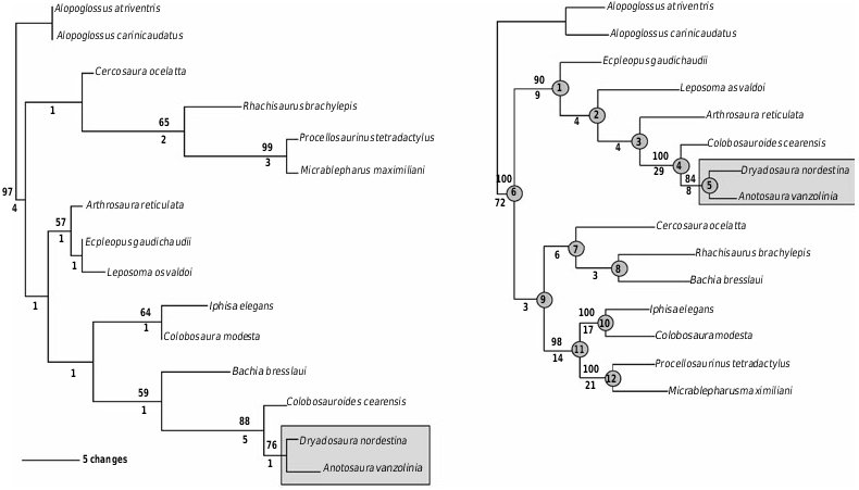
Parsimony analysis to determine the phylogenetic placement of Dryadosaura based on 37 morphological characters (all informative, Table 1) with all states coded as unordered, produced a single most parsimonious tree of 86 steps (CI = 0.60, RI = 0.72) ( Fig. 5A View Figure 5 ). ( Colobosauroides cearensis ( Anotosaura vanzolinia + Dryadosaura nordestina )) are recovered in a well supported clade ( BS = 88; Bremer value = 5.0), with the ( D. nordestina + A. vanzolinia ) clade supported at a level of BS = 76 and Bremer value = 1.0. The Ecpleopini clade was not recovered by the morphological partition alone, but there is no support ( BS <50%; Bremer value = 1.0) for the large clade that includes the ecpleopines C. cearensis , A. vanzolinia , D. nordestina , Arthrosaura reticulata , Ecpleopus gaudichaudii , Leposoma osvaldoi , plus the cercosaurinae Bachia bresslaui and the heterodactylines Iphisa elegans and Colobosaura modesta ( Fig. 5A View Figure 5 ).
Combined analyses of the morphological and molecular partitions resulted in a single most parsimonious tree of 2413 steps and 632 parsimony-informative characters (CI = 0.54, RI = 0.44) ( Fig. 5B View Figure 5 ). The ecpleopines are recovered as a strongly supported clade (node 1: BS = 90; Bremer value = 9; Table 2) related to Cercosaurini, with higher nodal support both for the ( Colobosauroides ( Anotosaura + Dryadosaura nordestina )) clade (node 4: BS = 100; Bremer value = 29), than the nested ( D. nordestina + A. vanzolinia ) clade (node 5: BS = 84; Bremer value = 8).
These analyses also recovered the sister-group relationship between Heterodactylini and Gymnophthalmini (node 11: BS = 98; Bremer value = 14) as well the monophyly of each (node 10: BS = 100; Bremer value = 17 and node 12: BS = 100; Bremer value = 21, respectively; Table 2). The monophyly of Cercosaurini was not recovered, but support for the alternative topology (( Rhachisaurus + Bachia ) Cercosaura ) was very low (node 7: BS <50; Bremer value = 6 and node 8: BS <50; Bremer value = 3). Although the Ecpleopini was recovered as a highly supported monophyletic group (node 1), this was not the case for its sister group when assembling the taxa included in node 9 ( BS <50; Bremer value = 3; Fig. 5B View Figure 5 ).
| T |
Tavera, Department of Geology and Geophysics |
| MZUSP |
Museu de Zoologia da Universidade de Sao Paulo |
No known copyright restrictions apply. See Agosti, D., Egloff, W., 2009. Taxonomic information exchange and copyright: the Plazi approach. BMC Research Notes 2009, 2:53 for further explanation.
|
Kingdom |
|
|
Phylum |
|
|
Class |
|
|
Order |
|
|
Family |
|
|
Genus |