
Echinoderes dalzottoi, Grzelak & Sørensen, 2022
|
publication ID |
https://doi.org/ 10.5852/ejt.2022.844.1949 |
|
publication LSID |
lsid:zoobank.org:pub:193EDD91-B24D-455C-B8AA-8133586A00A1 |
|
DOI |
https://doi.org/10.5281/zenodo.7225402 |
|
persistent identifier |
https://treatment.plazi.org/id/CFE372C3-2BAF-4A6B-B3C4-A0A14551747F |
|
taxon LSID |
lsid:zoobank.org:act:CFE372C3-2BAF-4A6B-B3C4-A0A14551747F |
|
treatment provided by |
Felipe |
|
scientific name |
Echinoderes dalzottoi |
| status |
sp. nov. |
Echinoderes dalzottoi View in CoL sp. nov.
urn:lsid:zoobank.org:act:CFE372C3-2BAF-4A6B-B3C4-A0A14551747F
Figs 8–10 View Fig View Fig View Fig ; Tables 6–7
Diagnosis
Echinoderes with spines in middorsal position on segments 4 and 6, and spines in lateroventral positions on segments 6 to 9. Tubes present in subdorsal, sublateral (might be missing in some specimens) and ventrolateral positions on segment 2, lateroventral positions on segment 5, sublateral positions on segment 8, and laterodorsal positions on segment 9. Sexually dimorphic tubes furthermore present in laterodorsal positions on segment 10 in males; females with fringe-like structure in midlateral positions. Minute scales present on segments 2 to 10, but regular cuticular hairs absent throughout trunk
Etymology
The species is named after Dr Matteo Dal Zotto in recognition of his contributions to kinorhynch taxonomy and ecology.
Material examined
Holotype
NEW ZEALAND • ♀; Pahaua Canyon, stn TAN1004/31; 41.4962° S, 175.6828° E; 730 m b.s.l.; Apr. 2010; NIWA TAN1004 Voyage; soft sediment; NIWA-159403 . Mounted for LM in Fluoromount G on HS slide. GoogleMaps
Paratypes
NEW ZEALAND • 1 ♂; Pahaua Canyon, stn TAN1004/27; 41.4983° S, 175.7043° E; 1013 m b.s.l.; Apr. 2010; NIWA TAN1004 Voyage; soft sediment; NHMD-917147 . Mounted as holotype GoogleMaps • 1 ♂; Honeycomb Canyon, stn TAN1004/58; 41.4080° S, 175.8977° E; 670 m b.s.l.; Apr. 2010; NIWA TAN1004 Voyage; soft sediment; NIWA-159404 . Mounted as holotype GoogleMaps .
Additional material
NEW ZEALAND • 1 ♂; same collection data as for holotype; personal reference collection of MVS. Mounted for SEM GoogleMaps • 1 ♂; Pahaua Canyon, stn TAN1004/27; 41.4983° S, 175.7043° E; 1013 m b.s.l.; Apr. 2010; NIWA TAN1004 Voyage; soft sediment; personal reference collection of MVS. Mounted for GoogleMaps SEM • 1 ♀; Honeycomb Canyon, stn TAN1004/58; 41.4080° S, 175.8977° E; 670 m b.s.l.; Apr. 2010; NIWA TAN1004 Voyage; soft sediment; personal reference collection of MVS. Mounted for SEM GoogleMaps .
Description
GENERAL. Adults with head, neck and eleven trunk segments ( Figs 8–10 View Fig View Fig View Fig ). Overview of measurements and dimensions in Table 6. Distribution of cuticular structures, i.e., sensory spots, glandular cell outlets, spines and tubes, summarized in Table 7. Head morphology could not be examined in detail in any of available specimens.
NECK. Consists of 16 placids. Midventral placid broadest, 13 µm in width and 15 µm in length, whereas all others narrower, measuring 7 µm in width at their bases ( Fig. 8 View Fig ). The trichoscalid plates are well developed ( Fig. 9D View Fig ).
SEGMENT 1. Consists of complete cuticular ring. Sensory spots present in subdorsal, laterodorsal and ventromedial positions. Sensory spots relatively large and without marginal hairs, located on anterior half of segment ( Figs 8A–B View Fig , 9C–D View Fig , 10B–C, E View Fig ). Glandular cell outlet type 1 present in middorsal position and in ventrolateral positions. Cuticular hairs or perforation sites not present. Posterior segment margin almost straight, forming pectinate fringe with short, sawtooth-like fringe tips ( Fig. 10B–C View Fig ).
SEGMENT 2. Consists of complete cuticular ring, with tubes located in subdorsal, sublateral and ventrolateral positions ( Figs 8A–B View Fig , 9C–D View Fig , 10B–C, E View Fig ); sublateral tubes missing in one paratype and two SEM specimens; no sexual or developmental differences explain presence or absence of tubes. Sensory spots of similar sizes as on preceding segment, present in middorsal, laterodorsal and ventromedial positions; ventromedial ones with long marginal hair. Unpaired glandular cell outlet type 1 present in middorsal position and as pair in ventromedial positions. Pachycyclus of anterior segment margin of regular thickness, interrupted in middorsal position. Secondary pectinate fringe present near anterior segment margin of this and following segments, but usually covered by preceding segment. This and following nine segments completely hairless. Cuticular hairs reduced to minute scales distributed around segment ( Fig. 10B–C, E–H View Fig ), emerging through perforation sites; perforation sites easily visible in LM ( Fig. 9C–K View Fig ). Posterior segment margin almost straight, but with rounded midventral extension ( Fig. 10D–E View Fig ); pectinate fringe tips as on preceding segment, except midventral area with slightly narrower fringe tips.
SEGMENT 3. Present segment, and eight remaining ones, consist of one tergal and two sternal plates ( Figs 8A–B View Fig , 9A, D View Fig ). Sensory spots present in subdorsal and midlateral positions. Sensory spots on this and following segments smaller than on preceding segments, all with one long marginal hair. Glandular cell outlets type 1 as on preceding segment. Perforation sites appear as band around segment, interrupted in middorsal and laterodorsal areas and in central part of sternal plate on this and following five segments ( Figs 8A–B View Fig , 9C–D, F–G View Fig , 10F–G View Fig ). Posterior segment margin straight, terminating in pectinate fringe with slightly more slender fringe tips along ventral margin than on preceding segments, otherwise as on preceding segment.
SEGMENT 4. With spine in middorsal position; spine relatively long (51 µm), reaching posterior margin of segment 5 ( Figs 8A View Fig , 10A, F View Fig ). Glandular cell outlets type 1 present in paradorsal and ventromedial positions. No other traits observed. Segment otherwise as segment 3.
SEGMENT 5. With tubes in lateroventral positions ( Figs 8B View Fig , 9D View Fig , 10G View Fig ). Sensory spots present in subdorsal, midlateral and ventromedial positions ( Figs 8A–B View Fig , 9C View Fig , 10F View Fig ). Glandular cell outlets type 1 present in middorsal and ventromedial positions. Perforation sites, secondary fringe and posterior segment margin as on preceding segment.
SEGMENT 6. With spines in middorsal and lateroventral positions ( Fig. 8A–B View Fig ). Middorsal spine, as on segment 4, relatively long (99 µm), reaching well beyond posterior segment margin of segment 8 ( Fig. 10A, I View Fig ). Sensory spots present in paradorsal, subdorsal, midlateral and ventromedial positions ( Figs 8A–B View Fig , 10F–G View Fig ). Glandular cell outlets type 1 present in paradorsal and ventromedial positions. Segment otherwise as segment 5.
SEGMENT 7. With spines in lateroventral positions, and sensory spots in paradorsal, midlateral and ventromedial positions ( Figs 8A–B View Fig , 9G View Fig , 10F–G View Fig ). Glandular cell outlets type 1 present in middorsal position and as pair in ventromedial positions. Tips of pectinate fringe of posterior segment margin slightly shorter and more slender than on preceding segments. Segment otherwise as segment 6.
SEGMENT 8. With spines in lateroventral positions, and tubes in sublateral positions ( Figs 8A–B View Fig , 9E View Fig , 10H View Fig ). Sensory spots present in paradorsal and subdorsal positions. Glandular cell outlets type 1 present in paradorsal and ventromedial positions. Band of perforation site patches interrupted in subdorsal area instead of laterodorsally as on preceding segments. Segment otherwise as segment 7.
SEGMENT 9. With spines in lateroventral positions. Tubes present in laterodorsal positions, but very close to midlateral line ( Figs 8A View Fig , 9E View Fig , 10H–I View Fig ). Sensory spots located in paradorsal, subdorsal and ventrolateral positions ( Figs 8A–B View Fig , 9J View Fig , 10I View Fig ). Glandular cell outlets type 1 present paradorsally and ventromedially. Small, rounded sieve plates located in sublateral positions ( Fig. 9K View Fig ). Perforation sites, secondary fringe and posterior segment margin as on preceding segment.
SEGMENT 10. With well-developed laterodorsal tubes, present in males only, located near posterior segment margin and close to midlateral line ( Figs 8C View Fig , 9H View Fig , 10K View Fig ). Females without tubes, but with fringelike structures present in midlateral positions ( Figs 8A View Fig , 10J View Fig ). Sensory spots present in subdorsal and ventrolateral positions ( Figs 8A–B View Fig , 10J–K View Fig ); subdorsal pair located rather close to paradorsal area. Glandular cell outlets type 1 present as middorsal one, and in ventromedial positions. Perforation sites restricted to paradorsal area, lateral sides of tergal plate, and lateral halves of sternal plates. Posterior segment margin of tergal plate straight, without fringe tips ( Fig. 10J–K View Fig ); margins of sternal plates concave, reaching posterior margin of terminal segment, with short and narrow fringe tips.
SEGMENT 11. With lateral terminal spines ( Figs 8A–B View Fig , 9A View Fig , 10D View Fig ). Females with lateral terminal accessory spines ( Figs 8A–B View Fig , 9I View Fig , 10A, D View Fig ); males with three penile spines ( Fig. 10K View Fig ). Dorsal and ventral penile spines slender and tubular, with dorsal ones much longer than ventral; median spines very stout, coneshaped ( Figs 8C View Fig , 10K View Fig ). Sensory spots present in subdorsal positions. Glandular cell outlet type 1 present middorsally. Segment devoid of characteristic perforation site patches, but very short cuticular hair-like structures covering paradorsal area. Very short fringes covering margins of tergal and sternal plates. Tergal extensions triangular ( Figs 8A, C View Fig , 10J View Fig ). Sternal extensions rounded, not extending beyond tergal extensions ( Figs 8B, D View Fig , 9K View Fig ).
Distribution
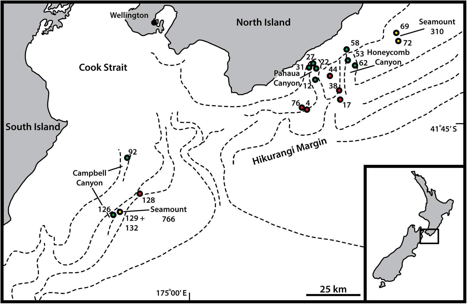
Canyons: Pahaua, Honeycomb, 670–1013 m b.s.l. See Fig. 1 View Fig for a geographic overview of stations and Table 1 View Table 1 for station and specimen information.
Taxonomic remarks on Echinoderes dalzottoi sp. nov.
See taxonomic remarks for E. dalzottoi sp. nov. below, together with remarks for E. leduci sp. nov.


No known copyright restrictions apply. See Agosti, D., Egloff, W., 2009. Taxonomic information exchange and copyright: the Plazi approach. BMC Research Notes 2009, 2:53 for further explanation.
|
Kingdom |
|
|
Phylum |
|
|
Class |
|
|
Order |
|
|
Family |
|
|
Genus |