
Echinoderes multiporus, Yamasaki & Neuhaus & George, 2018
|
publication ID |
https://doi.org/ 10.11646/zootaxa.4387.3.8 |
|
publication LSID |
lsid:zoobank.org:pub:DA75D56E-22BB-487E-8BA3-42C5C98F9E53 |
|
DOI |
https://doi.org/10.5281/zenodo.5952903 |
|
persistent identifier |
https://treatment.plazi.org/id/03CD87E0-2454-FFC8-3D86-2F1009C7FDAF |
|
treatment provided by |
Plazi |
|
scientific name |
Echinoderes multiporus |
| status |
sp. nov. |
Echinoderes multiporus sp. nov.
( Figs 2–7 View FIGURE 2 View FIGURE 3 View FIGURE 4 View FIGURE 5 View FIGURE 6 View FIGURE 7 ; Tables 2, 3)
Diagnosis. Echinoderes with middorsal acicular spines on segments 4, 6, and 8; ventrolateral tubes on segment 2; lateroventral tubes on segment 5; lateroventral acicular spines on segments 6–9; midlateral tubes on segment 10; type-2 glandular cell outlets in subdorsal position on segment 2 and in laterodorsal position on segments 4–9.
Etymology. The species name is composed of the Latin multi (many) and Latin porus (pore), referring to the type-2 glandular cell outlets on most segments.
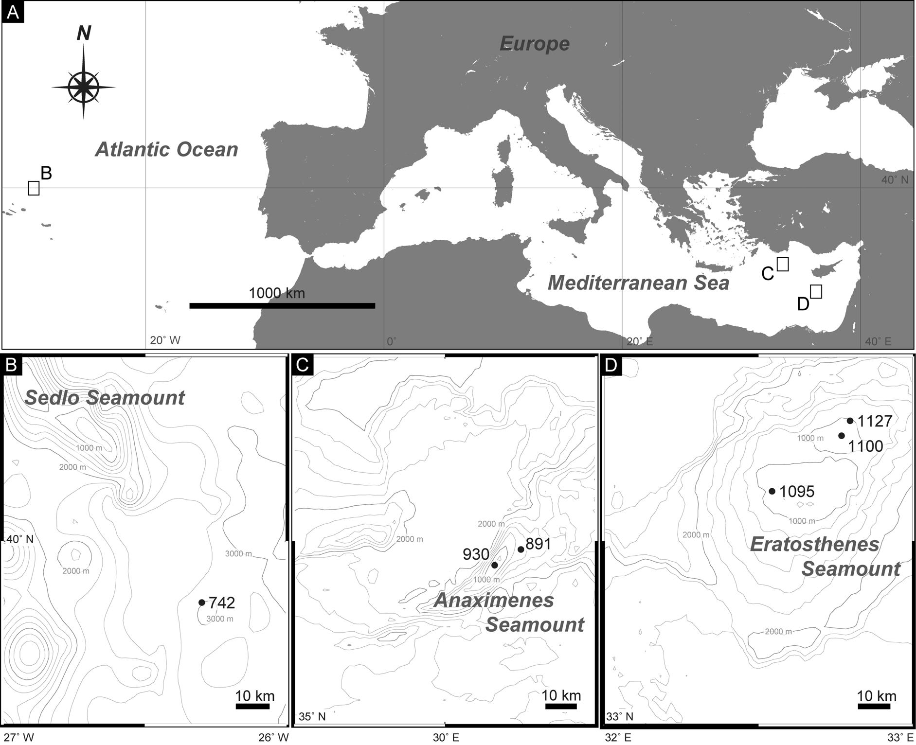
Material examined. Holotype: Adult male (ZMB 11590), collected at station 1127 on the Eratosthenes Seamount ( Fig. 1A, D View FIGURE1 ; Table 1), mounted as glycerol-paraffin slide on a Cobb aluminum frame.
Paratypes: four adult females and two adult males ( ZMB 11591a–11591f), collected at station 1095 on the Eratosthenes Seamount; one adult female and one adult male ( ZMB 11592a, 11592b), collected at station 1100 on the Eratosthenes Seamount ( Fig. 1A, D View FIGURE1 ; Table 1). All paratypes mounted as glycerol-paraffin slides on Cobb aluminum frames.
Additional material: one adult female and three adult males, collected at station 1095 on the Eratosthenes Seamount ( Fig. 1A, D View FIGURE1 ; Table 1), mounted on an aluminum stub for SEM observations (ZMB 11591g –11591j).
Type locality. Eratosthenes Seamount (33°49'49.80"N, 32°47'49.20"E), 991 m depth ( Fig. 1A, D View FIGURE1 ; Table 1).
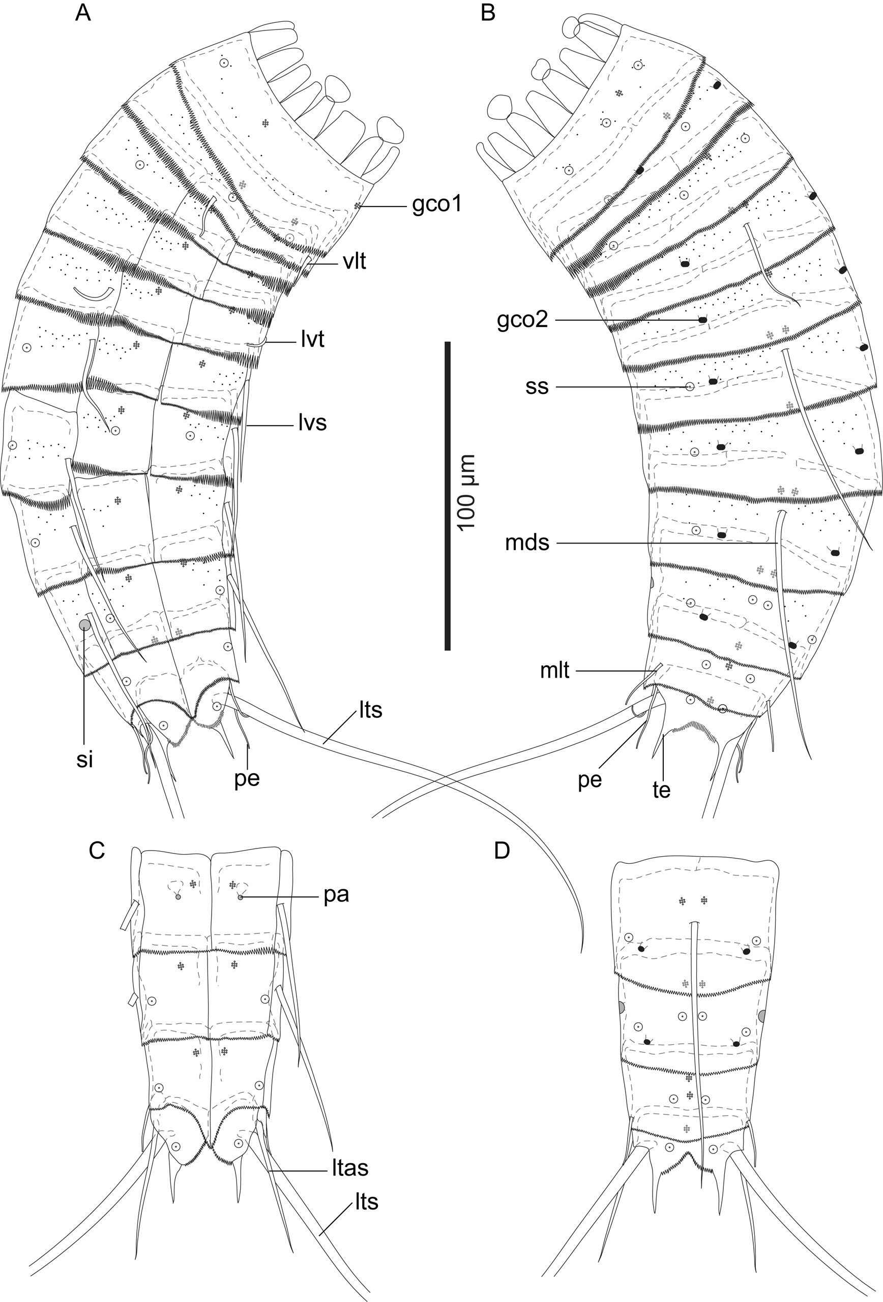
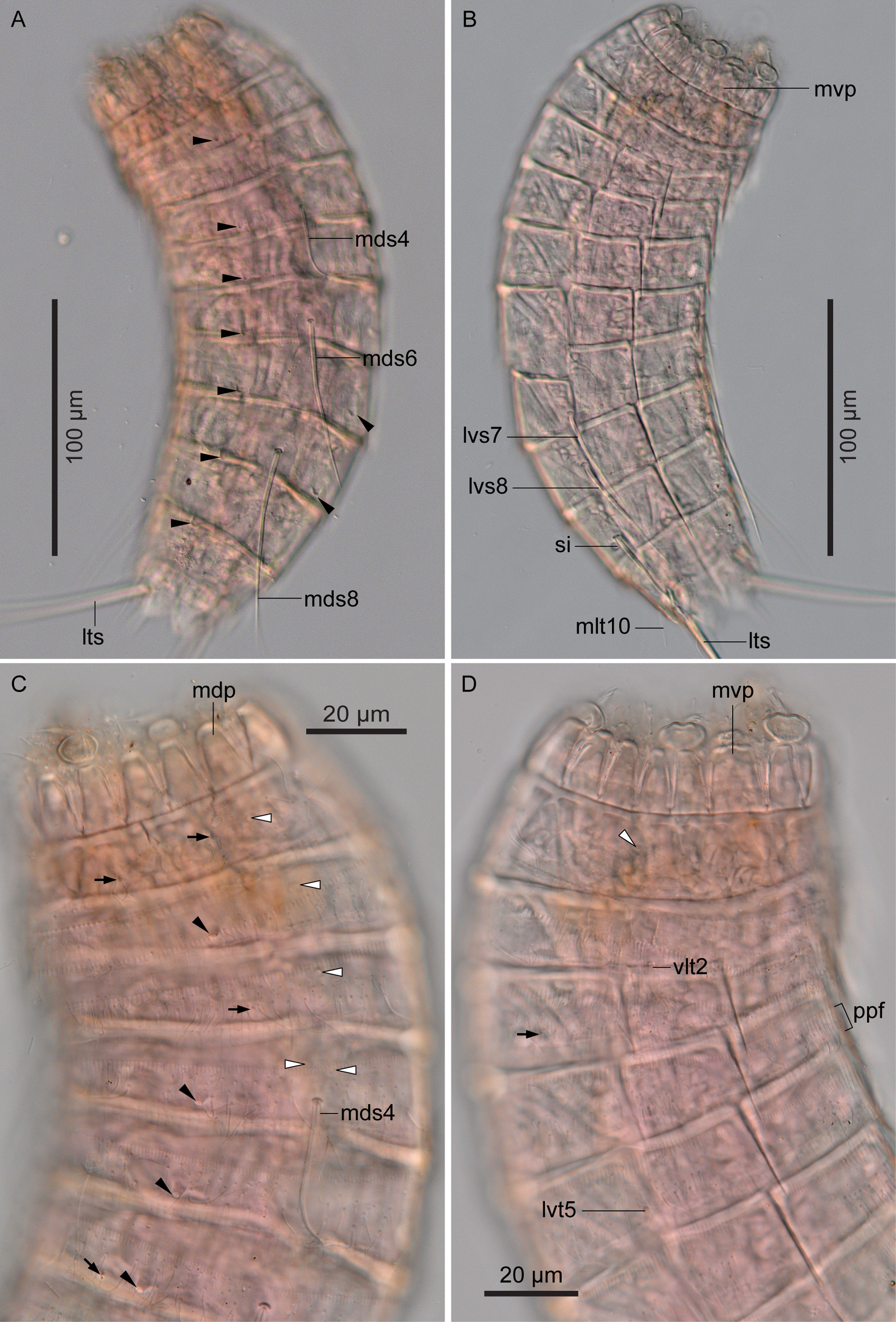
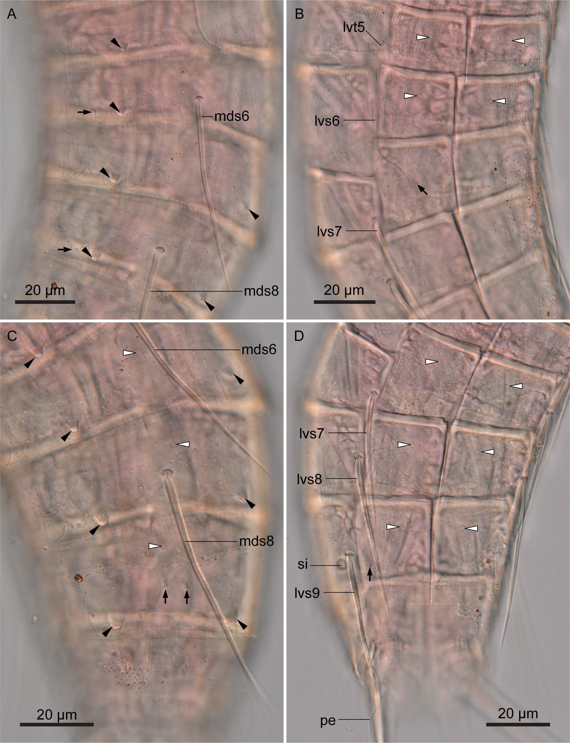
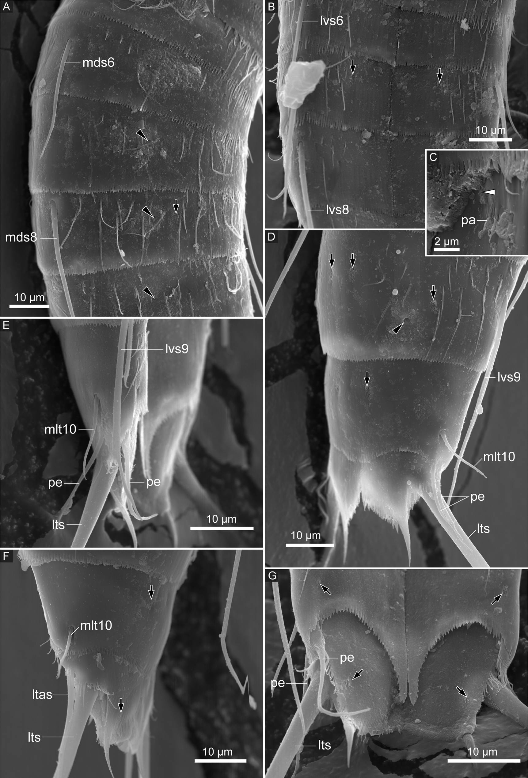
Description. Adult with head, neck, and eleven trunk segments ( Figs 2A, B View FIGURE 2 , 3A View FIGURE 3 , 4A, B View FIGURE 4 ). See Table 2 for measurements. Table 3 indicates the positions of cuticular structures (sensory spots, glandular cell outlets, spines, tubes, and sieve plates).
Head consisting of retractable mouth cone and introvert. Mouth cone with inner oral styles and nine outer oral styles. Introvert composed of one ring of primary scalids, several rings of spinoscalids, and one ring of trichoscalids. Exact number and arrangement of inner and outer oral styles and scalids not traceable because of withdrawn head in all examined specimens.
Neck with 16 placids ( Figs 2A, B View FIGURE 2 , 3B View FIGURE 3 , 4C, D View FIGURE 4 ). Midventral placid broadest. Remaining placids similar in size. Two trichoscalid plates present ventrally and four dorsally.
Segment 1 consisting of complete cuticular ring. This and following nine segments with thick pachycyclus at anterior margin of each segment ( Figs 2A–D View FIGURE 2 , 4A, B View FIGURE 4 ). Pachycyclus interrupted middorsally in segments 2–10 as well as at tergosternal junctions in segments 3–10. Sensory spots located in subdorsal and laterodorsal position ( Figs 2A, B View FIGURE 2 , 3B View FIGURE 3 , 4C View FIGURE 4 ). A couple of hairs present beside each sensory spot ( Fig. 3D View FIGURE 3 ). Several additional hairs sparsely distributed. Type-1 glandular cell outlets present in middorsal and lateroventral position ( Figs 2A, B View FIGURE 2 , 3C View FIGURE 3 , 4C, D View FIGURE 4 ). Posterior part of this and following nine segments with primary pectinate fringe ( Figs 2A–D View FIGURE 2 , 3C–D View FIGURE 3 , 4D View FIGURE 4 , 5A View FIGURE 5 ). Pectinate fringe teeth of primary pectinate fringe on this segment medium in length.
Segment 2 with complete cuticular ring as segment 1. Ventrolateral tubes present ( Figs 2A View FIGURE 2 , 3C View FIGURE 3 , 4D View FIGURE 4 , 5A View FIGURE 5 ). Few cuticular hairs rising from perforation sites in central to posterior area of this and following seven segments ( Figs 3C–D View FIGURE 3 , 4C, D View FIGURE 4 , 5A View FIGURE 5 , 6A–C View FIGURE 6 , 7A, B, D View FIGURE 7 ). Sensory spots present in middorsal, laterodorsal and ventromedial position ( Figs 2A, B View FIGURE 2 , 3C–E View FIGURE 3 ). Type-1 glandular cell outlet present in middorsal and ventromedial position ( Figs 2A, B View FIGURE 2 , 4C View FIGURE 4 ). Type-2 glandular cell outlets in subdorsal position ( Figs 2B View FIGURE 2 , 4A, C View FIGURE 4 ). Primary pectinate fringe on this and following six segments similar to fringe on segment 1, but with slightly longer pectinate fringe teeth in midlateral to ventrolateral area ( Figs 2A View FIGURE 2 , 3C, E View FIGURE 3 ).
Segment 3 and following eight segments consisting of one tergal and two sternal plates. Sensory spots present in subdorsal and sublateral position ( Figs 2A, B View FIGURE 2 , 3E View FIGURE 3 , 4C, D View FIGURE 4 ). Type-1 glandular cell outlets situated in middorsal and ventromedial position ( Figs 2A, B View FIGURE 2 , 4C View FIGURE 4 ).
Segment 4 with middorsal acicular spine ( Figs 2B View FIGURE 2 , 3E View FIGURE 3 , 4A, C View FIGURE 4 ). No sensory spots present. Type-1 glandular cell outlets present paradorsal and ventromedial position ( Figs 2A, B View FIGURE 2 , 4C View FIGURE 4 ). Type-2 glandular cell outlets present laterodorsally ( Figs 2B View FIGURE 2 , 3E, F View FIGURE 3 , 4A, C View FIGURE 4 ).
Segment 5 with lateroventral tubes ( Figs 2A View FIGURE 2 , 4D View FIGURE 4 , 5A View FIGURE 5 , 6B View FIGURE 6 ). Sensory spots absent. Type-1 glandular cell outlets present in middorsal and ventromedial position ( Figs 2A View FIGURE 2 , 6B View FIGURE 6 ). Type-2 glandular cell outlets present in laterodorsal position ( Figs 2B View FIGURE 2 , 4A, C View FIGURE 4 , 6A View FIGURE 6 ).
Segment 6 with middorsal and lateroventral acicular spines ( Figs 2A, B View FIGURE 2 , 4A View FIGURE 4 , 5A View FIGURE 5 , 6A–C View FIGURE 6 , 7A, B View FIGURE 7 ). Sensory spots present in midlateral position ( Figs 2A, B View FIGURE 2 , 6A View FIGURE 6 ). Type-1 glandular cell outlets present paradorsally and ventromedially ( Figs 2A, B View FIGURE 2 , 6B View FIGURE 6 ). Type-2 glandular cell outlets present in laterodorsal position ( Figs 2B View FIGURE 2 , 4A, C View FIGURE 4 , 6A, C View FIGURE 6 ).
Segment 7 with lateroventral acicular spines ( Figs 2A View FIGURE 2 , 4B View FIGURE 4 , 6B, D View FIGURE 6 ). Sensory spots present in ventromedial position ( Figs 2B View FIGURE 2 , 6B View FIGURE 6 , 7B View FIGURE 7 ). Midlateral sensory spots confirmed for six out of twelve specimens. Type-1 glandular cell outlets present middorsally and ventromedially ( Figs 2A, B View FIGURE 2 , 6C, D View FIGURE 6 ). Type-2 glandular cell outlets present in laterodorsal position ( Figs 2B View FIGURE 2 , 4A View FIGURE 4 , 6A, C View FIGURE 6 , 7A View FIGURE 7 ).
Segment 8 similar to segment 6, except for the presence of ventromedial papillae in females ( Figs 2A–D View FIGURE 2 , 4A, B View FIGURE 4 , 5B View FIGURE 5 , 6A, C, D View FIGURE 6 , 7A–C View FIGURE 7 ). Papillae visible only in SEM, but appearing as funnel-shaped subcuticular structures without large pore visible below papillae in LM ( Fig. 5B View FIGURE 5 ).
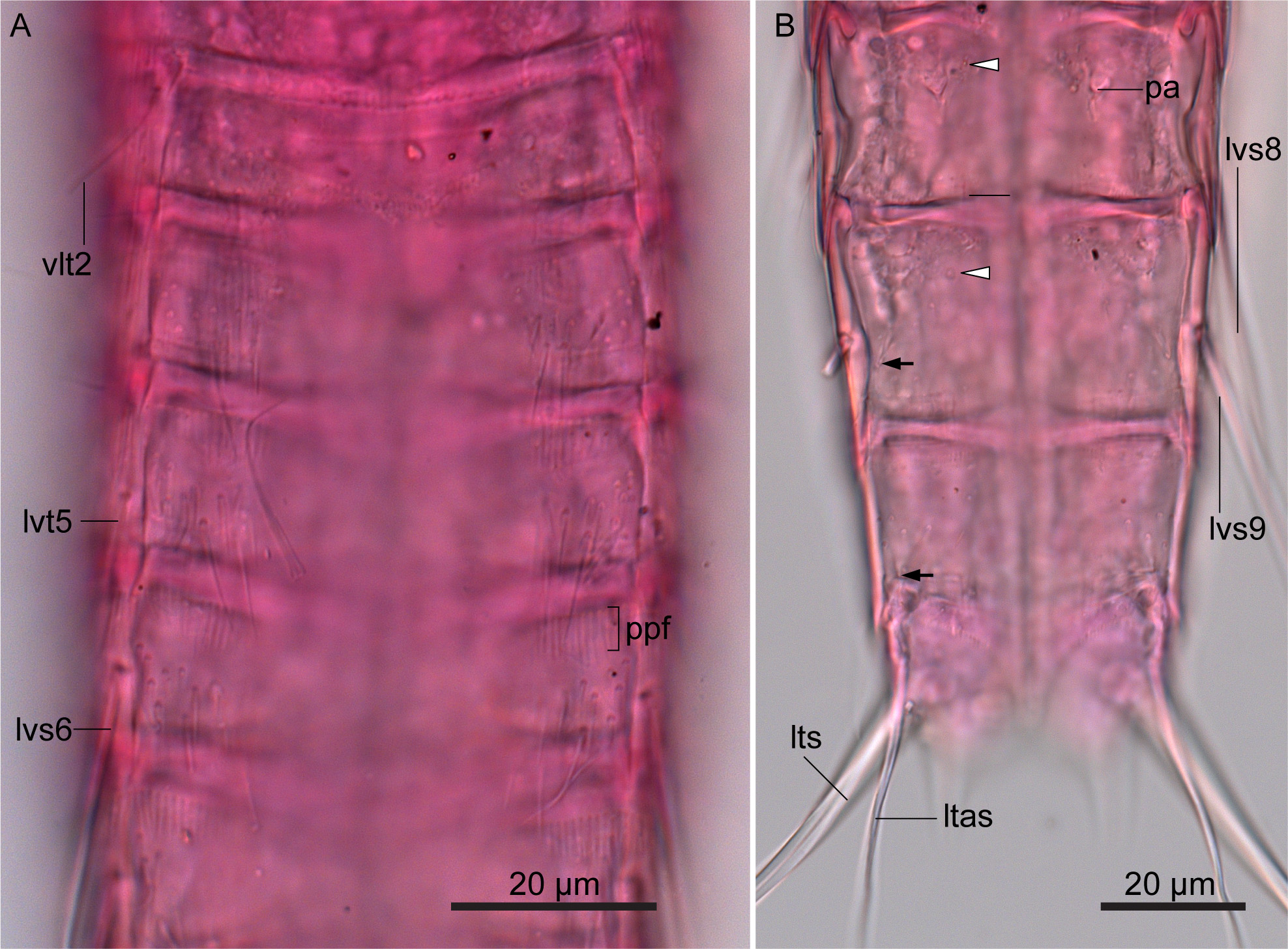
Segment 9 with lateroventral acicular spines ( Figs 2A, C View FIGURE 2 , 5B View FIGURE 5 , 6D View FIGURE 6 , 7D, E View FIGURE 7 ). Paradorsal, midlateral, and ventrolateral sensory spots present ( Figs 2A–D View FIGURE 2 , 5B View FIGURE 5 , 6C, D View FIGURE 6 , 7D View FIGURE 7 ). Type-1 glandular cell outlets present in paradorsal and ventromedial position ( Figs 2A–D View FIGURE 2 , 5B View FIGURE 5 , 6C, D View FIGURE 6 ). Type-2 glandular cell outlets present laterodorsally ( Figs 2B, D View FIGURE 2 , 4A View FIGURE 4 , 6C View FIGURE 6 , 7A, D View FIGURE 7 ). Small rounded sieve plates present in lateral accessory position ( Figs 2A, D View FIGURE 2 , 4B View FIGURE 4 , 6D View FIGURE 6 ). Primary pectinate fringe with quite shorter pectinate fringe teeth than fringe on preceding segment.
Segment 10 with midlateral tubes ( Figs 2B, D View FIGURE 2 , 4B View FIGURE 4 , 7D–F View FIGURE 7 ). No cuticular hairs present. Subdorsal and ventrolateral sensory spots present ( Figs 2A–D View FIGURE 2 , 5B View FIGURE 5 , 7D, F, G View FIGURE 7 ). Two type-1 glandular cell outlets aligned middorsally ( Fig. 2B, D View FIGURE 2 ). Additional pair of type-1 glandular cell outlets present in ventromedial position. Primary pectinate fringe similar to preceding segment.
Segment 11 with lateral terminal spines ( Figs 2A–D View FIGURE 2 , 4A, B View FIGURE 4 , 5B View FIGURE 5 , 7D–G View FIGURE 7 ). Two pairs of thin and long penile spines present in males, and one pair of lateral terminal accessory spines present in females ( Figs 2A–D View FIGURE 2 , 5B View FIGURE 5 , 6D View FIGURE 6 , 7D–G View FIGURE 7 ). Subdorsal and ventromedial sensory spots present ( Figs 2A–D View FIGURE 2 , 7F, G View FIGURE 7 ). Type-1 glandular cell outlet present middorsally ( Fig. 2B, D View FIGURE 2 ). Posterior edges of sternal plates rounded with thin pectinate fringe teeth ( Figs 2A, C View FIGURE 2 , 7G View FIGURE 7 ). Posterior edge of tergal plate protruding subdorsally, forming long pointed tergal extensions ( Figs 2B, D View FIGURE 2 , 7D, F, G View FIGURE 7 ).
Remarks. Based on the spine and tube pattern having middorsal acicular spines only on segments 4, 6, and 8, lateroventral acicular spines on segments 6–9, ventrolateral tubes on segment 2, lateroventral tubes on segment 5, and midlateral tubes on segment 10, and lacking lateral accessory tubes on segment 8, Echinoderes multiporus sp. nov. is similar to E. bermudensis Higgins, 1982 , E. joyceae Landers & Sørensen, 2016 , and Echinoderes apex Yamasaki et al., 2018 ( Higgins 1982; Landers & Sørensen 2016; Yamasaki et al. in press). However, the new species can be easily distinguished from the above mentioned three species by the presence of type-2 glandular cell outlets on segments 2 and 3–9 (type-2 glandular cell outlets are present on segments 2, 6, and 8 in E. joyceae and the undescribed species, but absent in E. bermudensis ). Echinoderes multiporus sp. nov. is the only Echinoderes species with the laterodorsal type-2 glandular cell outlets on most of the segments.
The presence of only two pairs of penile spines found in males of Echinoderes multiporus sp. nov. is also the rare character among the congeners. Males of Echinoderes species usually have three pairs of penile spine, whereas two pairs of penile spines have been found in the following ten species together with E. multiporus sp. nov.: Echinoderes . aureus Adrianov et al., 2002 ; Echinoderes . astridae Sørensen, 2014; Echinoderes . cavernus Sørensen et al., 2000; Echinoderes kristenseni Higgins, 1985 ; Echinoderes lanceolatus Chang & Song, 2002; Echinoderes newcaledoniensis Higgins, 1967 ; Echinoderes . marthae Sørensen, 2014; Echinoderes maxwelli (Omer-Cooper, 1957) ; Echinoderes remanei (Blake, 1930) ; and Echinoderes . rex Lundbye et al., 2011 (see Higgins 1964, 1967, 1985; Sørensen et al. 2000; Adrianov et al. 2002; Chang & Song 2002; Lundbye et al. 2011; Sørensen 2014). Despite the presence of only two pairs of penile spines in these species, the species do not agree in most of the other morphological characters, i.e., the shape and the patterns of spines, tubes, and type-2 glandular cell outlets. It can be supposed that only two pairs of penile spines evolved repeatedly independently within the genus. However, the morphological evolution of Echinoderes is still open to question due to the lack of knowledge about the phylogenetic relationships within the genus.
| ZMB |
Museum f�r Naturkunde Berlin (Zoological Collections) |
No known copyright restrictions apply. See Agosti, D., Egloff, W., 2009. Taxonomic information exchange and copyright: the Plazi approach. BMC Research Notes 2009, 2:53 for further explanation.