
Enochrus (Holcophilydrus) simulans ( Sharp, 1873 )
|
publication ID |
https://doi.org/ 10.5281/zenodo.4272324 |
|
DOI |
https://doi.org/10.5281/zenodo.4334966 |
|
persistent identifier |
https://treatment.plazi.org/id/039A87CB-FFA2-4955-FEAF-FB84FEA9EDA0 |
|
treatment provided by |
Felipe |
|
scientific name |
Enochrus (Holcophilydrus) simulans ( Sharp, 1873 ) |
| status |
|
Enochrus (Holcophilydrus) simulans ( Sharp, 1873) View in CoL
( Figs. 1B View Fig , 13–18 View Fig View Fig View Fig View Fig View Fig View Fig , 28 View Fig A–C, 66A, C, 67A)
Material examined. JAPAN: HONSHÛ: Shimane-ken : 1 L2, Hii-kawa river , Nishidai-bashi bridge, Hikawa-chô, 14.x.2007, MH ; 1 L1, 1 L2, mouth of Kando-gawa river , Izumo-shi, 10.ix.2007, MH ; 1 L3, Nakanosu , Nadabunchô, Izumo-shi (fallow paddy field), 23.vii.2007, MH ; 2 L3, Okinoshima , Sono-chô, Izumo-shi (artificial pond for conservation of the biotope), 11.v.2007, MH ; 1 L3, same locality, 23.vii.2007 (fixed), MH leg. & reared ; 1 L3, same locality, 13.ix.2007, MH ; 2 L3, Okinoshima , Sono-chô, Izumo-shi (paddy field), 10.vi.2007, MH ; 37 L1, 4 L2, same locality, 8.v.2008, MH ; 1 L2, Shakunouchi-kôen , Kisuki-chô, Un-nan-shi, 23.ix.2007, MH .
General morphology. Third instar. Colour. Head and sclerotised parts light yellowish brown; membranous parts milky white ( Fig. 1B View Fig ).
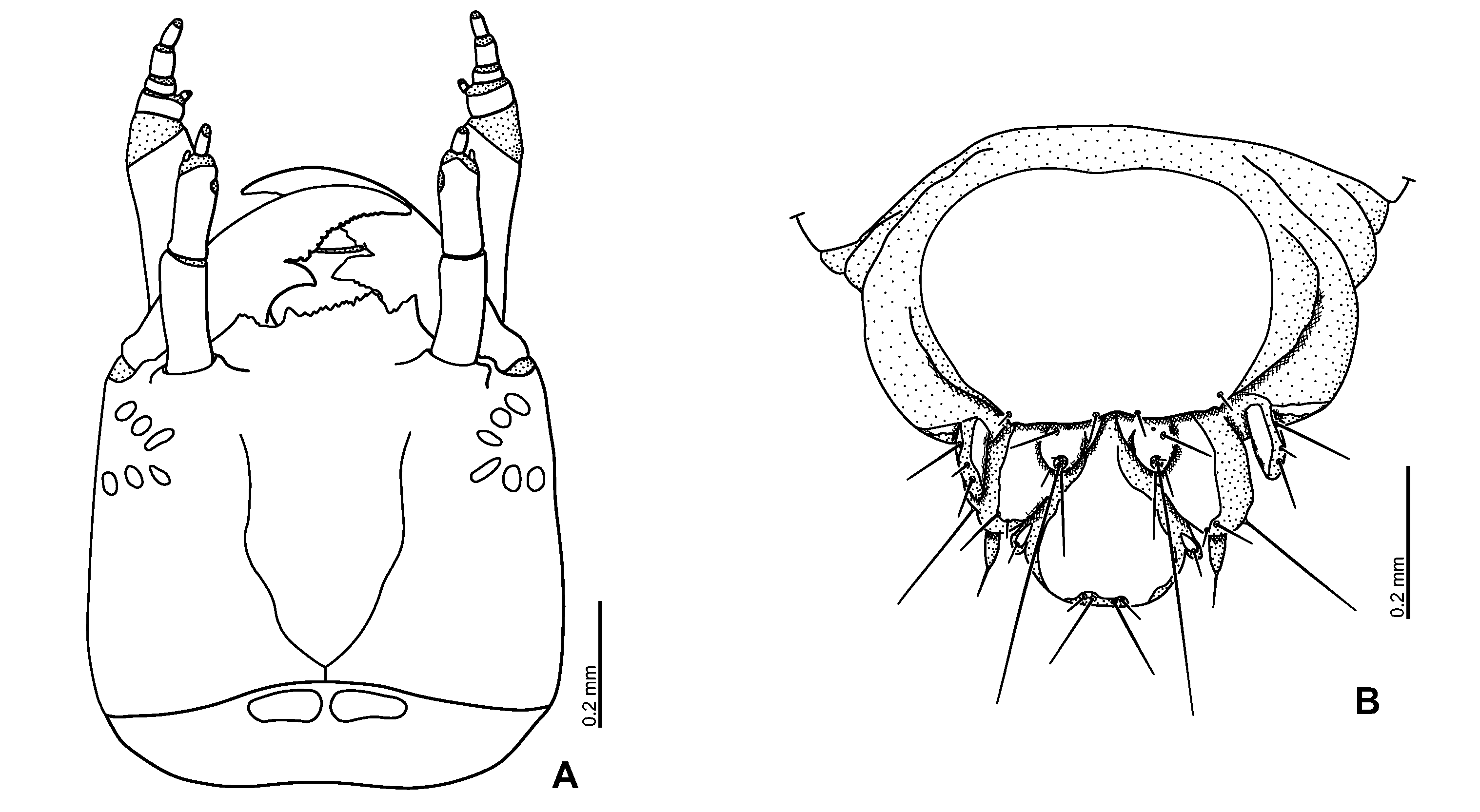
Head ( Figs. 16A View Fig , 17A View Fig ). Frontal lines almost V-shaped. Nasale serrate and with one tooth on left side. Right epistomal lobe strongly projecting, projecting as far as nasale; left lobe weakly projecting than right lobe, not projecting further than nasale. Lateral portion of anterior margin of epistome emarginate.
1 Based on all instars of E. (H.) simulans and E. (M.) japonicus , and third instar of E. (H.) umbratus . Enochrus (Lumetus) bicolor ( Fabricius, 1792) was excluded from the key.
Antenna ( Fig. 17B View Fig ) short, rather slender. Scape about as long as pedicel. Pedicel with inner membranous area completely surrounded by sclerite on apical third.
Mandibles ( Figs. 17 View Fig C–D) asymmetrical; inner edges of distal tooth and anterior part serrate; right mandible with two large inner teeth, left mandible with one large inner tooth.
Maxilla ( Figs. 17 View Fig E–F): Maxillary palpomere 1 about as long as palpomere 4, palpomere 2 the shortest, palpomere 3 longer than palpomere 4; palpomere 2 moderately wider than palpomere 3.
Labium ( Figs. 17 View Fig G–H): Mentum with small cuticular spines on basal half of dorsal surface. Labial palpi longer than prementum, covered with narrow cuticular spines on intersegmental membrane between palpomeres 1 and 2, and on apical and outer parts of palpomere 2.
Abdomen. Abdominal segments 2 to 7 similar to segment 1 except anteromedian part with only one small sclerite, lateral sclerites narrow, very small; segments 3 to 7 with spinose prolegs, spines of prolegs stout, strongly curved apically ( Fig. 28C View Fig ).
Spiracular atrium ( Fig. 16B View Fig ): Segment 8 with large, oval dorsal plate, posterior edge of plate almost rounded, bearing four very weak projections, each projection with one moderately short stout seta; procercus incompletely sclerotised, with two rather long and one short setae.
Second instar. Similar to third instar larva, more weakly sclerotised than third instar larva.
Head. Frontal lines more distinct than those in third instar.
Antenna short, slightly more slender than in third instar ( Fig. 15B View Fig ).
Maxilla: Maxillary palpomere 3 slightly shorter than palpomere 4 ( Figs. 15 View Fig F–G).
First instar. Similar to second instar larva, more weakly sclerotised than second instar larva.
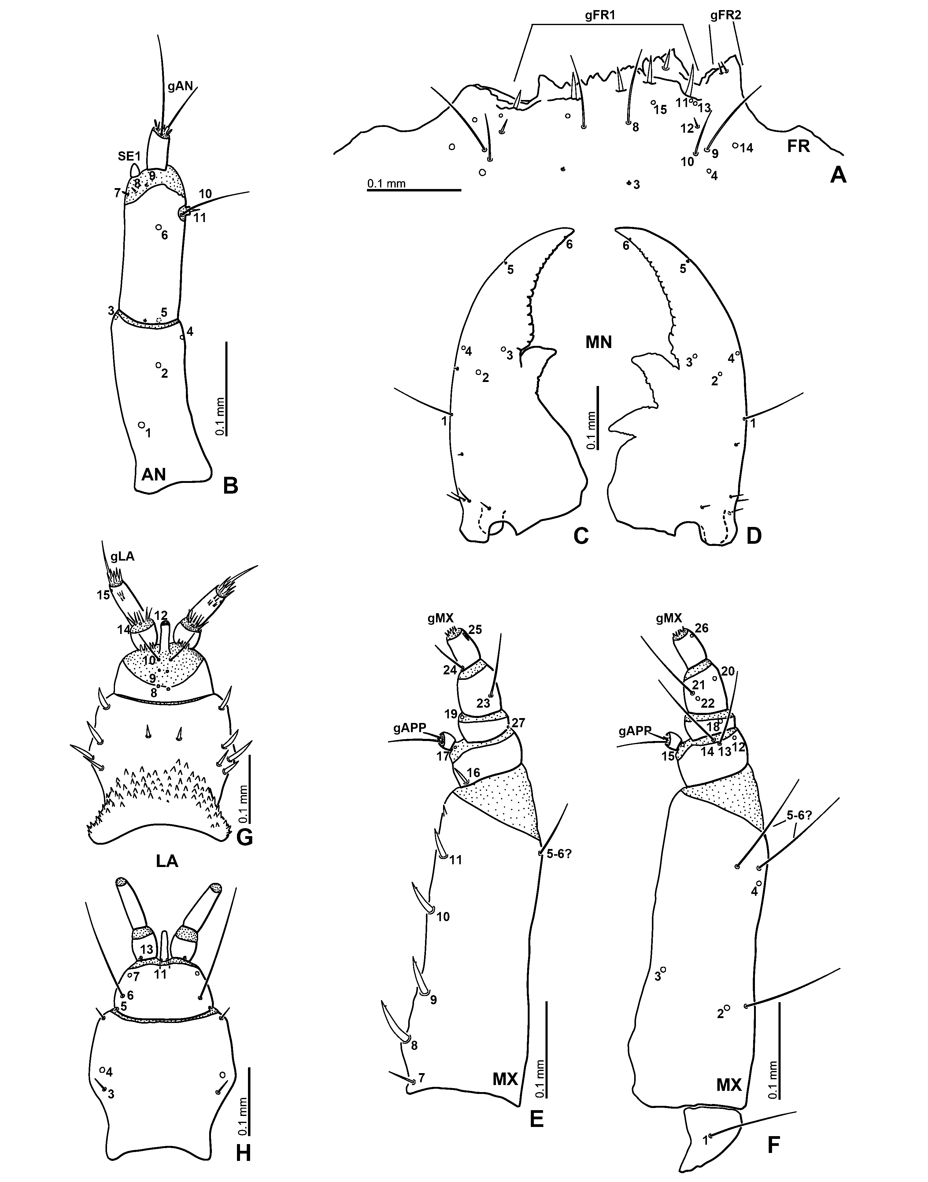
Head ( Fig. 13 View Fig ). Right epistomal lobe projecting further than nasale; left epistomal lobe weakly projected.
Antenna short, stout ( Fig. 14A View Fig ). Scape shorter than pedicel.
Maxilla ( Figs. 14 View Fig D–E): Maxillary palpomere 3 about as long as palpomere 4.
Labium ( Figs. 14 View Fig F–G): Mentum subquadrate, slightly wider than prementum; small cuticular spines on dorsal surface smaller than those of second and third instars.
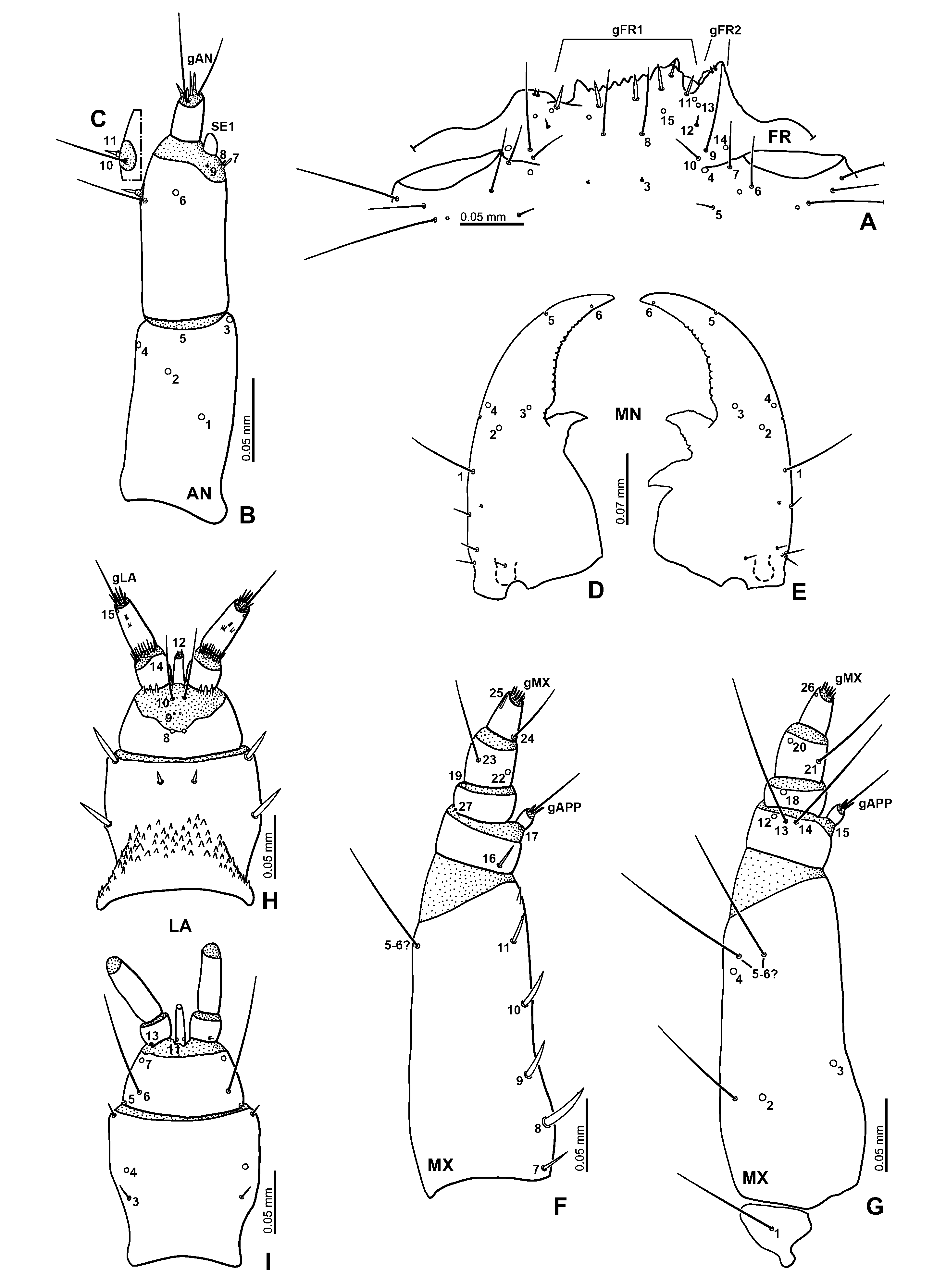
Primary chaetotaxy of head. Frontale altogether with 42 sensilla ( Figs. 13A, C View Fig ). Short seta FR 5 and moderately long seta FR 6 behind antennal socket. Second seta from right one of gFR1 shorter than other ones. Anterior margin of epistomal lobes with six setae altogether (gFR2); right lobe with two short setae; left lobe with four short setae, the setae shorter than those on right lobe. Two setae ( FR 9 and FR 10) and one pore-like sensillum ( FR 14) situated medioanteriorly to antennal socket; FR 9 long; FR 10 short. Each epistomal lobe with three sensilla; location of setae asymmetrical; FR 12 between FR 11 and FR 13 on left side; FR 13 between FR 12 and FR 11, FR 12 behind FR 13 on right side.
Parietale ( Figs. 13 View Fig A–B): PA 1–2 and PA 4–5 forming a longitudinal row; PA 3 located laterally of line connecting PA 1 and PA 5. PA 10 situated laterally of line connecting PA 7 and PA 8. PA 11 short seta. PA 14 rather short seta; PA 13, PA 16 and PA 18 rather long setae. PA 23 more distant from PA 24–25 than the latter from each other.
Antenna ( Fig. 14A View Fig ): AN 2 on distal 0.18 of antennomere 1. Setae AN 10–11 on inner membranous area surrounded by sclerite. Apical sensilla (gAN) with one long seta, two rather long setae, and a few short sensilla.
Maxilla ( Figs. 14 View Fig D–E): MX 2 located ventrally on basal 0.31 of sclerite; MX 3 on basal 0.38 of inner face. MX 23 at midlength of outer face of sclerite; MX 20 on lateral face of apical part; MX 21–22 on inner part of sclerite; MX 22 behind MX 21.
Labium ( Figs. 13B View Fig , 14 View Fig F–G): LA 8 situated dorsally at midwidth on posterior third of prementum, on borderline of sclerite and membranous area between prementum and palpi.Apical membranous area of palpomere 2 with one rather long setae and several short setae (gLA).
Secondary chaetotaxy of head. Second instar. Frontale ( Figs. 18 View Fig A–C): One rather short secondary seta located anteriorly to FR 1.
Parietale ( Figs. 18 View Fig A–C): One very small secondary sensilla close to PA 5, sometimes absent. One to three rather short secondary setae along frontal line, between PA 6 and PA 7 but mesally of line connecting PA 6 and PA 7. Two rather short secondary setae behind outer part of antennal socket; one between PA 8 and PA 9, close to PA 8; another one situated more medially than PA 8. One pore-like secondary sensillum and one rather short secondary seta close to outer margin of antennal socket, seta situated more mesally than pore-like sensillum. One short secondary seta behind PA 21; one short secondary seta behind PA 15; one rather long secondary seta situated slightly mesally to PA 13; two rather short secondary setae situated anteriorly to PA 16–17; one rather long secondary seta located anteriorly to PA 18; one rather long secondary seta situated ventrally between PA 18 and PA 28, close to PA 18.
Antenna: Antennal sensorium ( SE 1) proportionally smaller than in first instar ( Fig. 15B View Fig ).
Mandible ( Figs. 15 View Fig D–E): Outer face of mandible with two small secondary setae; one on median part, close to MN 4; one on basal fourth; four to five rather short secondary setae in basal part of mandible; one in basal fourth, others located more basally.
Maxilla ( Figs. 15 View Fig F–G): Outer face of stipes with two long secondary setae; one on apical part of sclerite, the other on basal third of sclerite.
Labium: Dorsal surface ( Fig. 15H View Fig ) with two rather short, stout secondary setae on lateral face; one pair of short, stout secondary setae situated anteromedially, close to distal margin; ventral surface ( Fig. 15I View Fig ) with one short secondary seta in each anterior corner.
Third instar. Similar to second instar larvae.
Parietale ( Figs. 18 View Fig D–F): Three to five rather short secondary setae between PA 6 and PA 7, on line connecting PA 6 and PA 7.
Antenna ( Figs. 17B View Fig ): Pedicel with one very small, indistinct secondary sensillum situated dorsally on basal margin of sclerite. Antennal sensorium ( SE 1) proportionally smaller than in second instar.
Labium: Dorsal surface of mentum ( Fig. 17G View Fig ) with four to five rather short, stout secondary setae laterally; one pair of short, stout secondary setae medioanteriorly.
Habitat. Standing water. Larvae were found in water, they seem to prefer shallow and muddy areas ( HAYASHI 2009a).
Identification. Only the adults of E. simulans occurred in the sampling sites, no other closely related species of Enochrus or other Acidocerini was found during the intensive survey by the second author. Therefore, we identified the Enochrus larvae collected as these sites as E. simulans .


| MN |
Museu Nacional, Universidade Federal do Rio de Janeiro |
No known copyright restrictions apply. See Agosti, D., Egloff, W., 2009. Taxonomic information exchange and copyright: the Plazi approach. BMC Research Notes 2009, 2:53 for further explanation.